Laser Surgery for Glaucoma: Types, Benefits, Risks & Recovery
What laser treatment does, who may benefit, and how ophthalmologists select between trabeculoplasty, iridotomy, and cyclophotocoagulation.
What is laser surgery for glaucoma?
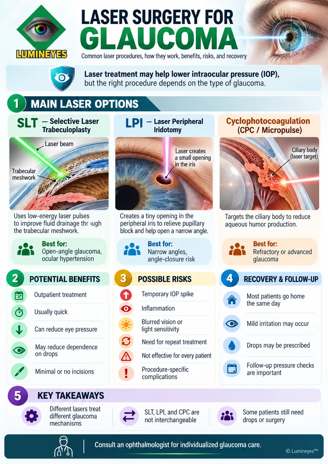
Laser surgery for glaucoma uses focused laser energy to lower intraocular pressure (IOP) by improving aqueous humor drainage, relieving a specific anatomical blockage, or reducing fluid production. The main procedures are selective laser trabeculoplasty (SLT), laser peripheral iridotomy (LPI), and cyclophotocoagulation (CPC). Treatment depends on glaucoma type, anatomy, severity, and target pressure.
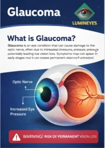
Glaucoma is a group of optic neuropathies characterized by progressive damage to the optic nerve, frequently associated with elevated IOP. Some patients develop glaucoma even when measured pressure is within the statistical normal range. The goal of treatment is to preserve remaining vision by achieving an individualized target pressure—not to restore optic nerve tissue that has already been lost.
Aqueous humor normally passes through the anterior chamber and leaves primarily through the trabecular meshwork at the drainage angle. Different laser procedures address different parts of this system. They are not interchangeable treatments.
For foundational patient education, see the American Academy of Ophthalmology's overview of glaucoma, including its explanation of laser treatment and long-term monitoring.
Main types of glaucoma laser surgery
The distinction between procedures begins with the tissue being treated. A laser that improves trabecular outflow is not the same as a laser that creates an iris opening or acts on the ciliary body.
Selective Laser Trabeculoplasty
SLT treats pigmented structures in the trabecular meshwork to improve aqueous outflow and lower IOP. It may be offered as first-line treatment or used alongside glaucoma medications.
Typical use: Primary open-angle glaucoma and ocular hypertension; selected secondary open-angle glaucomas require individualized assessment.
Laser Peripheral Iridotomy
LPI creates a small opening in the peripheral iris to relieve pupillary block. This helps equalize pressure across the iris and may permit a previously crowded drainage angle to open.
Typical use: Pupillary-block angle closure and selected eyes at risk of angle closure. Indication depends on detailed angle examination.
Cyclophotocoagulation
CPC directs laser energy toward the ciliary body to reduce aqueous production. Continuous-wave and micropulse approaches have different treatment characteristics and clinical considerations.
Typical use: Selected eyes needing additional pressure control, often including refractory or advanced glaucoma. Indications vary by technique and patient.
How the laser techniques differ
| Procedure | Main target | Principal objective | Typical consideration |
|---|---|---|---|
| SLT | Trabecular meshwork | Enhance conventional aqueous outflow | Open-angle disease with an accessible drainage angle |
| LPI | Peripheral iris | Relieve pupillary block | Pupillary-block angle closure or appropriately selected narrow angles |
| CPC | Ciliary body | Reduce aqueous humor production | Cases where a ciliary-body laser approach is clinically appropriate |
Some patients need lens extraction, minimally invasive glaucoma surgery (MIGS), trabeculectomy, drainage-device surgery, or a combination of approaches instead of—or after—laser treatment. These are distinct interventions selected according to angle anatomy, optic nerve damage, IOP targets, and prior response.
Which glaucoma laser procedure is best?
The best option is determined by the diagnosis, not by which laser technology is newest. The ophthalmologist evaluates the drainage angle and optic nerve, establishes a target IOP, and weighs expected pressure reduction against the patient's overall risk profile.
Before recommending laser treatment
- Glaucoma subtype: Open-angle, angle-closure, secondary glaucoma, or ocular hypertension.
- Angle anatomy: Gonioscopy assesses whether the trabecular outflow structures are accessible or blocked.
- Baseline and target IOP: Current pressure, trends, and treatment goals are considered together.
- Optic nerve status: Clinical examination, imaging, and visual field findings help determine severity.
Factors that change the plan
- Prior pressure response to eye drops or laser procedures.
- Inflammation, angle pigmentation, corneal clarity, and past ocular surgery.
- Lens-related crowding, peripheral anterior synechiae, or other anatomical mechanisms.
- Adherence to medications and ability to complete follow-up measurements.
The AAO's patient guide to laser iridotomy provides further information about how LPI works and when ophthalmologists consider it.
What happens during glaucoma laser treatment?
Many glaucoma laser procedures are performed in an outpatient setting using anesthetic drops and a contact lens or other specialized delivery system when indicated. The experience differs by the laser type, individual eye, and whether treatment takes place in an office or operating-room setting.
Confirm the diagnosis, pressure goal, prior history, angle findings, and planned procedure.
Instill the necessary eye drops and explain fixation, possible light flashes, and pressure sensations.
The ophthalmologist directs laser energy to the specified target tissue. Treatment time varies by method.
Check early eye pressure when appropriate, review symptoms, and schedule follow-up based on the procedure.
Recovery and when results appear
Patients commonly return home the same day after office-based SLT or LPI. Mild discomfort, temporary blurring, or light sensitivity can occur. Eye drops may be prescribed according to the procedure and examination findings. The pressure-lowering effect of SLT may develop over several weeks; LPI is aimed primarily at relieving a defined anatomical blockage. CPC recovery and pressure response may require a different follow-up schedule.
Follow-up remains essential even when the eye feels normal. Pressure measurement, angle status, medication review, and optic nerve surveillance determine whether additional treatment is required.
What are the benefits and risks of glaucoma laser surgery?
Glaucoma laser surgery may lower IOP, reduce reliance on medications, or address a specific obstruction in the aqueous drainage pathway. Benefits vary by laser type, disease stage, baseline pressure, and patient response. Laser treatment does not cure glaucoma, and some eyes still require medication or incisional surgery.
Potential benefits
- Lowering pressure toward a patient-specific target.
- Offering a medication-sparing approach in selected patients.
- Addressing pupillary block in an appropriately selected angle-closure eye.
- Providing additional pressure-control options when other treatments are insufficient.
Possible complications
- Transient or occasionally significant postoperative IOP elevation.
- Inflammation, discomfort, and temporary changes in vision.
- Insufficient response or later loss of pressure control.
- Procedure-dependent complications, including rare sustained inflammation or injury to adjacent ocular structures.
Success in glaucoma management means sustained protection of the optic nerve—not simply a reassuring pressure reading on the treatment day.
Is laser iris depigmentation the same as glaucoma laser surgery?
No. Glaucoma laser surgery targets mechanisms of aqueous humor drainage or production to protect the optic nerve. Cosmetic laser iris depigmentation (LID) changes visible iris pigmentation and is not a glaucoma treatment. Their indications, target tissues, procedures, and evidence bases must be evaluated separately.
Within this broader category, the Lumineyes™ laser iris depigmentation method is described by its developer as a staged, individualized approach intended to produce controlled pigment release with ophthalmic monitoring and adjustments based on clinical response. This is a protocol-specific account, not a description that can automatically be applied to every procedure called LID. Lumineyes™ laser eye color change procedure is conceptualized as selective stromal melanin modulation delivered through response-guided staged treatment, with controlled treatment-related pigment response
In the available Lumineyes practice records, the treating ophthalmologist reports no documented chronic or progressive IOP elevation or pigmentary glaucoma attributable to the protocol within the recorded observations, while temporary pressure changes have been managed clinically. The Lumineyes clinical-data overview explains the observational dataset and its follow-up limitations. Absence of a documented event in retrospective records is not the same as a universal zero-risk estimate.
Conversely, reports involving other cosmetic iris laser protocols cannot be assumed to establish the complication rate of Lumineyes without adequate procedural comparability. Patient selection, laser–tissue interaction, staging, examination findings, and follow-up all matter. This distinction is developed in the eye-color-change safety guide and the early IOP monitoring framework.
Laser surgery for glaucoma: frequently asked questions
Open a question for a concise, clinically focused answer. The right decision always depends on your individual examination.
01Can laser surgery cure glaucoma permanently?
02Is SLT better than glaucoma eye drops?
03Is laser iridotomy the same as SLT?
04How long does it take to recover after laser treatment?
05Can glaucoma laser surgery be repeated?
06Does glaucoma laser surgery restore lost vision?
07Is laser iris depigmentation a treatment for glaucoma?
08What symptoms after a glaucoma laser procedure need urgent attention?
The clinical bottom line
Laser surgery is a valuable component of modern glaucoma care, but SLT, LPI, and CPC solve different problems. An ophthalmologist should match the treatment to the diagnosis, anatomy, target pressure, and long-term visual prognosis. Follow-up—not the laser session alone—is central to protecting sight.
Medical reading: American Academy of Ophthalmology — Glaucoma.