What Is Vision Loss and Can Eye Color Change Procedures Affect It?
Vision loss represents a profound, frequently irreversible decline in ocular function caused by structural or neural disruption anywhere along the optical pathway. Evaluating how cosmetic anterior segment procedures impact this delicate system requires deep anatomical scrutiny.
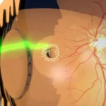
Vision loss is not a standalone diagnosis; it is the functional manifestation of tissue breakdown. The visual apparatus operates as an integrated optical and neurological circuit: light must refract clearly through the cornea and crystalline lens, traverse the vitreous humor without scatter, focus precisely on the photoreceptors of the retina, and transmit through the retinal ganglion cell axons of the optic nerve directly to the visual cortex of the brain. A pathological interruption at any point along this axis precipitates visual failure.
In clinical practice, the central challenge is that neural ocular tissue—specifically the sensory neuroretina and the optic nerve—possesses zero regenerative capacity. Once retinal ganglion cells undergo apoptotic death from elevated intraocular pressure, ischemia, or mechanical insult, the resulting visual loss is permanent.

The Pathophysiological Mechanisms of Vision Loss
Visual degradation manifests in several distinct clinical patterns depending on the underlying anatomical site of injury:
- Central Vision Loss (Scotoma & Macular Failure): Involves damage to the fovea centralis or papillomacular bundle. Patients lose visual acuity, color discrimination, and the ability to recognize faces or read, despite maintaining peripheral navigational sight.
- Peripheral Field Constriction ("Tunnel Vision"): Characterized by concentric narrowing of the visual field. Most commonly induced by chronic glaucoma, end-stage retinitis pigmentosa, or bilateral occipital infarctions sparing the macula.
- Sudden, Painless Visual Occlusion: An acute vascular or retinal crisis, such as Central Retinal Artery Occlusion (CRAO), ischemic optic neuropathy, or acute macula-off retinal detachment.
- Progressive Optical Opacification: Gradual light dispersion and scatter caused by corneal dystrophies, endothelial decompensation, or mature crystalline lens cataracts.
Among chronic systemic drivers, microvascular damage from diabetic retinopathy and hypertensive retinopathy represents a major global burden. In the posterior pole, diseases of the retina, macula, and yellow spot directly starve specialized photoreceptors, transforming high-definition daylight vision into irregular blind spots.
Glaucoma: The Asymptomatic Threat to the Optic Nerve
Glaucoma remains one of the leading etiologies of irreversible bilateral blindness worldwide. The pathology centers on progressive retinal ganglion cell (RGC) axon death at the level of the lamina cribrosa, frequently—though not exclusively—driven by elevated intraocular pressure (IOP).
The physiological danger of open-angle glaucoma lies in its absolute silence during early and moderate stages. The central visual acuity often measures a deceptive 20/20 on the Snellen chart while peripheral receptive fields are silently excavated. Because the brain compensates for gradual field deficits through binocular overlapping, patients frequently do not notice visual field constriction until over 40% to 50% of the optic nerve fibers have been permanently lost. Routine tonometry, gonioscopy, and optical coherence tomography (OCT) of the retinal nerve fiber layer (RNFL) are the only reliable defenses against this irreversible decline.
Can Eye Color Change Procedures Cause Vision Loss?
The direct, unequivocal medical answer is yes. Unregulated, anatomically aggressive, or poorly engineered eye color change procedures carry a verified, documented risk of causing severe, irreversible vision loss and legal blindness.
However, grouping all cosmetic eye color techniques into a single risk category is clinically inaccurate. The ultimate danger to vision depends strictly on which ocular layer is manipulated, how aqueous humor dynamics are handled, and whether the intervention respects corneal endothelial and trabecular cell biology.
⚠ The High-Risk Spectrum: Why Method Selection Dictates Visual Survival
Cosmetic anterior segment interventions differ vastly in their biological invasiveness. Surgical insertions of foreign plastic materials or indiscriminate injection of industrial pigments pose catastrophic risks to the eye's delicate drainage apparatus and endothelial pumps. Understanding the exact mechanical etiology of failure for each modality is vital for patient safety.
1. Cosmetic Silicone Iris Implants: High-Velocity Ocular Destruction
Cosmetic iris implants (such as NewColorIris or BrightOcular) involve surgically inserting an artificial, colored silicone diaphragm through a large corneal incision into the anterior chamber, seating it directly over the patient's living iris.
From an anterior segment perspective, placing a non-flexible synthetic disc in the anterior chamber creates an anatomical catastrophe:
- Uveitis-Glaucoma-Hyphema (UGH) Syndrome: The constant micromovement of the silicone plate against the vascularized iris tissue causes repetitive mechanical friction. This leads to chronic anterior uveitis, micro-hyphemas (blood pooling in the anterior chamber), and massive pigment shedding.
- Secondary Intractable Glaucoma: Shed pigment, erythrocytes, and inflammatory proteins physically clog the trabecular meshwork. This causes refractory intraocular pressure spikes (often exceeding 45–50 mmHg) that crush the optic nerve head within weeks, demanding emergency trabeculectomies or glaucoma drainage implants.
- Endothelial Decompensation (Bullous Keratopathy): The implant rubs against the delicate endothelial monolayer of the posterior cornea. Unlike epithelial cells, human corneal endothelial cells cannot regenerate. Once cell density drops below critical survival levels (under 500–800 cells/mm²), the cornea becomes permanently edematous and opaque, requiring a full-thickness corneal transplant (penetrating keratoplasty or DMEK).
For these reasons, the global ophthalmic community considers artificial color iris implants medically unsafe for cosmetic purposes.
2. Keratopigmentation (Corneal Tattooing): Stromal Micro-Distortion
Keratopigmentation uses a femtosecond laser to dissect a circumferential circular pocket within the clear corneal stroma, followed by the manual injection of synthetic or mineral pigments to physically mask the natural iris color underneath.
While keratopigmentation avoids direct contact with the anterior chamber angle and iris, it creates permanent structural and functional compromises within the cornea:
- Optical Aberrations and Severe Night Glare: In low-light environments, the human pupil naturally dilates beyond the artificial clear visual aperture of the tattoo. The pupil margin overlaps with the dense pigment ring, inducing massive higher-order aberrations, severe halos, light scattering, and disabling night myopia.
- Permanent Loss of Ophthalmic Visualisation: The opaque pigment in the cornea forms an irreversible visual barrier. In the event that the patient later develops a retinal break, peripheral retinal tear, or needs cataract surgery, the operating surgeon’s slit-lamp and surgical visualization of the anterior chamber angle and extreme retinal periphery is permanently obstructed.
- Corneal Biomechanical Instability: Delaminating deep stromal lamellae to force dense pigment layers into the cornea impairs fluid dynamics and risks chronic stromal melt, sterile keratitis, or persistent foreign-body inflammatory responses.
3. Laser Eye Color Change: Physics, Energy Control, and Trabecular Safety
Non-invasive laser eye color change operates under completely different biological principles. It does not introduce foreign silicone discs, and it does not carve permanent pockets inside the optical corneal window. Instead, it utilizes targeted light energy to initiate the natural metabolic clearance of melanin from the anterior stromal surface of the iris.
However, the clinical question remains: Can an eye color laser cause vision loss?
The answer is: Yes, if performed with incorrect wavelengths, excessive pulse energy, or uncontrolled protocols.
When unqualified clinics attempt eye color modification using off-the-shelf industrial lasers, high-fluence Q-switched Nd:YAG systems, or cutaneous dermatological lasers, the outcome is catastrophic. Excessive energy induces thermal shockwaves that rupture cellular membranes indiscriminately, causing intense anterior chamber fibrinous uveitis, posterior synechiae (iris sticking to the crystalline lens), and massive acute pigment dumping that overwhelms the trabecular meshwork, resulting in severe pigmentary glaucoma.
Preventing vision-threatening complications requires adherence to the biophysics of selective iris depigmentation. Under Dr. Mustafa Mete's patented protocol, low-energy, highly specific light wavelengths are applied in titrated, fractional sessions. This allows macrophage-mediated phagocytosis to clear microscopic melanin granules gradually, preventing sudden pigment saturation in Schlemm's canal.
Furthermore, patient safety is anchored by our proprietary early IOP monitoring framework, which tracks aqueous outflow dynamics and corneal endothelial integrity before, during, and after treatment to eliminate pressure spikes. To understand the exhaustive medical safeguards against complications, review our comprehensive analysis of laser eye color change costs and risks.
Comparative Risk Analysis of Cosmetic Eye Color Methods
| Procedure | Primary Anatomical Target | Mechanism of Vision Threat | Visual Prognosis / Reversibility |
|---|---|---|---|
| Cosmetic Iris Implants | Anterior Chamber Angle & Iris Root | UGH syndrome, endothelial cell loss, secondary intractable angle-closure glaucoma. | Catastrophic; frequently causes permanent vision loss requiring corneal or glaucoma surgery. |
| Keratopigmentation | Deep Corneal Stroma | Severe night glare, chromatic halos, loss of peripheral field, permanent blockage of fundus examination. | Irreversible; pigment remains trapped permanently in the visual axis; cannot be surgically washed out. |
| Unregulated / Copycat Lasers | Iris Stroma (High Fluence) | Thermal coagulative necrosis, severe acute pigmentary glaucoma, chronic anterior uveitis. | High risk of irreversible optic nerve cupping if IOP spikes are mismanaged. |
| MyLumineyes® Selective Laser | Superficial Iris Melanin Only | Mitigated via low-fluence selective photodisruption, staged fractionation, and continuous IOP surveillance. | High clinical safety profile; non-invasive, preserves corneal biomechanics and trabecular function. |
Clinical Strategy for Preventing Vision Loss
The preservation of human vision requires unwavering adherence to fundamental medical rules, whether dealing with natural degenerative diseases or elective anterior segment procedures. To protect optical function across a lifetime:
- Mandatory Preoperative Biomicroscopy & Gonioscopy: Any anterior segment procedure must be preceded by open-angle verification (Shaffer Grade III or IV), baseline endothelial specular microscopy (confirming cell density > 2000 cells/mm²), and baseline RNFL-OCT mapping.
- Reject Invasive Artificial Implants: Never undergo surgical placement of non-therapeutic artificial colored devices within the anterior chamber. The long-term complication rate approaches near certainty over time.
- Continuous Intraocular Pressure Surveillance: Following any laser or intraocular manipulation, tonometric monitoring at scheduled post-procedure intervals is crucial to detect early subclinical outflow resistance.
- Systemic Health Management: Tight glycemic control in diabetes and blood pressure regulation are non-negotiable baselines to prevent retinal capillary microaneurysms and non-perfusion.
According to foundational research endorsed by the World Health Organization (WHO), roughly 80% of visual impairment globally is completely avoidable or curable if clinical interventions occur prior to permanent structural and neurological cell destruction.
Final Medical Perspective
Vision is the most sophisticated sensory modality of human life. Altering the cosmetic appearance of an eye must never take precedence over preserving its biological integrity. If you are exploring aesthetic ocular changes, your primary consideration must not be convenience or novelty, but the surgical qualifications of the ophthalmologist, the physical properties of the technology employed, and whether the procedure respects the micro-anatomy of the anterior chamber.
Frequently Asked Questions: Vision Loss & Eye Color Change
Can an eye color change procedure cause permanent blindness?
Yes. Invasive techniques like cosmetic silicone iris implants have caused permanent blindness in numerous patients worldwide due to severe secondary glaucoma and corneal endothelial failure. Unregulated, high-energy generic lasers can also induce blinding glaucoma if performed by non-specialists without trabecular safety protocols.
Why do ophthalmologists strongly advise against cosmetic iris implants?
Cosmetic iris implants rest directly inside the anterior chamber, where they rub against the living iris tissue and the delicate posterior corneal endothelium. This induces chronic inflammation (uveitis), dangerous hyphemas (bleeding), endothelial cell death, and rapid, medication-resistant spikes in intraocular pressure that crush optic nerve fibers.
Does keratopigmentation (corneal tattoo) affect night vision?
Yes. In low-light environments, your pupils naturally enlarge to let in more light. When the pupil dilates past the edge of the central clear zone left by the corneal tattoo, light passes through the pigment border, causing severe light scatter, halos, reduced contrast sensitivity, and compromised night driving vision.
How does the MyLumineyes® laser avoid vision-threatening glaucoma?
The MyLumineyes technique uses custom, low-fluence wavelengths targeted selectively at stromal melanin without bursting cellular membranes violently. By fractionating treatments over multiple sessions and utilizing an early IOP monitoring framework, microscopic pigment is cleared gradually by the body's natural macrophages without clogging the trabecular meshwork.
Medical Disclaimer: This comprehensive clinical guide is authored by Dr. Mustafa Mete for educational purposes and does not substitute for an individual ophthalmic examination. Any sudden reduction in vision, pain, or eye redness requires emergency evaluation by a board-certified eye surgeon.