How Vision, Eyesight, and Color Perception Work
Vision is a complex cognitive and physiological process in which the visual organs and the brain collaborate to interpret light reflected from the surrounding environment. To understand how we see, we must examine how light travels through the structure of the eye, how the brain processes these signals, and how photopigments create our perception of color.
The Mechanism of Vision: How Eyesight Functions
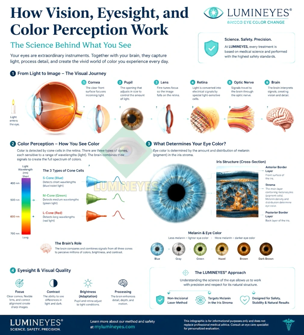
For an object to be visible, it must either emit light or reflect light from an external source. The process of sight occurs through a precise sequence of optical refraction and neurological transmission:
Light Entry and Refraction: Light rays reflected from an object first strike the cornea (the transparent outer layer of the eye), where the initial and primary refraction of light occurs.
Pupil Regulation: The refracted light passes through the pupil, the aperture controlled by the iris to adjust for lighting conditions.
Lens Focus: Light then passes through the crystalline lens, undergoing a second refraction to fine-tune focus.
Traversal of the Vitreous Body: The focused light travels through the clear, gel-like vitreous body toward the back of the eye.
Retinal Processing (Macula / Fovea Centralis): The light lands on the retina, specifically focusing on the macula (often referred to as the yellow spot). Here, an inverted, real image is formed.
Signal Transduction: Photoreceptor cells in the retina convert light energy into electrical impulses.
Brain Interpretation: The optic nerve transmits these electrical signals to the primary visual cortex (occipital lobe) of the brain. The visual cortex interprets and flips the inverted image upright, creating the conscious visual perception of our surroundings.
If any of these optical structures—cornea, lens, or axial length—cannot focus light precisely onto the retina, refractive errors occur, resulting in blurred vision.
How Does the Human Eye Perceive Colors?
Color perception depends on the physical properties of light waves and specialized sensory receptors in the retina.
The Visible Light Spectrum
Human visual perception is limited to wavelengths on the electromagnetic spectrum between 400 nanometers (nm) and 700 nm:
700–600 nm: Red light
600–500 nm: Green light
500–400 nm: Blue light
Photoreceptors and Color Processing
The retina contains two main types of photoreceptors: rods (responsible for vision in low light) and cones (responsible for color and high-acuity vision).
Humans typically possess trichromatic vision, meaning we have three types of cone photoreceptors sensitive to short (blue), medium (green), and long (red) wavelengths. The brain integrates simultaneous signals from these three cone types to perceive millions of distinct color shades.
Color Blindness: Deficiencies or mutations in the photopigment of specific cone receptors—most commonly the green or red cones—lead to color vision deficiencies (such as red-green color blindness), making it difficult to distinguish specific hues.
Green Perception: The human eye is evolutionarily tuned to be most sensitive to shades of green, allowing for high detail recognition within green spectral ranges.
How Object Colors Are Perceived
An object’s apparent color depends on which wavelengths of white light it absorbs and reflects:
White: An object reflecting 100% of visible light appears white.
Black: An object absorbing all visible light appears black.
Colored Objects: An object that absorbs blue and green wavelengths while reflecting red wavelengths appears red to the human observer.
The Genetics of Eye Color and Iris Pigmentation
Eye color is a polygenic trait determined primarily by the amount, type, and distribution of melanin within the iris stroma—not a genetic disease or anomaly.
Melanin Types and Distribution
Eumelanin: Brown to black pigment. High concentrations result in dark brown eyes.
Pheomelanin: Reddish-yellow pigment. Varying combinations with low eumelanin levels contribute to green, hazel, or amber eyes.
Light / Blue Eyes: Blue eyes contain minimal melanin in the anterior stroma. The blue appearance is not caused by blue pigment, but by Rayleigh scattering of light within the fibrous iris tissue.
Up to 16 genes (including OCA2 and HERC2) interact to regulate melanin synthesis, transport, and storage in the eye.
Infant Eye Color Changes
At birth, infant eyes often appear lighter or blue because melanocytes have not yet been fully exposed to light to trigger complete melanin synthesis.
3 to 6 Months: The most significant changes in iris pigmentation occur during this window.
1 to 3 Years: Pigmentation stabilizes for most children. Light eyes may darken into hazel or brown, but dark brown eyes do not naturally turn blue.
Can You Change Your Eye Color Permanently?
While temporary factors—such as pupil dilation, ambient lighting, contrasting clothing, or scleral inflammation—can alter perceived eye color, true anatomical changes require clinical intervention.
For individuals seeking permanent iris color alteration, advanced ophthalmological techniques exist:
Laser Eye Color Change: The Lumineyes procedure uses low-energy, targeted laser wavelengths to selectively break down dark melanin pigments in the front layer of the iris. Over time, the body naturally clears these pigments, revealing the underlying natural blue, green, or hazel tones. It is widely recognized as the safest and most non-invasive method when performed by experienced specialists.
Keratopigmentation: Pigments are mechanically placed into the corneal stroma to mask the underlying iris color.
Iris Implants: Artificial silicone implants placed over the iris, though associated with high risks of severe ocular complications such as glaucoma, corneal failure, and uveitis.
Visual Sensitivity and UV Protection
Individuals with lighter eye colors (blue, green, gray) have less protective melanin in the iris and retinal pigment epithelium. Consequently, light eyes are more vulnerable to UV radiation and photo-sensitivity (photophobia).
Tips for Maintaining Long-Term Ocular Health:
Wear broad-spectrum UV400 sunglasses in bright outdoor environments.
Maintain a comfortable distance when reading or working on digital screens to minimize eye strain.
Avoid direct exposure to high-intensity light sources to protect the macula and delicate photoreceptors.
Schedule regular comprehensive eye exams with an ophthalmologist.