Color Blindness Test (Ishihara Plates): Clinical Administration, Photopigment Genetics & Diagnostic Limitations
Pseudoisochromatic color vision testing represents one of the most widely deployed diagnostic screenings in ophthalmic medicine. Designed to evaluate congenital red-green dyschromatopsia, understanding the optics, plate architecture, and physiological limitations of the Ishihara test is vital for accurate clinical interpretation.
Congenital color vision deficiency (dyschromatopsia) affects roughly 8% of biological males and 0.5% of females worldwide. Colloquially termed "color blindness," true achromatic vision (seeing strictly in grayscale) is an extraordinary rarity. The vast majority of affected individuals possess trichromatic or dichromatic vision with reduced sensitivity across specific overlapping spectral bandwidths.
First introduced in 1917 by Dr. Shinobu Ishihara at the University of Tokyo, the Ishihara Pseudoisochromatic Test remains the clinical benchmark for detecting red-green deficits. However, its simplicity often obscures the precise optical physics required for its execution, leading to frequent misinterpretations when performed outside standardized clinical settings.
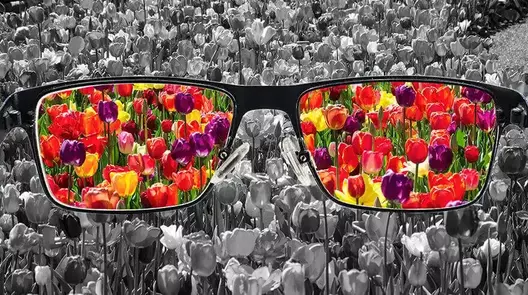
Clinical evaluation: Pseudoisochromatic plates isolate chromatic confusion lines without providing luminance clues.
The Pseudoisochromatic Principle: How Ishihara Plates Function
The Ishihara test relies on the concept of pseudoisochromatism ("false equal color"). An individual with intact trichromacy uses both chromaticity (hue and saturation) and luminance (brightness) to segment visual scenes. A color-deficient eye, lacking functional L-cones (erythrolabe) or M-cones (chlorolabe), relies heavily on subtle brightness variations to differentiate confusing hues.
To prevent brightness-based guessing, Ishihara plates are constructed using matrices of circular dots that vary randomly in:
- Dot Diameter: Sizes range from small to large to disrupt spatial edge detection.
- Luminance / Value: Dots are assigned varying lightness levels, ensuring that neither the figure nor the background provides a consistent luminance contrast.
- Confusion Hues: Foreground and background colors are selected along established color confusion lines derived from CIE chromaticity diagrams, where dichromatic eyes perceive distinctly different wavelengths as identical.
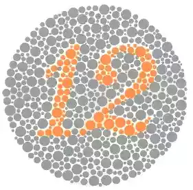
Plate 1 (Demonstration): Designed with extreme luminance and chromatic contrast to be readable by individuals with complete color blindness.
The 4 Distinct Morphologies of Ishihara Plates
A complete 38-plate or 24-plate Ishihara clinical album contains four engineered plate designs, each serving a distinct diagnostic purpose:
| Plate Classification | Optical Design Mechanism | Normal Trichromat Response | Red-Green Deficient Response |
|---|---|---|---|
| Demonstration Plates (e.g., Plate 1) | Constructed with severe luminance contrast independent of hue. | Reads the designated numeral (e.g., "12"). | Reads the designated numeral accurately (rules out malingering or total optical opacity). |
| Transformation Plates | Contains two overlapping figure paths; one defined by hue, one by luminance. | Reads the chromatic numeral (e.g., "74"). | Reads the alternative, secondary numeral (e.g., "21"). |
| Vanishing Plates | Figure dots lie strictly on confusion lines relative to background dots. | Easily identifies the embedded numeral (e.g., "6" or "45"). | Fails to perceive any numeral; sees only a random field of dots. |
| Hidden Digit Plates | Luminance differences define a figure visible only when chromatic noise is neutralized. | Sees a confusing, uniform pattern of dots with no distinct number. | Easily reads the hidden numeral (e.g., "2"), as normal color cues do not distract them. |
What Ishihara CANNOT Diagnose: Major Clinical Limitations
While exceptional for occupational screenings (such as aviation, maritime, and railway certifications), the Ishihara test has pronounced diagnostic limitations that require supplementary instrumentation:
- Blind to Tritan Defects (Blue-Yellow): The Ishihara test contains zero plates capable of assessing S-cone (cyanolabe) deficiency. It cannot evaluate tritanopia, tritanomaly, or early acquired maculopathies. Screening for blue-yellow defects requires Hardy-Rand-Rittler (HRR) plates or the Farnsworth D-15 test.
- Cannot Quantify Severity: Ishihara plates are qualitative, not quantitative. They indicate the presence of a red-green deficit but cannot reliably differentiate mild anomalous trichromacy from severe complete dichromacy. Precise quantification requires a Nagel Anomaloscope using Rayleigh color-matching equations ($589\text{ nm} = 546\text{ nm} + 670\text{ nm}$).
- Pediatric & Illiterate Limitations: Standard numeral plates cannot be administered to young children or illiterate patients. In these cases, pediatric tracing plates (following continuous winding paths with a soft brush) must be substituted.
Why Online Color Blindness Tests Are Clinically Unreliable
Self-administered digital Ishihara tests accessible via smartphone screens and web browsers provide recreational value but fail clinical standards. Digital testing introduces uncontrollable optical variables:
⚠ The Optical Failure of Digital Screen Screenings
Administering pseudoisochromatic tests requires rigid physical parameters that consumer digital displays cannot satisfy:
- Color Space Discrepancies: Computer monitors operate in sRGB or DCI-P3 color spaces, which compress gamut boundaries and distort calibrated printed pigment reflections.
- Display Gamma & Brightness: Uncalibrated screen backlight intensities introduce artificial luminance cues, allowing color-deficient users to "cheat" plates via brightness contrasts.
- Ambient Illuminant Failure: Clinical Ishihara testing mandates standard daylight illumination—specifically CIE Standard Illuminant C or D65 (approx. 500–600 lux). Testing under domestic tungsten bulbs, yellowish LEDs, or with software blue-light filters (Night Shift/f.lux) invalidates test geometry entirely.
Acquired vs. Congenital Dyschromatopsia: When Color Loss Means Pathology
Congenital color blindness is stationary, bilateral, symmetric, and genetically determined by X-linked recessive mutations on chromosome Xq28. If a patient experiences a change in color perception later in life, it is an ocular alarm bell.
Acquired Dyschromatopsia indicates progressive damage to the retina, optic nerve, or intracranial pathways, frequently conforming to Köllner’s Rule:
- Blue-Yellow (Tritan) Failure: Typically indicates disease of the outer retina, photoreceptors, or media opacities (e.g., age-related macular degeneration, diabetic macular edema, central serous chorioretinopathy).
- Red-Green (Protan/Deutan) Failure: Characteristically indicates pathology of the inner retina, retinal ganglion cells, or optic nerve axons (e.g., demyelinating optic neuritis, compressive pituitary lesions, open-angle glaucoma).
- Toxic Neuropathies: Medications like ethambutol, chloroquine/hydroxychloroquine, and heavy metal exposure produce progressive color desaturation long before visual acuity drops on a standard eye chart.
The Scientific Reality of "Color Blindness Glasses"
Commercial spectacles marketed to "cure" color blindness (such as EnChroma) have achieved massive internet visibility. From an ophthalmic perspective, patients must understand what these optical devices actually accomplish:
These lenses are passive multiband notch filters. They contain specialized absorptive dyes that block narrow wavelengths of light precisely where the spectral sensitivity curves of red (L) and green (M) cones overlap (around 580 nm). By filtering out this ambiguous wavelength zone, the glasses increase chromatic contrast between reds and greens for anomalous trichromats.
Laser Eye Surgery & Color Perception: Anatomical Independence
A persistent misconception among patients is whether corneal refractive surgery (like LASIK or PRK) or anterior segment laser modifications can either cause or cure color vision defects:
The anatomical answer is absolute: Refractive and cosmetic eye lasers have zero interaction with retinal color vision.
Human color perception is mediated entirely by photopigments embedded in cone outer segments located in the posterior sensory retina and interpreted by the primary visual cortex, as detailed in our analysis of how human color vision works.
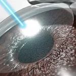
Conversely, refractive laser surgery reshapes the anterior stroma of the clear cornea, while our proprietary MyLumineyes® laser eye color change procedure selectively targets superficial melanin within the anterior border of the iris. Neither laser penetrates the posterior chamber, contacts the neurosensory retina, nor influences the X-linked genetics governing cone opsins. To explore the genetic loci that govern external eye appearance, review our clinical guide to eye color genetics and inheritance.
Frequently Asked Questions: Color Blindness & Testing
Can you fail an online color blindness test but still have normal vision?
Yes. Online tests are heavily distorted by uncontrolled screen brightness, display color gamuts (sRGB vs. OLED), ambient room lighting, and software blue-light filters. An accurate diagnosis requires an in-person examination using standardized, physical Ishihara or HRR plate albums under calibrated D65 illumination.
Can LASIK or laser eye surgery fix color blindness?
No. Laser refractive surgery operates strictly on the external cornea to correct refractive errors like myopia, hyperopia, and astigmatism. Color blindness is caused by missing or altered photopigments inside the cone cells of the retina at the back of the eye, which cannot be modified by corneal or anterior segment lasers.
Why does the Ishihara test miss blue-yellow color blindness?
The Ishihara test was engineered specifically to detect red-green photopigment defects (protan and deutan deficiencies). Its confusion lines do not intersect the blue-yellow (tritan) axis. Evaluating blue-yellow deficits requires Hardy-Rand-Rittler (HRR) plates or the Farnsworth-Munsell 100-Hue test.
Is color blindness progressive over time?
Inherited, congenital color vision deficiency is genetically stable and remains completely unchanged throughout your entire life. If you notice your color perception deteriorating over weeks or months, it indicates an acquired medical condition—such as optic nerve inflammation, glaucoma, or macular disease—demanding immediate ophthalmic evaluation.
Medical Disclaimer: This diagnostic overview is authored by Dr. Mustafa Mete for educational purposes and does not replace a clinical examination. If you experience sudden color desaturation, progressive difficulty identifying traffic signals, or visual decline, consult a board-certified ophthalmologist immediately.