Prosthetic Eye Surgery: Ocular Prosthesis, Enucleation & Anophthalmic Socket Reconstruction
An ocular prosthesis is far more than a cosmetic shell. In reconstructive ophthalmic surgery, managing an anophthalmic socket requires precise volume replacement, preservation of extraocular muscle dynamics, and meticulous conjunctival fornix reconstruction to restore facial symmetry and structural integrity.
The loss of an eye represents both a profound physiological transition and a significant aesthetic challenge. When disease, trauma, or intractable ocular pathology necessitates surgical globe removal, reconstructive oculoplastic surgery focuses on restoring an anatomically stable, comfortable, and cosmetically lifelike socket.
A modern ocular prosthesis does not transmit visual signals to the brain, but when paired with an integrated porous orbital implant, it tracks conjugate eye movements, supports the palpebral fissure, and prevents deep orbital structural collapse.
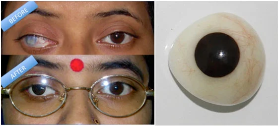
Reconstructive ocular rehabilitation: Handcrafted acrylic prosthesis fitted over a vascularized orbital implant.
Clinical Indications for Globe Removal
Surgical removal of an eye is an irreversible decision considered only after all conservative, medical, and globe-salvaging therapies have been exhausted. Indications include:
- The Painful Blind Eye: End-stage refractory conditions—such as neovascular glaucoma, absolute glaucoma, chronic ciliary spasm, or chronic ocular ischemia—where complete loss of light perception (NLP) is accompanied by debilitating, unmanageable ocular pain.
- Severe Open-Globe Trauma: Extensive lacerations with massive posterior uveal prolapse, disruptions where retinal architecture cannot be salvaged, or high-velocity injuries carrying a risk of sympathetic ophthalmia in the contralateral eye.
- Intraocular Malignancy: Primary intraocular tumors—predominantly large uveal (choroidal or ciliary body) melanomas or pediatric retinoblastomas—where globe-sparing treatments (such as plaque brachytherapy or proton beam radiation) are oncologically contraindicated.
- End-Stage Panophthalmitis: Overwhelming bacterial or fungal intraocular infections that penetrate the scleral shell and threaten adjacent orbital soft tissues or intracranial pathways despite intensive systemic and intravitreal antimicrobial therapy.
- Phthisis Bulbi: A shrunken, disfigured, non-functioning atrophic globe resulting from historical trauma, long-standing retinal detachment, or chronic low intraocular pressure (hypotony).
Surgical Techniques: Evisceration vs. Enucleation vs. Exenteration
The selection of the surgical procedure dictates subsequent implant placement, prosthetic motility, and complication risk profiles:
| Surgical Modality | Anatomical Scope of Resection | Muscle & Scleral Preservation | Primary Clinical Indications |
|---|---|---|---|
| Evisceration | Removal of intraocular contents (cornea, iris, retina, lens, vitreous); optic nerve remains untouched. | Preserved: The outer scleral shell, Tenon's capsule, and all extraocular muscle insertions remain fully intact. | Painful blind eyes (non-tumoral), endophthalmitis, phthisis bulbi, and trauma without suspected intraocular neoplasms. |
| Enucleation | Complete excision of the intact eyeball (globe) with transection of the optic nerve. | Reattached: Sclera is removed; the four rectus muscles are isolated, preserved, and sutured directly to an artificial orbital implant. | Primary intraocular tumors (uveal melanoma, retinoblastoma), severe trauma risking sympathetic ophthalmia, or phthisis with unclear margins. |
| Exenteration | Radical resection of the globe, extraocular muscles, orbital adipose tissue, and variable amounts of eyelids and periosteum. | Resected: All orbital soft tissue contents are removed down to the bony orbital walls. | Locally invasive malignancies, such as adenoid cystic carcinoma of the lacrimal gland, advanced orbital rhabdomyosarcoma, or invasive cutaneous carcinomas. |
1. Evisceration: Technique and Advantages
Evisceration is widely considered the procedure of choice when malignancy has been definitively ruled out. Through a 360-degree peritomy, the cornea is excised (keratectomy), and the intraocular contents are meticulously curetted and aspirated until the white scleral shell is entirely clean.
Because the extraocular muscle insertions into the sclera remain undisturbed, evisceration offers superior postoperative implant motility, minimal disruption of delicate orbital fat architecture, and a drastically reduced incidence of implant migration or extrusion.
2. Enucleation: Technique and Extraocular Re-anchoring
In cases of suspected uveal melanoma or retinoblastoma, cutting into the eye is contraindicated due to the danger of malignant cell seeding. The globe must be removed entirely intact. The four rectus muscles (medial, lateral, superior, inferior) are carefully hooked, isolated, and secured with absorbable double-armed sutures prior to disinsertion from the sclera.
The optic nerve is clamped and transected posterior to the globe—aiming for an optic nerve stump clearance of 8 to 12 mm in tumor cases. An engineered spherical orbital implant is then introduced deep into the muscle conus, and the rectus muscles are sutured directly to the implant's covering material or integrated pores to restore dynamic movement.
Orbital Implants & Biomaterials: Restoring Conal Volume
The human eye displaces approximately 6.5 to 7.0 milliliters of volume within the orbit. If an eye is removed without volume restoration, the patient develops Post-Enucleation Socket Syndrome (PESS), characterized by severe deep superior sulcus hollows, backward sinkage (enophthalmos), lower eyelid sagging (ectropion), and ptosis.
To prevent PESS, an orbital implant—typically 18 to 22 mm in diameter—must be placed immediately inside the socket:
- Porous Bio-Integrated Implants (Hydroxyapatite & Porous Polyethylene / Medpor): Modern porous spheres feature interconnecting micro-channels (averaging 200–500 microns) that permit fibrovascular ingrowth from host orbital tissue over several months. This biological incorporation anchors the implant firmly, drastically lowering the risk of long-term migration or late extrusion.
- Non-Porous Implants (Silicone & Polymethyl Methacrylate / PMMA): Chemically inert, smooth spherical implants. While cost-effective and presenting very low friction, they do not integrate with host fibrovascular tissue, relying entirely on anterior fascial closure layers to prevent exposure.
Scleral Shells: The Non-Surgical Alternative for Atrophic Eyes
Globe removal is not universally necessary for every visually impaired or cosmetically compromised eye. When an eye has become shrunken or discolored (as in non-painful phthisis bulbi or congenital microphthalmia) but remains quiescent and structurally stable, a Scleral Shell Prosthesis serves as an alternative.
A scleral shell is an ultra-thin (1.5 to 2.5 mm) custom-molded acrylic shell fabricated to fit directly over the patient's existing, desensitized cornea and sclera like a large contact lens. It requires no surgical resection, preserves native anatomy, and provides natural ocular motility driven by the patient's own intact muscles. However, if the cornea retains high tactile sensation, a protective conjunctival flap (Gundersen flap) may be surgically placed over the cornea first to eliminate discomfort.
Prosthetic Eye Fabrication & Motility Dynamics
Following surgery, a clear plastic spacer known as a conformer is placed behind the eyelids. The conformer maintains the depth of the upper and lower conjunctival fornices and prevents socket shrinkage while postoperative edema resolves.
Between 6 to 8 weeks post-surgery, the patient visits a specialized ocularist for prosthetic design:
- Socket Impression: An alginate or silicone cast of the healed socket is taken to capture the precise contours of the conjunctival surface and implant projection.
- Acrylic Curing: Medical-grade polymethyl methacrylate (PMMA) is cured, carved, and smoothed to optical perfection.
- Artisan Iris Painting: The ocularist hand-paints the iris striations, pupil aperture, and limbal blending using fine pigments, matching the contralateral biological eye down to microscopic details. Fine red silk fibers are embedded into the scleral acrylic to replicate conjunctival micro-vasculature.
- Conjugate Movement: Movement of the prosthesis relies on frictional contact between the deep conjunctival surface and the posterior face of the acrylic shell. While absolute extremes of gaze are slightly restricted compared to a biological eye, standard conversational saccades move in unison with the healthy eye.
⚠ Essential Medical Distinction: Ocular Prosthesis vs. Cosmetic Iris Surgery
It is vital not to confuse an ocular prosthesis with cosmetic anterior segment procedures. An ocular prosthesis is designed exclusively for non-seeing, anophthalmic sockets or atrophic blind globes.
For functional, sighted eyes:
- Invasive cosmetic implants—such as artificial iris implant surgery—placed into the anterior chamber of healthy seeing eyes carry catastrophic risks of pigmentary glaucoma, hyphema, and corneal blindness.
- For patients with intact ocular structures seeking permanent aesthetic iris lightening, the only medically validated, non-incisional method is targeted low-fluence laser eye color change, which works strictly on superficial stromal melanin without surgical entry.
Long-Term Prosthetic Maintenance and Socket Hygiene
Maintaining a healthy anophthalmic socket requires measured, gentle hygiene:
- Avoid Excessive Removal: The prosthesis should not be removed daily. Removing the shell too frequently introduces contaminants, stretches the lower eyelid, and irritates the conjunctival lining. Most patients need to remove and clean the prosthesis with mild soap and sterile saline only once every few weeks or months.
- Annual Professional Polishing: Over time, tear-film proteins, lipids, and micro-scratches roughen the acrylic surface, leading to giant papillary conjunctivitis (GPC) and excessive mucoid discharge. The prosthesis should be professionally buffed and polished by an ocularist once a year.
- Annual Ophthalmic Evaluation: Routine examinations with an ophthalmologist are mandatory to monitor the integrity of the conjunctiva over the orbital implant, confirm there is no subclinical implant exposure, and check the intraocular pressure and retinal health of the solitary functioning eye.
Frequently Asked Questions: Prosthetic Eye Surgery
Does a prosthetic eye restore any vision?
No. A prosthetic eye is an artificial cosmetic device made of medical-grade acrylic. It does not contain optical components, light sensors, or neural connections to the optic nerve. Its sole function is to restore facial symmetry, orbital volume, and cosmetic appearance after globe removal.
Can a prosthetic eye move naturally with the other eye?
Yes. Modern surgical techniques—particularly evisceration or enucleation paired with porous bio-integrated implants (such as porous polyethylene)—preserve extraocular muscle attachments. When the opposite eye moves, the implant moves within the orbit, transferring motion to the prosthetic shell for natural conversational tracking.
What is the difference between evisceration and enucleation?
Evisceration removes only the internal contents of the eye while preserving the patient's natural white sclera and muscle attachments, providing superior motility. Enucleation removes the entire eyeball and transects the optic nerve, which is mandatory when treating primary intraocular tumors like melanoma to prevent systemic spread.
How often does an artificial eye need to be replaced?
While the internal orbital implant remains permanently in the socket for life, the external acrylic prosthesis typically requires replacement every 5 to 7 years. Over time, subtle changes in orbital fat, eyelid elasticity, and facial tissue volume alter socket geometry, requiring a newly molded shell for optimal fit and symmetry.
Medical Disclaimer: This comprehensive guide to anophthalmic socket reconstruction is authored by Dr. Mustafa Mete for educational purposes. Any painful blind eye, structural trauma, or suspected ocular tumor requires an urgent, individualized evaluation by a board-certified ophthalmic surgeon.