
Heterochromia Treatment: Types, Causes & Clinical Correction
Different-colored eyes can be a lifelong, harmless trait—or a new sign that requires an ophthalmic examination. This clinical guide explains complete, sectoral and central heterochromia, diagnostic considerations, available correction options, and the original heterochromia-focused clinical work behind the Lumineyes™ approach.
What Is Heterochromia?
Heterochromia is a difference in iris color between the two eyes or within distinct regions of one iris. It may involve complete, sectoral or central patterns and can be present from birth or acquired later. Many congenital cases are harmless, while new or progressive differences require ophthalmic evaluation.
Heterochromia does not automatically require treatment. When an underlying eye disease causes the color difference, that condition takes priority. Cosmetic options include properly fitted colored contact lenses; selected adults may explore physician-assessed iris pigment modification, but outcomes, safety and long-term color symmetry cannot be guaranteed.

What Are the Three Main Types of Heterochromia?
The three commonly described patterns are complete, sectoral and central heterochromia. Complete heterochromia means the two irises differ overall in color. Sectoral heterochromia affects a defined portion of one iris. Central heterochromia describes a contrasting ring near the pupil; not every normal central color variation represents disease.
Complete Heterochromia
Each iris has a different overall color, such as one blue eye and one brown eye. It may be congenital or acquired.
Sectoral Heterochromia
Also called partial heterochromia, it is a wedge or defined area within a single iris that differs from the remaining iris color.
Central Heterochromia
A contrasting central ring encircles the pupil while the peripheral iris shows another color. A central ring can also be a normal pigment pattern.
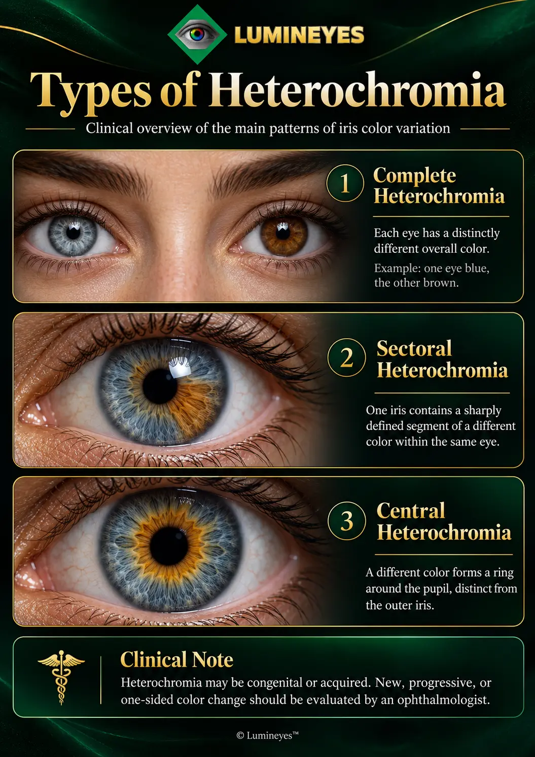
What Is Sectoral Heterochromia?
Sectoral heterochromia is a localized difference in pigmentation within one iris, often appearing as a distinctly colored wedge or segment. It may be inherited, associated with a developmental variation, or acquired through ocular conditions. Because the difference is spatial rather than uniform, careful sector-by-sector observation is important.
Sectoral heterochromia is especially relevant to the original Lumineyes clinical work described below. For detailed structures beneath the visible color pattern, explore the iris anatomy guide and the separate iris stroma reference.
Does Central Heterochromia Need Treatment?
Usually not. A stable central amber or brown ring within a lighter iris often represents a normal appearance rather than a treatable disease. A newly appearing, progressively changing, inflamed or otherwise unusual iris pattern should be assessed to exclude acquired pathology before any aesthetic decision.
What Causes Heterochromia in One or Both Eyes?
Heterochromia can result from inherited pigment patterns, developmental differences or acquired eye conditions. Congenital cases may remain unchanged for life. New color differences can accompany inflammation, injury, iris tissue loss, pigment disorders, selected medications or other ocular and neurological findings that warrant individualized investigation.
Congenital Heterochromia
Appears at birth or during early development. It can be an isolated trait, but a pediatric examination is appropriate when combined with abnormal pupils, eyelid asymmetry or other clinical features.
Acquired Heterochromia
Develops after previously similar iris colors. Possible explanations include iris inflammation, trauma, medication effects, pigment dispersion and melanocytic abnormalities. New changes deserve clinical assessment.
Which Medical Conditions Can Change Iris Color?
- Fuchs uveitis syndrome: Chronic intraocular inflammation can produce iris atrophy, color differences and associated cataract or glaucoma.
- Horner syndrome: When congenital or acquired early, altered sympathetic input can be associated with a lighter iris and may accompany ptosis and miosis.
- Iris nevus or melanoma: Focal pigmentation or a changing iris lesion requires proper evaluation; a photograph is not sufficient for diagnosis.
- Trauma or postoperative change: Injury or treatment may alter iris tissue and coloration.
- Pigment dispersion syndrome: Pigment release and associated anatomical changes require a pressure and anterior-segment assessment, not cosmetic treatment by default. Read the dedicated pigment dispersion syndrome and heterochromia discussion.
When Should a New Eye Color Difference Be Examined?
Seek prompt ophthalmic evaluation for sudden or progressive eye color change, a new iris spot or mass, pain, redness, light sensitivity, blurred vision or unequal pupils. A new drooping eyelid with pupil asymmetry, particularly with head or neck pain or neurological symptoms, requires urgent medical evaluation.
How Does an Ophthalmologist Diagnose Heterochromia?
- History and chronology: Confirm whether the difference is congenital, stable, newly acquired or progressive.
- Visual function and pupils: Assess vision, pupil responses, ocular alignment and any associated neurological features.
- Slit-lamp examination: Evaluate iris texture, stromal loss, inflammation, pigment deposits, corneal condition and lens findings.
- Pressure and angle assessment: Measure intraocular pressure; consider gonioscopy, anterior-segment imaging, optic nerve evaluation or other tests where appropriate.
- Clinical decision: Treat any relevant ocular disease first. Consider aesthetic options only after a clear diagnosis and suitable risk discussion.
How Did Heterochromia Research Influence the Lumineyes Method?
Dr. Mustafa Mete identifies the challenge of asymmetric iris pigmentation as an early clinical motivation behind Lumineyes. This work emphasized differences in pigment distribution between eyes and individual iris sectors, rather than assuming that the entire iris presents one uniform treatment target.
In the original MyLumineyes account, early sectoral cases informed a more individualized clinical philosophy: examine iris anatomy, assess regional pigment patterns, observe response over time and reconsider the next step before additional exposure. This is presented as the physician's first-party development record, not independent proof of a worldwide priority claim.
The historical sequence, selected documentation and distinctions between earlier clinical work and the later 8G XTRA generation are explored in the history of laser eye color change and Lumineyes development. A broad treatment description belongs on the Laser Eye Color Change pillar; this page remains dedicated to heterochromia.
Clinical authorship and scientific status: An early clinical achievement and a documented case are meaningful parts of a method's history. Claims such as “world's first,” unique exclusivity or guaranteed permanent correction require independently reviewable, dated source records and appropriate comparison with the published literature. The clinical photographs below are original first-party documentation.
What Do the Original MyLumineyes Heterochromia Cases Show?
The existing MyLumineyes archive documents visible changes in iris color patterns following physician-guided laser treatment. The images include sectoral presentations and a congenital heterochromia case labeled with a three-month postoperative follow-up. They illustrate individual clinical observations, not a controlled trial or a guarantee of identical outcomes.
Sectoral Heterochromia: Early Follow-up
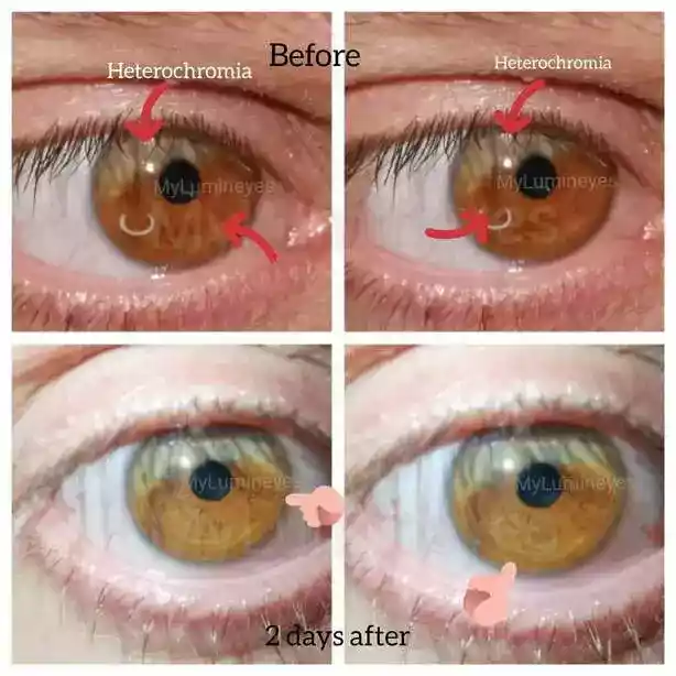
The original page identifies this as sectoral heterochromia and describes an image captured approximately two days following treatment. The series illustrates a short-term visible change in a localized pigmented region.
Early appearance can be affected by treatment response, lighting and ocular conditions. It should not be treated as proof of final pigment stability or absence of adverse events.
Sectoral Heterochromia: Documented Visual Progression
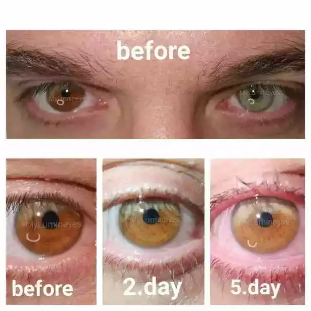
This photograph was previously published as a day-by-day progression of localized iris color change. It is useful for explaining why a regional pigmentation difference cannot be assessed only as a uniform blue-to-brown spectrum.
The timing and pattern illustrated are specific to the documented patient. They should not be generalized into a universal treatment rate, dose or safety conclusion.
Congenital Heterochromia: Three-Month Follow-up
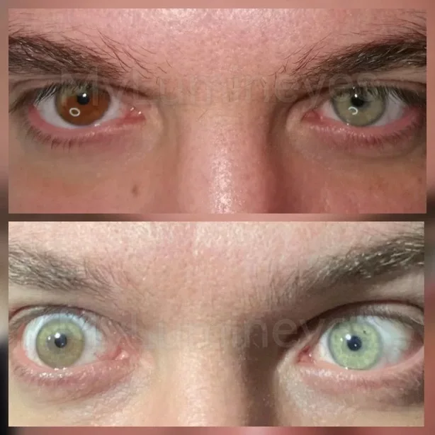
The original article describes this image as a three-month postoperative observation in a congenital heterochromia case. The visible appearance suggests a more balanced iris color pattern at that observation point.
Three months of photographic follow-up is not equivalent to lifelong stability. Longer-term claims require serial clinical assessments, consistent photography and ocular health measurements.
Why the originals remain important: These are clinical patient photographs already published on this page, rather than generic illustrations. The new infographic explains the three pattern types, while these preserved photographs document actual cases. No clinical image has been removed or replaced.
For broader first-party longitudinal observations—not necessarily limited to heterochromia—consult the MyLumineyes clinical data archive. Its reported sample, follow-up duration and study limitations must be interpreted separately from these individual cases.
How Is Laser Iris Depigmentation Considered for Heterochromia Correction?
Laser-based iris depigmentation aims to reduce selected visible iris pigment rather than implant a new color or deposit dye in the cornea. In a heterochromic iris, the intended clinical challenge is spatial selectivity: whether different pigmentation regions can be addressed while preserving acceptable ocular function and managing procedure-related risk.
Within the Lumineyes clinical framework, the target concept concerns the contribution of anterior iris melanin to visible color and its relationship to stromal anatomy. These concepts are developed in Selective Stromal Melanin Modulation (SSMM), a conceptual clinical framework rather than an independently validated guarantee of cellular selectivity.
What Happens to Pigment After Laser Exposure?
Treatment-related pigment modification can be followed by redistribution, cellular handling, possible aqueous-outflow interaction and variable optical changes over time. The eye's inflammatory state and intraocular pressure must be assessed as part of this process; the presence of pigment fragments alone does not show whether they will clear without complication.
- Initial examinationDocument pigmentation, sector boundaries, pupil findings, intraocular pressure, anterior chamber and angle status.
- Individual planningConsider the location of the visible disparity, the clinical diagnosis, alternatives and whether treatment is suitable at all.
- Staged treatmentWhere selected, physician-directed treatment is followed by observation rather than an assumed universal endpoint.
- Early monitoringAssess IOP, inflammation, corneal status, visual function and response before considering further stages.
- Response reviewDocument changing iris appearance and residual pigment without promising exact color symmetry.
- Long-term follow-upEvaluate clinical stability and the durability of the observed change beyond the initial photograph.
The Biological Fate of Pigment research synthesis explains why altered endogenous iris pigment and deposited artificial pigment have different biological pathways. The Early IOP Monitoring Framework examines the pressure-monitoring rationale after exposure.
How Does LIPS Grade V Describe Sectoral Heterochromia?
LIPS Grade V describes a spatially heterogeneous iris pigmentation phenotype, not an extreme grade of pigment density. In the proposed Lumineyes Iris Pigmentation Scale, Grades I–IV describe an ordered spectrum of diffuse apparent pigmentation; Grade V is a separate, non-ordinal category for marked regional variation.
Spatially Heterogeneous Iris Pigmentation
Regional pigmentation patterns, including some heterochromic presentations, may be described in this category. Grade V is not darker than Grade IV by definition and does not specify a universal laser setting or predict treatment response.
Explore the complete LIPS clinical framework →For quantitative research into regional iris color and architecture, read the proposed L-OAT iris phenotyping framework. These research owner pages provide the scientific details without duplicating their full content here.
What Are the Treatment Options for Heterochromia?
The correct approach depends on whether heterochromia is a harmless trait, a sign of disease or an aesthetic concern. Stable, benign heterochromia usually needs no treatment. Medical causes require diagnosis and disease-directed care. Cosmetic options include contact lenses and, in selected adult contexts, procedures affecting different ocular structures.
| Clinical question | Laser iris pigment modification | Artificial iris implantation | Keratopigmentation | Colored contact lenses |
|---|---|---|---|---|
| Where does the method act? | Native iris pigment, primarily in the anterior iris treatment concept. | Introduces an intraocular artificial structure. | Places pigment within the corneal stroma. | Colors the appearance through a removable lens on the eye surface. |
| Intended appearance | Reduce the contribution of natural pigment; variable and not exact-color predictable. | Mask or alter visible iris appearance with a prosthetic device. | Create the appearance of another iris color through corneal pigment. | Temporarily cover the existing appearance with a chosen lens color. |
| Main considerations | Pigment release, inflammation, IOP, outflow, iris/corneal health and uncertain long-term generalizability. | Intraocular surgery and serious device-associated risks, including glaucoma, corneal damage and inflammation. | Corneal surgical and optical considerations, pigment behavior and future ophthalmic assessment. | Fitting, hygiene, oxygenation and infection risks with improper use. |
| Reversibility | Intended pigment reduction may be long-lasting and cannot be assumed reversible. | May require surgical explantation, which carries further risk. | Deposited corneal pigment is not simply removable without further intervention. | Removable; natural iris pigmentation remains unchanged. |
| Role in heterochromia | Individualized consideration only after examination; clinical outcome and eligibility vary. | Not a routine cosmetic solution for a healthy eye; functional iris reconstruction is a separate indication. | Cosmetic camouflage in selected contexts after detailed counseling. | Non-surgical appearance management when medically fitted and used properly. |
Clinical comparison of anatomical targets and relevant risks—not a claim of universal comparative safety or a recommendation to undergo elective surgery. The source page's distinctive green/gold comparison-table design is retained in an updated, more balanced form.
Should a Healthy Person Treat Heterochromia?
Not necessarily. Many people with lifelong heterochromia have healthy eyes and require no correction. A cosmetic preference should be separated from a medical indication. In particular, invasive options should not be treated as interchangeable with a removable colored contact lens or with the treatment of an underlying eye disease.
For broader methods and terminology, the clinical owner is the main laser eye color change guide. For the meaning and limitations of durable results, consult the Permanent Eye Color Change resource.
Is Heterochromia Laser Treatment Permanent?
Laser iris pigment reduction is intended to create a long-lasting change in appearance, but permanent and perfectly symmetrical results cannot be guaranteed. The stability of a treated iris depends on the initial pigment pattern, biological response, residual pigmentation, treatment course and duration of follow-up.
The original three-month case image records appearance at that visit; it is not sufficient by itself to establish lifelong symmetry. Meaningful long-term evidence combines sequential comparable photography with visual acuity, IOP, anterior-segment observations and appropriate longitudinal clinical records.
Research into permanence, stromal biology and clinical endpoints is discussed in Biological Basis and Limitations of Permanence. Where safety is discussed, monitoring and stopping decisions are considered in the L-SAFE research framework, which is conceptual and not yet a validated scoring instrument.
What Evidence Supports Clinical Heterochromia Correction?
Photographic cases demonstrate observed visible changes, and the first-party Lumineyes archive provides a record of clinical development and follow-up. These evidence types should be distinguished from independent prospective studies. The published literature on cosmetic iris depigmentation remains limited and methodologically varied, so different laser protocols should not be assigned identical efficacy or safety profiles.
Frequently Asked Questions About Heterochromia and Treatment
Is heterochromia harmful?
Most isolated congenital heterochromia is harmless and does not reduce vision. However, a new, progressive or symptomatic color difference may reflect inflammation, injury, medication effects or another ocular disorder. An examination determines whether the finding is a normal trait or a sign requiring treatment.
Can sectoral heterochromia be corrected?
Sometimes its appearance can be modified, but correction is not medically necessary for every patient. Colored contact lenses offer a removable cosmetic option. Selected adult patients may explore physician-assessed pigment modification, although exact regional symmetry, safety and durability cannot be promised.
What is the difference between complete and sectoral heterochromia?
Complete heterochromia describes two irises that differ in overall color. Sectoral heterochromia describes a sharply contrasting portion of a single iris. Both patterns may be congenital or acquired, but their anatomical distribution and clinical assessment are not identical.
Is central heterochromia a disease?
Usually not. A stable ring of different pigmentation surrounding the pupil often represents a normal iris appearance. New asymmetry, inflammation, visual symptoms or a changing pigment lesion should prompt examination rather than an assumption that the pattern is harmless.
Can one brown eye and one blue eye be normal?
Yes. Two differently colored eyes may represent isolated congenital complete heterochromia and coexist with normal visual function. The finding should still be considered in the patient's medical history, especially if it developed recently or accompanies other eye or neurological symptoms.
What causes acquired heterochromia in adults?
Acquired heterochromia may follow intraocular inflammation, injury, certain medications, pigmentary disorders or focal iris lesions. Because causes vary considerably, evaluation may include slit-lamp examination, pressure measurement and assessment of the iris, ocular angle, lens and pupil findings.
Can pigment dispersion syndrome cause eye color differences?
Yes, pigment dispersion can be associated with changes in iris appearance, but it is a clinical condition involving pigment release and potential pressure complications—not simply a cosmetic pattern. Suspected cases require assessment of intraocular pressure, the angle and the optic nerve. Read the clinical guide.
Does laser heterochromia correction preserve natural iris texture?
Laser iris pigment modification acts on natural iris tissue rather than inserting artificial pigment, so preserving native anatomy is an intended principle of the approach. That intention does not prove that surrounding tissues are unaffected. Clinical examination and follow-up remain essential, and the final appearance varies.
How long does heterochromia laser correction take?
There is no universal treatment time or session count for heterochromia. Planning depends on the pigment distribution, iris anatomy, ocular condition and response between stages. The visible result may continue evolving after a session, and individual clinical reassessment determines whether any further intervention is appropriate.
Is heterochromia treatment permanent?
Some pigment-related changes may remain stable for a long period, but a specific final shade, complete symmetry or lifelong stability cannot be guaranteed. Long-term follow-up is necessary to distinguish early photographic change from durable biological and clinical stability.
Can children undergo cosmetic heterochromia treatment?
No cosmetic iris depigmentation is proposed for infants or children within the MyLumineyes patient approach. Congenital differences should first receive appropriate pediatric ophthalmic assessment when indicated. Any eye disease or visual-development issue takes priority over cosmetic appearance.
Who should seek an ophthalmologist immediately?
Anyone with sudden color change accompanied by pain, redness, blurred vision, a new pigmented lesion or unequal pupils needs prompt assessment. A new drooping eyelid and pupil difference with severe head or neck pain or neurological symptoms warrants emergency evaluation.
How Does MyLumineyes Approach Heterochromia Assessment?
The core principle is diagnosis before cosmetic intervention. The ophthalmologist must establish the type and history of heterochromia, distinguish stable congenital variation from acquired disease, assess ocular structures and pressure, and then discuss whether any proposed appearance modification has an acceptable clinical rationale for that individual.
Request an Individual Ophthalmic Assessment
Patients seeking information about heterochromia or its cosmetic management can request a physician-led eligibility discussion. Photos help illustrate visible color patterns but cannot independently establish a diagnosis or treatment suitability.
Contact MyLumineyes →
Dr. Mustafa Mete
Ophthalmologist · Developer of the Lumineyes™ clinical approach
Clinical focus: iris pigmentation, heterochromia assessment, anterior-segment laser procedures, response-guided treatment and longitudinal ophthalmic observation. This page distinguishes firsthand clinical documentation from externally validated scientific evidence.
Physician profile →Selected external medical context: AAO EyeWiki: Pigment Dispersion Syndrome and Pigmentary Glaucoma. External references on this page are restricted to AAO-owned resources; other scientific and clinical records are linked through the internal Research Library.
Medical notice: Educational information only. Cosmetic laser iris pigmentation modification is elective and has potential ocular risks, including inflammation, elevated intraocular pressure, pigment-related outflow effects and vision complications. No image, treatment history or conceptual framework substitutes for a clinical evaluation or establishes guaranteed safety, effectiveness, symmetry or permanent results.
Visual credits: Four original patient images from the existing heterochromia article and one new educational infographic © Lumineyes™. The clinical photographs are retained unaltered. Image reproduction and reuse are governed by the Copyright & Content Use Policy.