Clinical Reference Guide · Procedures · Risks · Evidence
Eye Color Change Surgery: Methods, Risks, Cost, Recovery & Alternatives
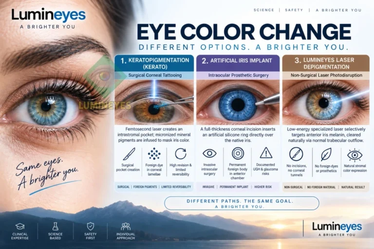
“Eye color change surgery” is a broad search term that can refer to very different procedures. Keratopigmentation changes appearance by placing pigment in the cornea; cosmetic iris implants place a device inside the anterior chamber; laser iris depigmentation reduces visible iris pigment without adding corneal dye or an implant. Because these approaches act on different tissues, their risks, recovery, reversibility, evidence base, and color-selection limits should be evaluated separately.
Publisher disclosure: MyLumineyes provides non-incisional laser iris depigmentation. This guide therefore separates external published evidence from descriptions of the Lumineyes™ methodology and does not present the comparison table as a ranking of procedures.
Can surgery permanently change eye color?
Yes, but the term includes fundamentally different interventions. Keratopigmentation adds pigment within the cornea; a cosmetic iris implant masks the natural iris with an intraocular device; and laser iris depigmentation reduces visible iris pigment. None should be described as risk-free, and a visually satisfactory result does not by itself establish normal intraocular pressure, corneal health, or long-term ocular safety.[1]
1. Types of Eye Color Change Procedures: What Actually Changes?
The visible color of the eye is mainly produced by the iris behind the clear cornea, but a procedure does not have to alter the iris itself to change apparent eye color. The most useful first question is therefore anatomical: which tissue or structure is being changed?
Keratopigmentation
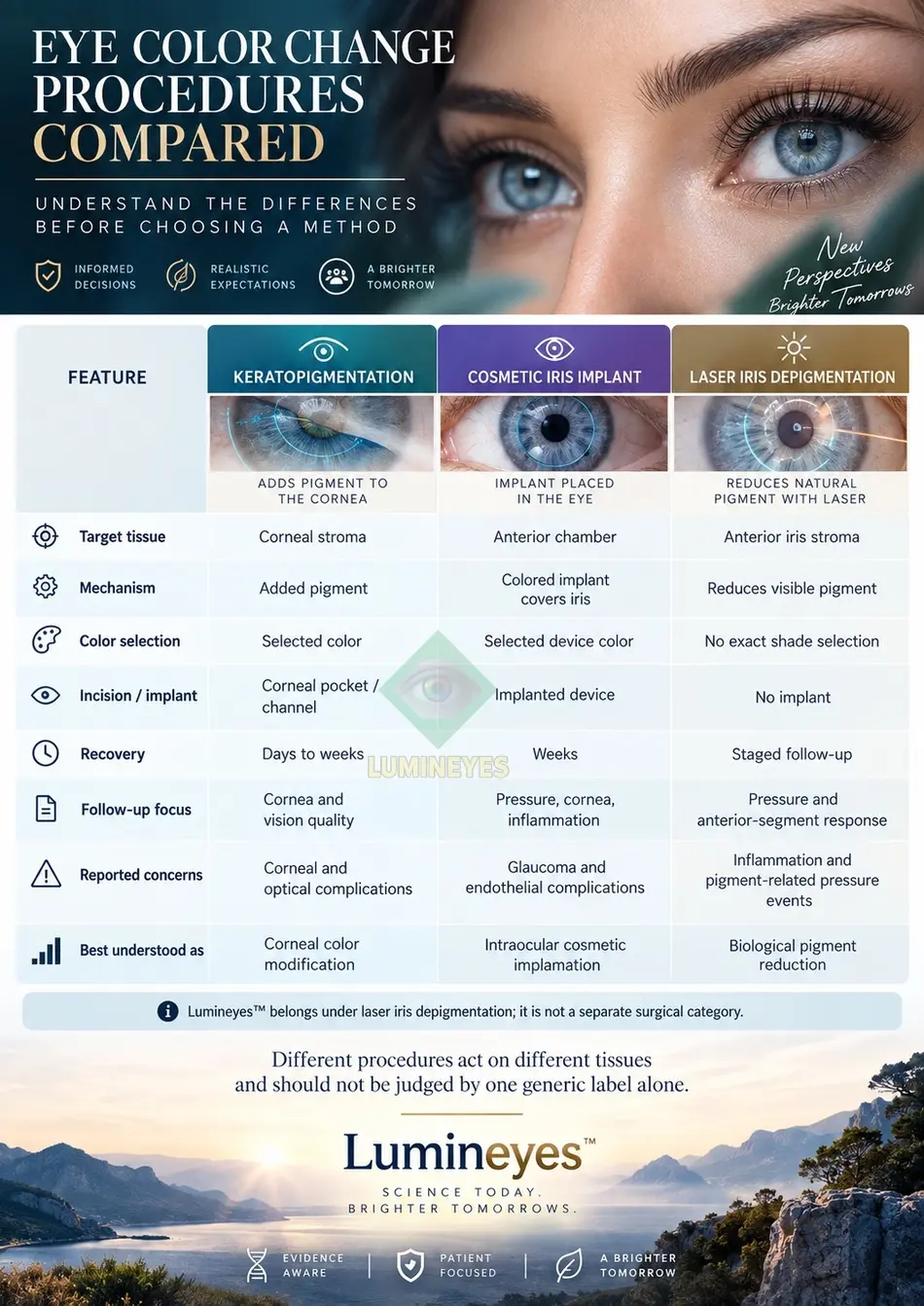
Femtosecond-assisted techniques create an intrastromal corneal pocket or channel and place pigment within the cornea, altering the appearance seen in front of the natural iris.
Cosmetic iris implants
A colored prosthetic diaphragm is inserted into the anterior chamber over the natural iris. Cosmetic implantation must be distinguished from medically indicated artificial-iris reconstruction for aniridia or major iris defects.
Laser iris depigmentation
Laser treatment acts on melanin-bearing structures in the anterior iris without adding a corneal pigment or placing an intraocular implant. Visible change depends on baseline phenotype and the biological response to treatment.
2. Eye Color Change Methods Compared
| Question | Keratopigmentation | Cosmetic iris implant | Laser iris depigmentation |
|---|---|---|---|
| Treatment site | Corneal stroma, anterior to the iris. | Anterior chamber, over the natural iris. | Melanin-bearing structures within the optically relevant anterior iris stroma. |
| Mechanism | Adds exogenous pigment within a corneal pocket or channel. | Adds a colored prosthetic device that masks the native iris. | Reduces visible iris pigment through protocol-specific laser–pigment interaction; the Lumineyes™ research terminology describes its intended target as Selective Stromal Melanin Modulation (SSMM). |
| Incision / implant | A corneal pocket or channel is created. | An incision is required for intraocular device insertion. | No corneal pigment pocket and no intraocular implant. |
| Color selection | Pigment can be selected, although the final appearance can vary. | Device color can be selected; selection does not establish safety. | No artificial shade is added. The visible result depends on existing iris structure, pigment distribution and biological response. |
| Reversibility | Not simply reversible; pigment removal or revision may be incomplete and may require further corneal intervention. | Explantation is another intraocular operation and does not guarantee reversal of prior damage. | Pigment reduction is intended to be lasting and should not be treated as readily reversible. |
| Key structures requiring follow-up | Corneal clarity, pigment position, corneal topography/tomography and endothelium as clinically indicated. | Corneal endothelium, anterior chamber, angle, intraocular pressure, lens and optic nerve as clinically indicated. | Intraocular pressure, anterior-chamber response, cornea, angle/outflow and other ocular findings according to the protocol and clinical response. |
| Published concerns | Dysphotopsia, pigment migration/fading, infection, corneal injury and other procedure-specific complications have been reported.[2] | Glaucoma, uveitis, hyphema, cataract, endothelial loss, corneal decompensation and vision loss are documented in complication series.[3] | Inflammation, pigment-related IOP elevation and severe pigmentary glaucoma have been reported; evidence is protocol-heterogeneous and long-term data remain limited.[5][6] |
The comparison above describes anatomy, mechanism and reported concerns. It does not assign an overall safety ranking. Different studies involve different operators, techniques, devices, laser protocols, populations and follow-up durations.
3. Is Eye Color Change Surgery Safe?
Safety must be considered for the specific procedure, protocol, patient and follow-up system. Terms such as “laser-assisted,” “non-incisional,” “minimally invasive,” or “permanent” describe procedural characteristics; they do not prove that an intervention is risk-free.
Keratopigmentation
Modern cosmetic keratopigmentation has published case-series data and medium- to long-term follow-up in selected populations, but corneal and optical complications remain possible. Pigment behavior, corneal integrity and visual symptoms must be interpreted within the exact technique used.[2][7]
Cosmetic iris implants
Published complication series document potentially severe anterior-segment damage, including glaucoma and corneal decompensation. Device removal may be necessary and does not ensure reversal of established damage.[3][8]
Laser iris depigmentation
Published evidence includes a large prospective single-center series using one defined protocol as well as adverse-event case reports involving severe pigmentary glaucoma and other complications. Neither type of evidence should be generalized automatically to every laser protocol.[5][6][9]
Evidence matching matters. A study of one device, pigment, corneal technique or laser protocol is not proof of another provider’s outcomes. Ask which published evidence actually matches the proposed treatment, how many eyes were studied, how long follow-up lasted, which adverse events occurred and how much follow-up data were missing.
Regulatory distinction. FDA approval of the CustomFlex Artificial Iris concerns treatment of full or partial aniridia; it does not represent approval for cosmetic color change in an otherwise healthy iris.[4]
4. How the Lumineyes™ Laser Methodology Is Framed
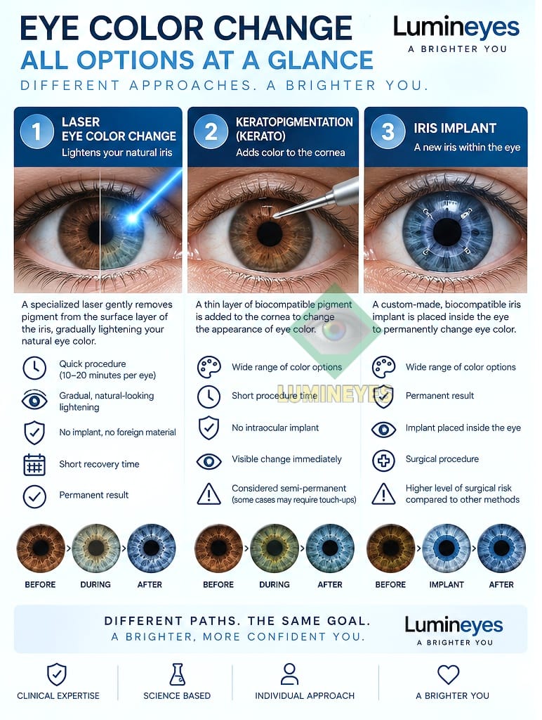
Lumineyes™ is a protocol-specific form of non-incisional laser iris pigment reduction. It should not be treated as interchangeable with every historical or published laser iris depigmentation technique. The methodology is described clinically as staged and response-guided, with treatment decisions based on observed ocular response rather than a fixed session count.
Three Lumineyes™ research terms used on this site
Selective Stromal Melanin Modulation (SSMM) describes the intended pigment-targeting concept; controlled treatment-related pigment response describes the biological response layer; and response-guided staged treatment describes the decision architecture used to reassess whether further exposure should be considered.
These are operational and conceptual terms. They do not, by themselves, prove histological selectivity, comparative superiority, or long-term safety.
The clinic’s reported real-world observations are maintained separately in the Lumineyes™ Clinical Data record. That page treats reported outcomes as retrospective observations and explicitly distinguishes a clinical archive from a prospectively designed trial.
5. How Much Does Eye Color Change Surgery Cost?
There is no single reliable global price for “eye color change surgery.” Costs vary by country, technique, surgeon or clinic, whether treatment involves one or both eyes, facility fees, number of treatment stages, postoperative care, and whether revision or complication management becomes necessary.
| Cost component | Keratopigmentation | Iris implant | Laser depigmentation |
|---|---|---|---|
| Pre-treatment testing | Ask whether corneal imaging and endothelial assessment are included. | Ask which anterior-segment, endothelial, pressure and lens assessments are included. | Ask which baseline ophthalmic tests and monitoring visits are included. |
| Procedure fee | Clarify pigment, femtosecond/facility fees and one- versus two-eye pricing. | Clarify device, operating-room, anesthesia and one- versus two-eye pricing. | Clarify whether the quote is a program, a fixed number of sessions, or staged according to response. |
| Follow-up | Ask which corneal and visual follow-up visits are included. | Ask which pressure, corneal, gonioscopic and intraocular follow-up visits are included. | Ask which early and later monitoring visits are included and how treatment pauses are handled. |
| Revision / additional care | Ask how touch-up, pigment revision or treatment of a corneal complication is billed. | Ask about explantation or additional glaucoma/corneal surgery costs. | Ask how additional evaluation, extended monitoring or a discontinued treatment course is handled. |
| Travel | For medical tourism, include accommodation, local stay, return travel and a plan for urgent ophthalmic care after returning home. | ||
For current clinic-specific information, use the dedicated laser eye color change cost and risk page. A current written quote is more useful than a generic internet price range.
6. Recovery and Follow-Up
“Recovery time” is not one number. Patients should separate at least three questions: when everyday activities can resume, when objective ocular findings are stable, and when the final cosmetic appearance can reasonably be assessed.
Keratopigmentation
Early care focuses on corneal healing, symptoms and infection prevention. Later review may include corneal clarity, pigment position, refraction, topography/tomography and endothelial assessment according to technique and findings.
Cosmetic iris implants
Because an intraocular device is present, follow-up may require ongoing evaluation of inflammation, intraocular pressure, angle, corneal endothelium, lens and other structures. Complication management can extend well beyond the initial postoperative period.
Laser iris depigmentation
No corneal pigment pocket or intraocular implant is created, but laser-induced pigment and inflammatory responses can affect the anterior segment. Pressure, anterior-chamber response, cornea and angle/outflow therefore remain clinically relevant during staged treatment.
Urgent symptoms: sudden or marked loss of vision, severe eye pain, rapidly worsening redness, significant photophobia, nausea with eye pain, or other acute visual symptoms after an eye procedure require urgent ophthalmic assessment rather than waiting for a routine cosmetic follow-up.
7. Results, Permanence and Reversibility
“Permanent” describes the intention to create a lasting change; it is not a guarantee that the appearance will never evolve, that a patient will remain satisfied, or that no later ocular issue will occur.
| Method | How appearance is created | Can an exact shade be selected? | Important limitation |
|---|---|---|---|
| Keratopigmentation | Selected pigment is added within the cornea. | A target pigment can be chosen, but the final appearance still depends on optics, technique and healing. | Fading, pigment movement, dysphotopsia or revision may occur. |
| Iris implant | A colored device masks the natural iris. | The device color is selected. | Explantation does not guarantee reversal of structural damage. |
| Laser depigmentation | Visible iris pigment is progressively reduced. | No. The procedure does not add a chosen dye; outcome depends on the individual iris and treatment response. | The original color should not be assumed to be readily restorable, and a precise final shade cannot be guaranteed. |
Before-and-after photographs are useful only when the procedure, time interval and imaging conditions are clear. The clinic’s laser before-and-after gallery shows Lumineyes™ cases; those images are not keratopigmentation or implant outcomes and cannot predict an individual result.
8. What Does the Published Evidence Show?
Study design matters. A complication referral series answers a different question from a prospective treatment series; a case report demonstrates that an event can occur but cannot determine its incidence; and a single-center favorable series cannot guarantee the same results in another protocol or clinic.
- Broad reviews: 2022 and 2025 reviews describe cosmetic iris implants, laser iris depigmentation and keratopigmentation as mechanistically distinct approaches with unequal evidence bases and different reported complications.[1][10]
- Keratopigmentation: published cosmetic series provide medium- and long-term outcome data in selected populations, while additional reports describe complications across broader indications and techniques.[2][7]
- Cosmetic iris implants: complication series document glaucoma, uveitis, endothelial damage, corneal decompensation and the need for further surgery. Referral-series data should not be misread as incidence among all recipients.[3][8]
- Laser iris depigmentation: a prospective report described outcomes in 1,176 eyes using one defined protocol, while separate case reports document severe pigmentary glaucoma and other complications. These bodies of evidence are complementary, not interchangeable.[5][6][9]
Why reviews can disagree. A 2025 narrative review favors keratopigmentation on the basis of the available literature, while also acknowledging that the comparative evidence is limited and largely single-center. This page reports that interpretation as the authors’ conclusion rather than reproducing it as an independent ranking by MyLumineyes.[1]
9. Lumineyes™ Research: Separating Clinical Data, Mechanism, Safety and Phenotype
One reason eye-color literature becomes confusing is that mechanistic hypotheses, clinical outcomes and monitoring frameworks are often discussed as though they were the same type of evidence. The Lumineyes™ Research Archive separates these functions.
For the wider methodological framework, see How Clinical Evidence Is Evaluated in Laser Eye Color Change and the Lumineyes™ Research Library.
10. What to Check Before Choosing Any Eye Color Procedure
- What exact procedure is proposed, and which tissue is altered?
- Who will perform it, and what ophthalmic credentials and complication-management resources are available?
- Which examination findings would make the procedure unsuitable?
- What evidence supports this exact device, pigment, laser protocol or surgical technique?
- What complications has the provider personally observed, and how were they managed?
- What is the expected follow-up schedule, and who will provide urgent care if you travel home?
- Which outcomes are measured objectively rather than judged only from photographs?
- What happens if the desired appearance is not achieved or if treatment must be stopped?
For people who want a temporary appearance change, professionally fitted colored contact lenses are an alternative that does not permanently alter ocular tissue. They still require correct prescribing, fitting and hygiene because contact lenses can cause corneal injury and infection.[11]
Eye Color Change Surgery: Frequently Asked Questions
What is the safest eye color change surgery?
No single procedure should be declared universally safest from the procedure name alone. Published reviews have favored keratopigmentation on the basis of the currently available literature, but the comparative evidence is limited and protocol-dependent. Individual risk also depends on ocular anatomy, technique, operator, patient selection and follow-up.[1]
Is laser eye color change surgery?
Laser iris depigmentation is commonly grouped with “eye color change surgery” in search and review literature, but it is non-incisional and does not involve an implanted device or a corneal pigment pocket. It is better described precisely as laser iris depigmentation or laser iris pigment reduction.
Can I choose an exact blue, green or hazel shade?
Keratopigmentation and colored implants add a selected artificial color, although the final appearance can still differ from a sample. Laser depigmentation does not add a chosen dye, so a precise final shade cannot be guaranteed.
Are cosmetic iris implants FDA-approved for changing healthy eye color?
No. FDA approval of the CustomFlex Artificial Iris concerns full or partial aniridia and should not be interpreted as approval for purely cosmetic color change in a healthy iris.[4]
Can eye color procedures damage vision?
Yes. Sight-threatening complications are documented in the literature, particularly with cosmetic iris implants, and serious adverse events have also been reported after laser iris depigmentation. Keratopigmentation can also produce corneal and optical complications. Risk must be discussed for the exact procedure and protocol.
Can keratopigmentation be reversed completely?
It should not be considered simply reversible. Pigment is placed within corneal tissue, and revision or removal may be incomplete and may itself require further intervention.
Can laser iris depigmentation be reversed?
It should not be presented as readily reversible. The treatment is intended to reduce visible iris pigment, and restoration of the original appearance cannot be assumed.
How much does eye color change surgery cost?
Prices vary substantially by country, method, provider, one- versus two-eye treatment, facility costs, number of stages, follow-up and possible revision. A current itemized quote is more reliable than a global price estimate.
Does laser iris depigmentation have long-term safety data?
The published literature remains limited and protocol-heterogeneous. Some prospective data exist for a specific protocol, while case reports document serious complications. MyLumineyes also maintains a separate retrospective clinical archive, but retrospective clinic observations are not equivalent to independent prospective validation.
Discuss eye health before making a cosmetic decision
MyLumineyes consultations concern its laser iris depigmentation service. An ophthalmic assessment can address suitability, expectations, objective findings, evidence and follow-up. It does not guarantee eligibility or a particular final shade.
Ask about an ophthalmic assessmentScientific References and External Evidence
- D'Oria F, Alio JL. Surgical Techniques for Cosmetic Eye Color Change: A Narrative Review. Ophthalmol Ther. 2025;14:1685–1694. doi:10.1007/s40123-025-01177-0. PubMed.
- Alio JL, et al. Cosmetic Keratopigmentation in Sighted Eyes: Medium- and Long-term Clinical Evaluation. Cornea. 2021. PubMed.
- Balal S, et al. Complications and Management of Cosmetic Iris Implants. Am J Ophthalmol. 2025. PubMed.
- U.S. Food and Drug Administration. CustomFlex Artificial Iris — PMA P170039. Approved indication concerns full or partial aniridia. FDA.
- Grimaldos Ruiz P. Photoablative cosmetic iridoplasty: effective, safe, and predictable-eye color change in 1176 eyes. Int Ophthalmol. 2021;41:1381–1393. doi:10.1007/s10792-021-01693-5. PubMed.
- Liu J, Korban S, Moster MR, Rhéaume MA, Wang Q. Bilateral severe iatrogenic pigmentary glaucoma following laser treatment for cosmetic iris color change. Am J Ophthalmol Case Rep. 2023;32:101927. doi:10.1016/j.ajoc.2023.101927. PubMed.
- Alio JL, et al. Keratopigmentation with micronised mineral pigments: complications and outcomes in a series of 234 eyes. Br J Ophthalmol. 2018. PubMed.
- Arthur SN, et al. Uveitis-glaucoma-hyphema syndrome and corneal decompensation in association with cosmetic iris implants. Am J Ophthalmol. 2009. PubMed.
- Jaber W, Nemet M, Waisbourd M. Laser Iris Depigmentation Resulting in Markedly Elevated Intraocular Pressure and Herpes Simplex Keratitis with Corneal Scarring: Case Report. Case Rep Ophthalmol. 2026;17:312–317. doi:10.1159/000551226. PubMed.
- D'Oria F, Abu-Mustafa SK, Alio JL. Cosmetic Change of the Apparent Color of the Eye: A Review on Surgical Alternatives, Outcomes and Complications. Ophthalmol Ther. 2022;11:465–477. doi:10.1007/s40123-022-00458-2. PubMed.
- American Academy of Ophthalmology. Four Risks of Colored Contacts. AAO.
Evidence note: The references include narrative reviews, prospective or retrospective case series, complication series, regulatory information and case reports. These designs answer different questions and should not be treated as equivalent levels of evidence.