Exogenous pigment deposition
Cosmetic keratopigmentation introduces pigment into the corneal stroma. Persistence of that introduced material within the target tissue contributes directly to the intended optical effect.

A mechanistic research synthesis examining how pigment origin, anatomical location, laser-related pigment liberation, biological processing and long-term persistence differ across major approaches to cosmetic eye-color modification.
Procedures that produce a similar visible eye color are not necessarily biologically equivalent. The relevant distinction begins with where the pigment is located, whether it is endogenous or introduced, how it interacts with tissue, and what happens to that pigment after treatment.
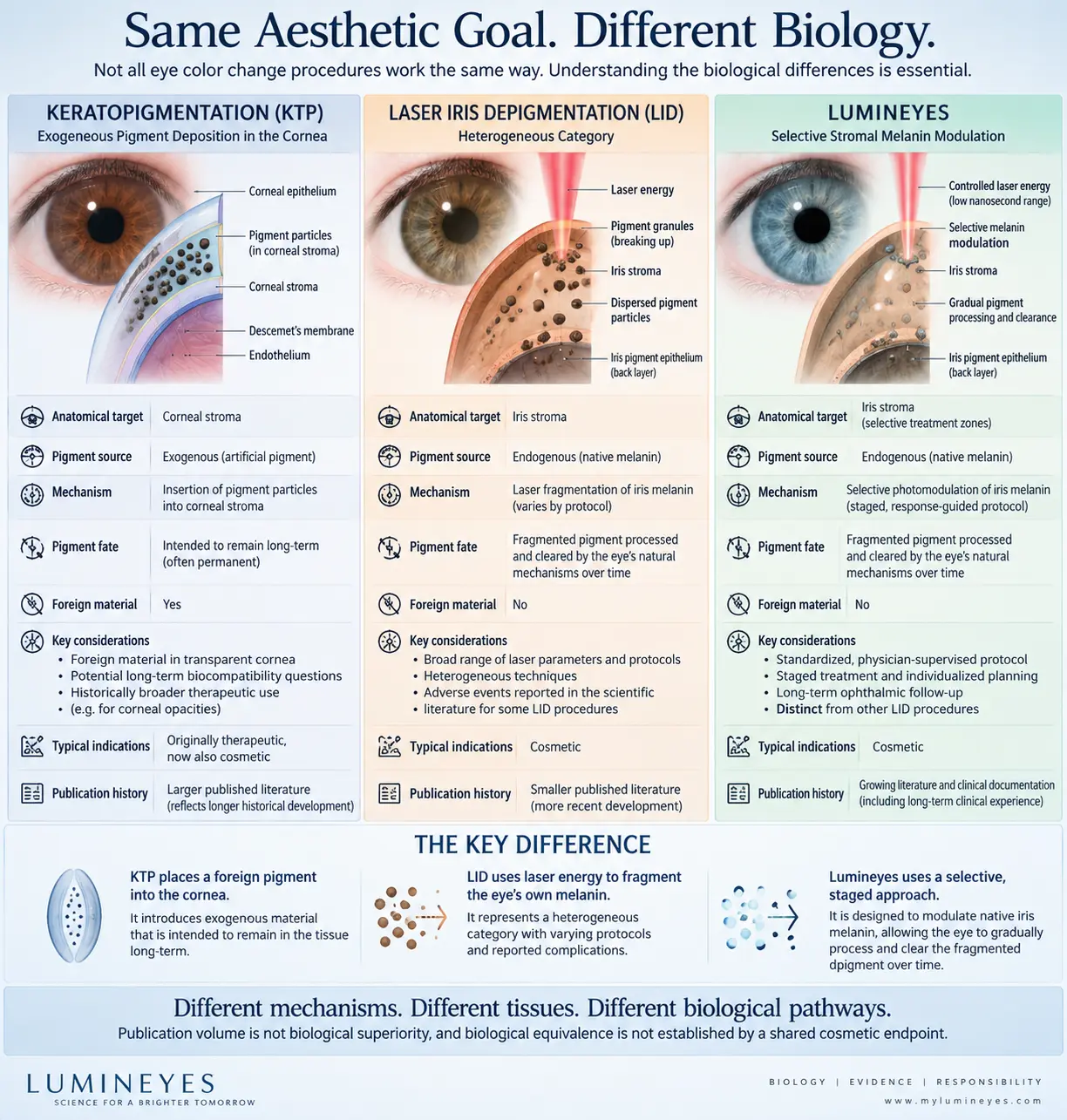
Cosmetic keratopigmentation relies on persistence of exogenous pigment within the corneal stroma. Cosmetic iris implants introduce an artificial structure into the anterior segment. Laser-based iris pigment modification instead interacts with native iris pigmentation and therefore raises a different biological question: how is altered or liberated endogenous pigment processed over time?
A shared cosmetic endpoint therefore does not establish biological equivalence.
Apparent eye color is an optical phenotype. It does not reveal the biological process that produced it. A lighter or differently colored eye may result from deposition of exogenous pigment, placement of an artificial structure, or modification of pigmentation that was already present within the iris.
Meaningful scientific comparison therefore begins with the anatomical compartment, pigment origin and intended biological fate, rather than with the final visible color alone.
Cosmetic keratopigmentation introduces pigment into the corneal stroma. Persistence of that introduced material within the target tissue contributes directly to the intended optical effect.
Cosmetic iris implantation changes appearance through placement of an artificial structure within the anterior segment rather than by modifying native iris pigment.
Iris-directed laser procedures interact with pigment already present within iris tissue. The subsequent biological question concerns pigment modification, redistribution, processing, outflow interaction and clearance.
Modification of endogenous iris pigment does not end biologically when laser exposure ends. Pigment-containing material may undergo redistribution within the anterior segment and may encounter cellular-processing and aqueous-outflow pathways before the ocular environment returns toward baseline.
The clinically relevant variables therefore extend beyond the fact that pigment has been altered. They include the quantity and characteristics of liberated material, the rate at which it appears, its spatial distribution, inflammatory activity, trabecular interaction, cellular processing and clearance kinetics.
This sequence is conceptual. Individual domains may overlap and should not be interpreted as a validated fixed biological timetable.
The presence of pigment-containing material within the aqueous environment does not, by itself, establish pathological accumulation. Biological significance depends on the relationship between release, redistribution, processing and clearance over time.
A transient pigment burden that is progressively processed represents a different biological state from persistent particulate accumulation accompanied by sustained inflammation, impaired outflow or pressure elevation.
This temporal distinction is developed further in the Temporal Response Architecture and the Pigment Clearance Kinetics research records.
Ophthalmology already contains established procedures in which pigmented anterior-segment tissues interact with laser energy or in which iris pigment and particulate material may enter the anterior chamber. These procedures are not equivalent to cosmetic iris pigment modification, but they provide useful mechanistic context.
SLT interacts with melanin-containing cells within the trabecular meshwork. Experimental work demonstrates inflammatory and cellular signaling, including recruitment of monocytes and macrophages, together with modulation of conventional aqueous outflow.
Peripheral iridotomy intentionally photodisrupts iris tissue. Pigment and other particulate material may enter the anterior chamber, and an early transient IOP rise is a recognized post-laser response.
These precedents do not transfer a safety profile to another procedure. They demonstrate a narrower physiological principle: anterior-segment pigment exposure must be interpreted through burden, kinetics, inflammation, outflow response and recovery.
Pigment presence ≠ pigment toxicity ≠ chronic outflow failure
Pigment dispersion is an observation. Persistent inflammation, sustained IOP elevation and structural outflow disease are clinical states. Their relationship cannot be inferred from pigment detection alone.
Multiple treatment sessions are not automatically equivalent to response-guided staged treatment. The biological rationale for staging is that an earlier exposure may change the pigment burden and ocular state that exist before any subsequent exposure.
Within the MyLumineyes framework, treatment progression is therefore conceptualized as:
Response-guided staging is a clinical framework, not a validated universal retreatment algorithm.
A scientifically useful comparison should not begin with the question “Which procedure changes eye color?” It should examine the biological intervention itself.
| Biological dimension | Keratopigmentation | Endogenous iris pigment modulation |
|---|---|---|
| Primary compartment | Corneal stroma | Iris stroma |
| Pigment origin | Exogenous | Endogenous |
| Primary optical strategy | Pigment deposition | Reduction / modulation of native pigment contribution |
| Artificial pigment introduced | Yes | No intended pigment addition |
| Desired pigment fate | Persistence within target tissue contributes to the optical result | Biological processing and reduction of the optical contribution of native pigment |
| Relevant biological question | How does deposited pigment interact with corneal tissue over time? | How is modified or liberated native pigment processed and cleared? |
| Post-treatment response | Tissue response to introduced material and surgical manipulation | Pigment redistribution, cellular response, outflow interaction and recovery |
| Long-term evidence base | More extensive published literature | Smaller published evidence base; continued systematic documentation required |
This table describes biological differences and should not be interpreted as a safety ranking.
The fact that two procedures involve different tissues, pigment sources and biological pathways is scientifically important. It does not, however, establish that one procedure has superior long-term safety.
Likewise, a larger published literature does not by itself establish biological superiority. Historical maturity, broader clinical adoption and a longer period of observation naturally create greater opportunity for publication.
Published LID reports include different wavelengths, pulse characteristics, energies, treatment geometries, treated areas, staging strategies, monitoring protocols and patient populations. Complications reported after one procedure therefore establish possibility within the broader literature but should not automatically be assigned to every other laser-based protocol.
Meaningful comparison requires procedural and biological comparability rather than classification by the word “laser” alone.
See the Laser Iris Depigmentation — Clinical Perspective for the protocol-specific discussion.
A pigment-fate model becomes clinically useful only when its proposed biological domains can be measured longitudinally in the same eyes.
Can treatment-related pigment redistribution and clearance be quantified reproducibly across serial examinations?
How do pigment burden, anterior-chamber activity and trabecular interaction relate to short- and long-term IOP behavior?
Does the direction of change over time distinguish transient biological processing from persistent or clinically significant pigment accumulation?
The sources below represent major evidence domains relevant to anatomical compartment, pigment deposition, laser iris procedures, trabecular response and anterior-segment laser biology.
A shared cosmetic endpoint does not establish biological equivalence.
Understanding eye-color modification requires attention to anatomical compartment, pigment origin, tissue interaction, pigment persistence or clearance, inflammatory response, aqueous-outflow interaction and longitudinal clinical outcome—not simply the final visible color.
Lumineyes Assistant
×