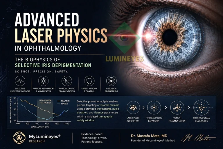
Laser Physics in Ophthalmology: A Scientific Review of Selective Iris Depigmentation
Selective iris depigmentation sits at the intersection of ophthalmic laser physics, tissue optics, pigment biology, anterior-segment physiology, and clinical decision-making. The central engineering problem is not simply how to deliver laser energy to the iris, but how to confine an optical interaction to melanin-rich anterior stromal targets while limiting unnecessary exposure of adjacent tissue and accounting for the biological consequences of liberated pigment.
Abstract
Laser iris depigmentation is a technically demanding application of biomedical optics because wavelength, pulse duration, pulse energy, fluence, spot geometry, beam profile, repetition pattern, treatment interval, and tissue pigmentation interact rather than operate independently. This review examines the physical principles relevant to selective stromal pigment targeting, including wavelength-dependent absorption, thermal confinement, selective photothermolysis, photomechanical and photoacoustic contributions, fluence, spot-size effects, and spatial beam homogeneity. It then connects these engineering variables with ocular biology: pigment fragmentation, inflammatory response, aqueous outflow, intraocular-pressure surveillance, and staged treatment planning.
The review also distinguishes well-established laser-tissue principles from the still-limited clinical evidence base for cosmetic laser iris depigmentation. Published reports confirm that laser-tissue interaction can be modeled using established principles of selective absorption and energy confinement, but they do not justify assuming that all devices, parameters, operators, or treatment protocols are clinically equivalent. In this context, the Lumineyes™ methodology is discussed as a specific response-guided, staged clinical framework rather than as a synonym for the heterogeneous laser iris depigmentation literature.
Quick Answer
- The target is melanin, not “eye color” itself. Apparent color change follows reduction and biological clearance of selected anterior stromal pigment.
- Energy alone does not define the tissue effect. Wavelength, pulse duration, fluence, spot size, beam profile, repetition pattern, and tissue state must be interpreted together.
- Physics does not replace clinical control. Pigment liberation can alter anterior-chamber inflammatory load and aqueous outflow; therefore staged exposure, clinical reassessment, and intraocular-pressure monitoring remain central to risk management.
Keywords: laser physics; selective photothermolysis; laser-tissue interaction; biomedical optics; ophthalmology; iris depigmentation; melanin; fluence; photomechanical effects; photoacoustic effects; anterior segment; laser engineering; staged treatment.
1. From Cosmetic Description to a Laser–Tissue Interaction Problem
Eye-color change is often described according to its visible endpoint, yet the clinically relevant process begins much earlier at the level of photon absorption, tissue architecture, pigment density, and biological response. In anterior-segment laser work, identical nominal pulse energies do not necessarily produce identical effects. The same energy can generate different local fluence when spot size changes; different absorption when wavelength changes; different thermal confinement when pulse duration changes; and different tissue response when pigment density, stromal geometry, or prior exposure differs.
For this reason, selective iris depigmentation is better understood as a controlled laser–tissue interaction problem than as a simple pigment-removal procedure. The goal is to produce a selective effect in melanin-rich superficial stromal structures while preserving the broader architecture of the iris and avoiding unnecessary energy transfer to adjacent ocular structures. This conceptual approach is consistent with the wider principles of ophthalmic laser engineering, tissue optics, and selective photothermolysis.
The clinical literature on cosmetic laser iris depigmentation remains substantially smaller and more heterogeneous than the foundational literature on laser–tissue interaction itself. Published reviews have specifically emphasized the limited number of independent clinical studies and the need for additional long-term evidence. Accordingly, the physical plausibility of selective pigment targeting should not be conflated with proof that every implementation of laser iris depigmentation is safe, standardized, or equivalent.
2. Terminological Framework
To separate the physical target, the biological consequence, and the clinical control strategy, the Lumineyes™ research framework uses three linked terms. These terms are intended to describe the logic of the methodology rather than to redefine established laser-physics terminology.
These three layers are intentionally distinct. A laser may create a physically selective interaction, yet the eye must still process the resulting pigment and inflammatory load. Likewise, a technically successful pulse does not by itself determine whether another exposure is appropriate. This is why engineering parameters and biological surveillance must be considered together.
3. The Core Physical Principle: Selective Photothermolysis
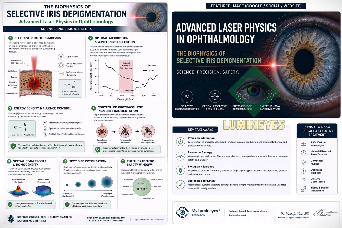
Anderson and Parrish's theory of selective photothermolysis established that appropriately chosen optical radiation can preferentially affect an absorbing target when wavelength and pulse duration are selected to favor the chromophore and to confine energy before substantial heat diffuses into surrounding tissue. Melanin is a broadband absorber across visible wavelengths, making it a biologically relevant chromophore for pigment-targeting laser systems.
In simplified form, thermal relaxation time can be related to target dimension and thermal diffusivity:
where d represents an effective target dimension and α the thermal diffusivity of the surrounding medium. The equation is useful as a conceptual model: smaller targets generally dissipate heat more rapidly than larger targets, and pulse duration therefore influences whether deposited energy remains locally confined or spreads into adjacent tissue.
The iris, however, is not a homogeneous slab of isolated pigment particles. It is a vascular, contractile, highly structured tissue with variable pigmentation, crypts, furrows, stromal thickness, and regional differences in optical accessibility. Consequently, thermal-relaxation concepts should guide—not oversimplify—the interpretation of ocular laser exposure.
| Parameter | Primary physical role | Why it matters clinically |
|---|---|---|
| Wavelength | Determines relative chromophore absorption and tissue penetration. | Influences how preferentially optical energy interacts with melanin compared with surrounding structures. |
| Pulse duration | Influences thermal confinement and the balance between photothermal and stress-related effects. | Too long a pulse may increase unwanted thermal spread; extremely short high-peak-power interactions may produce different mechanical effects. |
| Pulse energy | Total energy delivered in each pulse. | Cannot be interpreted without spot area, pulse duration, beam profile, and tissue absorption. |
| Fluence | Energy per unit area. | More directly represents local optical exposure than pulse energy alone. |
| Spot size | Defines irradiated area and modifies local energy density. | Small changes in diameter can materially change fluence for the same pulse energy. |
| Beam profile | Defines spatial energy distribution within the spot. | Central peaks or local hot spots may create non-uniform tissue exposure despite identical nominal energy. |
| Repetition pattern | Determines temporal accumulation of multiple exposures. | Biological burden depends on cumulative exposure and the interval available for tissue response. |
4. Wavelength Selection and Melanin Absorption
Wavelength is one of the first determinants of laser selectivity because biological chromophores differ in their absorption spectra. Melanin absorbs broadly across the visible and near-infrared spectrum, but its absorption is not constant across wavelengths. At the same time, ocular tissues contain other potential absorbers and scattering structures; therefore wavelength should never be evaluated in isolation.
The practical objective is not simply to choose a wavelength that melanin can absorb. It is to establish a wavelength–pulse–fluence combination in which the intended pigment interaction occurs with the lowest reasonable collateral exposure. General tissue-optics literature supports this wavelength-dependent framework, while the exact clinical behavior of a given iris-treatment platform must still be established by its own technical validation and clinical evidence.
This distinction is particularly important for cellular selectivity in laser eye-color change. A wavelength may be physically compatible with melanin targeting, yet the resulting clinical response still depends on tissue pigmentation, optical geometry, treatment density, and the cumulative amount of liberated pigment.
5. Fluence: Why Pulse Energy Alone Is an Incomplete Metric
Fluence, commonly expressed as energy per unit area, is a central quantity in laser dosimetry:
where E is pulse energy and A is the irradiated area. Because spot area changes with the square of radius, modest changes in spot diameter can produce disproportionately important changes in energy density. A pulse-energy value therefore becomes clinically meaningful only when interpreted together with spot geometry and beam profile.
The concept of a therapeutic window follows from this relationship. Exposure below the interaction threshold may produce little useful pigment effect, whereas excessive local exposure may increase tissue stress, inflammatory response, or disruption beyond the desired target. In practice, however, a single universal numerical window should not be assumed across different devices or protocols because optical delivery systems, pulse shape, beam homogeneity, focusing, and tissue characteristics differ.
6. Photothermal, Photomechanical and Photoacoustic Components
Laser–tissue interaction is often described using simplified labels, but pulsed irradiation can generate overlapping mechanisms. Depending on pulse duration, irradiance, absorption, and confinement, energy deposition may produce thermal effects, thermoelastic stress, pressure transients, plasma-mediated effects, or combinations of these phenomena. Vogel and Venugopalan's review of pulsed laser ablation illustrates why interaction mechanism must be inferred from the complete pulse regime rather than from wavelength alone.
For selective iris pigment treatment, the relevant objective is controlled disruption of melanin-containing structures without unnecessary structural damage. Describing this process as purely “photoacoustic” or purely “photothermal” can therefore be misleading unless the specific pulse characteristics and measured tissue effects justify the label. A more defensible engineering description is that the treatment seeks selective optical absorption with localized thermal and, depending on the pulse regime, stress-related mechanical contributions.
7. Beam Profile, Hot Spots and Spatial Homogeneity
Nominal pulse energy describes how much energy leaves the system, but not how that energy is distributed across the treatment spot. A Gaussian-like beam concentrates greater intensity near the center, whereas a flatter spatial profile distributes energy more uniformly. Optical aberrations, focusing errors, contamination, misalignment, and beam-shaping components can further alter local intensity.
This is clinically relevant because two systems with the same wavelength, spot diameter, and pulse energy may still expose tissue differently if one produces a pronounced central peak. Spatial beam homogeneity therefore belongs to the same safety conversation as wavelength and fluence. Calibration should address not only nominal output but also the reproducibility and geometry of the delivered beam.
| Beam characteristic | Potential optical consequence | Clinical relevance |
|---|---|---|
| Central energy peak | Higher local irradiance than the average fluence suggests. | May increase focal tissue stress or produce uneven pigment response. |
| Flat / homogenized profile | More uniform spatial energy distribution. | May improve reproducibility when the optical system is properly calibrated. |
| Irregular / distorted profile | Unpredictable local peaks and valleys. | Reduces confidence in nominal dose calculations. |
| Variable focus | Changes effective spot size at tissue. | Alters local fluence even if the selected energy remains unchanged. |
8. Spot Size as a Coupled Variable
Spot diameter determines more than treatment precision. Because spot area directly enters the fluence calculation, changing spot size changes local energy density even when pulse energy remains fixed. Smaller spots can increase spatial precision but also raise fluence unless energy is correspondingly adjusted. Larger spots distribute energy over a wider area but may alter optical coupling, treatment density, and the amount of pigment affected per exposure.
Spot size should therefore be treated as a coupled variable, not as an independent setting. Meaningful optimization requires consideration of beam diameter at the tissue plane, pulse energy, beam profile, focusing, pulse duration, target pigmentation, and treatment spacing.
9. The Biological Phase Begins When the Laser Pulse Ends
Pigment fragmentation is only the beginning of the biological process. Once pigment-containing structures are disrupted, the anterior segment must handle liberated pigment, cellular debris, and the associated inflammatory response. These events may involve phagocytic processing, redistribution of particulate material, and movement through aqueous pathways. The visible change in iris appearance therefore evolves over time rather than being fully determined at the instant of exposure.
This biological phase explains why the concepts of pigment clearance and biological permanence cannot be reduced to laser energy alone. Individual variability in pigment density, stromal architecture, inflammatory response, aqueous dynamics, and previous treatment exposure can alter both the pace and magnitude of visible change.
9.1 Anterior-chamber pigment load and intraocular pressure
Liberation of iris pigment is especially relevant to aqueous outflow. Pigment and inflammatory material can interact with the trabecular outflow pathway, making intraocular-pressure surveillance an important component of clinical management. Published laser-iris-depigmentation literature includes reports of transient pressure elevation as well as severe pressure-related complications in individual cases; these reports do not establish the incidence for every protocol, but they demonstrate why pressure cannot be treated as a peripheral variable.
A modern clinical framework should therefore connect laser exposure to post-treatment biological response rather than treating each session as an isolated technical event. When a clinically meaningful response is present, automatic re-exposure is difficult to justify solely because a preplanned session count has not yet been completed.
10. Response-Guided Staging Rather Than a Universal Session Count
A central principle of the Lumineyes™ methodology is that there is no scientifically defensible fixed number of laser sessions that can be applied to every iris. Treatment burden depends on baseline pigmentation, stromal architecture, regional heterogeneity, biological clearance, the magnitude of treatment-related pigment response, and the eye's recovery between exposures.
The logic of response-guided staged treatment is therefore to separate technical capability from clinical permission to continue. A device may be capable of delivering another exposure, yet the biological state of the eye may indicate that further exposure should be postponed or stopped. This distinction is particularly important in elective treatment, where the tolerance for preventable ocular risk must remain low.
The related clinical framework for laser eye-color change expands this concept by integrating ophthalmic examination, treatment response, intraocular pressure, and staged decision-making rather than relying on color outcome alone.
11. The 8G Xtra Laser System: A Proprietary Engineering Context
Within the Lumineyes™ development program, the 8G Xtra Laser System represents an engineering effort to control several interacting variables—optical delivery, pulse characteristics, treatment geometry, and staged exposure—within a unified clinical workflow. The design philosophy is based on the principle that no single specification can independently guarantee selective tissue interaction.
It is important to distinguish this design philosophy from independent clinical validation. The foundational literature cited in this review supports the underlying physics of wavelength-dependent absorption, energy confinement, beam geometry, and pulsed tissue interaction. It does not independently validate proprietary device performance. Device-specific claims should therefore be supported by technical characterization, prospective clinical data, and transparent reporting of treatment parameters where appropriate.
This distinction strengthens rather than diminishes the Lumineyes™ research position: the methodology should be evaluated on its own documented protocol, response-guided logic, safety monitoring, and clinical evidence rather than being assumed to be interchangeable with every historical form of laser iris depigmentation.
12. Clinical Evidence and Safety: What Physics Can—and Cannot—Prove
Selective photothermolysis is a well-established physical principle. The optical properties of biological tissues, including wavelength-dependent absorption and scattering, are extensively documented. Pulsed laser–tissue interactions have also been studied for decades. These bodies of evidence provide a strong theoretical foundation for selective pigment targeting.
The clinical evidence for cosmetic laser iris depigmentation is a separate question. Reviews published in 2022 and 2025 described the literature as limited and emphasized the need for broader independent validation and long-term follow-up. Case reports have documented adverse events including iris perforation, pigment-related pressure elevation, and more recently markedly elevated intraocular pressure with additional corneal complications after treatment performed elsewhere. Case reports cannot quantify population-level incidence, identify the risk of a different protocol, or establish causality for every component of a complex postoperative course; nevertheless, they are relevant safety signals.
For that reason, a scientifically responsible account of iris depigmentation should avoid two opposite errors. It should not dismiss the procedure solely because complications have been reported with heterogeneous techniques, but it should also not infer safety simply from the elegance of the underlying physics. Safety is a property of the complete clinical system: patient selection, device characteristics, calibration, exposure strategy, operator judgment, postoperative management, monitoring, and the decision to stop.
| Evidence layer | What it can support | What it cannot establish by itself |
|---|---|---|
| Foundational laser physics | Selective absorption, thermal confinement, fluence, tissue optics, pulsed interaction mechanisms. | Clinical safety of a particular iris-treatment protocol. |
| Device engineering | Output stability, pulse characteristics, beam profile, spot geometry, optical calibration. | Long-term patient outcomes without clinical follow-up. |
| Clinical case reports | Identification of possible complications and failure modes. | Population incidence or comparative safety ranking. |
| Prospective / longitudinal clinical data | More reliable estimates of efficacy, ocular response, and adverse-event patterns within a defined protocol. | Generalization to unrelated devices or protocols without external validation. |
13. Why Clinical Expertise Remains Essential
Laser engineering can improve precision, but it cannot substitute for ophthalmic judgment. The iris is part of a dynamic anterior-segment system. Appropriate assessment includes not only pigmentation but also anatomy, intraocular pressure, anterior-chamber findings, corneal status, prior ocular history, expected adherence, and the eye's response to previous exposure.
Clinical expertise is especially important because a technically “successful” pigment interaction may still be followed by a biological response that changes the risk of further treatment. Monitoring therefore should be designed to answer a clinical question: has the eye recovered sufficiently that another exposure can be considered? This is fundamentally different from asking whether more pigment remains visible.
Readers seeking broader clinical context can review the site's laser eye-color change overview, ocular safety discussion, and off-label treatment methodology.
14. Future Directions in Ophthalmic Laser Engineering
The next advances in selective iris treatment are likely to come from measurement and feedback rather than simply from higher power. Potential areas include high-resolution iris mapping, objective optical phenotyping, improved beam characterization, automated dosimetry, real-time imaging, anterior-segment OCT integration, treatment-density mapping, and software that records regional exposure over time.
A particularly important direction is the transition from fixed parameter tables toward closed-loop treatment systems. In such systems, the next exposure would depend on measured anatomy and biological response rather than on a predetermined number of sessions. This approach is consistent with the broader Lumineyes™ research program, which treats visible appearance, iris structure, laser exposure, and postoperative response as linked but separable data domains.
Future evidence should also improve transparency. Useful studies would report device type, wavelength, pulse duration, spot geometry, energy range, treatment density, session spacing, intraocular-pressure monitoring, anterior-segment findings, endothelial measurements where relevant, and long-term follow-up. Without these variables, apparently similar “laser iris depigmentation” studies may describe materially different procedures.
15. Conclusion
Selective iris depigmentation is best understood as a coupled optical–biological problem. Its physical foundation rests on well-established principles: wavelength-dependent absorption, selective photothermolysis, thermal confinement, pulsed laser–tissue interaction, fluence, spot geometry, and spatial beam distribution. Yet these principles describe how a selective interaction may be engineered; they do not independently prove the safety of a clinical protocol.
The most defensible clinical model therefore combines engineering precision with biological restraint. In the Lumineyes™ framework, this is expressed through selective stromal melanin modulation, observation of the controlled treatment-related pigment response, and response-guided staged treatment. The methodology is intentionally distinguished from the heterogeneous historical LID literature and should ultimately be judged by transparent protocol definition, prospective measurements, long-term outcomes, and reproducible safety data.
Progress in this field will depend less on claims of maximal energy or maximal speed than on better characterization of optical delivery, better measurement of tissue response, and stricter rules governing when further exposure should—or should not—occur.
References
- Anderson RR, Parrish JA. Selective photothermolysis: precise microsurgery by selective absorption of pulsed radiation. Science. 1983;220(4596):524–527. doi:10.1126/science.6836297.
- Jacques SL. Optical properties of biological tissues: a review. Physics in Medicine & Biology. 2013;58(11):R37–R61. doi:10.1088/0031-9155/58/11/R37.
- Mainster MA. Ophthalmic applications of lasers. Survey of Ophthalmology. 1997;42(2):101–120. doi:10.1016/S0039-6257(97)00031-8.
- Niemz MH. Laser-Tissue Interactions: Fundamentals and Applications. 5th ed. Springer; 2019. doi:10.1007/978-3-030-10520-5.
- Vogel A, Venugopalan V. Mechanisms of pulsed laser ablation of biological tissues. Chemical Reviews. 2003;103(2):577–644. doi:10.1021/cr010379n.
- Welch AJ, van Gemert MJC, eds. Optical-Thermal Response of Laser-Irradiated Tissue. 2nd ed. Springer; 2011. doi:10.1007/978-90-481-8831-4.
- D'Oria F, Abu-Mustafa SK, Alio JL. Cosmetic change of the apparent color of the eye: a review on surgical alternatives, outcomes and complications. Ophthalmology and Therapy. 2022;11(2):465–477. doi:10.1007/s40123-022-00458-2.
- Ferrari F. Case report of an iris perforation after YAG laser iris depigmentation for aesthetic reasons. Journal Français d'Ophtalmologie. 2021;44(5):e283–e285. doi:10.1016/j.jfo.2020.07.016.
- D'Oria F, Alio JL. Surgical techniques for cosmetic eye color change: a narrative review. Ophthalmology and Therapy. 2025;14(8):1685–1694. doi:10.1007/s40123-025-01177-0.
- Jaber W, Nemet M, Waisbourd M. Laser iris depigmentation resulting in markedly elevated intraocular pressure and herpes simplex keratitis with corneal scarring: case report. Case Reports in Ophthalmology. 2026;17(1):312–317. doi:10.1159/000551226.