Clinical Interpretation of Common Claims in Laser Eye Color Change: A Lumineyes™ Evidence Framework
Mechanism, iris biology, pigment response, IOP kinetics, procedure-specific risk, optical phenotype, and the boundaries between published evidence and clinical frameworks
Quick Answer
Laser eye color change cannot be interpreted responsibly through slogans such as “instant color conversion,” “pigment release always means glaucoma,” or “all eye-color procedures carry the same risk.” These statements collapse different biological endpoints and different procedures into categories that are too broad for clinical use.
The Lumineyes™ framework approaches laser iris depigmentation as a protocol-specific, staged interaction with native iris pigment. Its terminology separates the intended target (selective stromal melanin modulation), the observed biological response (controlled treatment-related pigment response), and the decision to re-expose (response-guided staged treatment). These concepts are presented as a clinical framework under continued evaluation, not as independent proof of superior safety or efficacy.
For the canonical overview of the procedure itself, see Laser Eye Color Change. This Research Archive article has a narrower purpose: to show how common claims should be separated into established biology, published procedure-specific evidence, first-hand clinical observation, and testable research hypotheses.

1. Why Evidence-Specific Interpretation Matters
Public discussion of cosmetic eye-color procedures frequently merges several different questions: Does the eye become lighter? What tissue is altered? What complications are biologically possible? How often do those complications occur under a specific protocol? Does a normal postoperative measurement prove long-term safety? These are not the same scientific question, and one cannot be answered reliably by evidence designed for another.
This distinction is especially important in laser iris depigmentation (LID). Published literature includes a prospective single-center series, narrative reviews, and case reports of significant complications. These sources are valuable, but they represent different evidence types and should not be pooled rhetorically into a single conclusion without accounting for device parameters, treatment density, session interval, patient selection, postoperative management, and follow-up duration.[1–6]
Evidence should be interpreted at the level at which it was generated. A case report can establish that an event is possible; a single-center series can describe outcomes under a defined protocol; a review can synthesize available literature; and a clinic archive can document first-hand experience. None of these, by itself, creates a universal risk rate for every laser iris depigmentation protocol.
| Evidence category | What it can contribute | What it cannot establish by itself | Example in this article |
|---|---|---|---|
| Established biology External anatomical / physiological literature | Defines iris pigmentation biology, aqueous-outflow physiology, glaucoma definitions, and general laser–tissue principles. | Does not validate the clinical performance of a specific proprietary LID protocol. | Human iris pigmentation studies; pigment dispersion / pigmentary glaucoma literature; selective photothermolysis. |
| Published LID evidence Series, reviews, case reports | Documents outcomes, complications, and limitations reported for specific clinical applications. | Does not automatically transfer incidence rates or outcomes across devices, schedules, or operators. | Ruiz 2021; D'Oria et al. 2022; Liu et al. 2023; D'Oria & Alió 2025; Jaber et al. 2026. |
| First-hand clinical archive Lumineyes™ documentation | Can preserve protocol history, longitudinal observations, decision logic, and real-world follow-up. | Is not equivalent to independently audited or peer-reviewed outcome evidence. | Clinical data pages, monitoring frameworks, case documentation, protocol evolution. |
| Conceptual framework Testable terminology / models | Creates explicit definitions, measurable hypotheses, and a structure for prospective research. | Does not become validated simply because it is clinically plausible or internally consistent. | SSMM, controlled treatment-related pigment response, response-guided staged treatment, L-SAFE, L-OAT. |
| Not established Claims beyond available evidence | Identifies questions that still require prospective or independent confirmation. | Should not be presented as a settled fact. | Universal protocol superiority, guaranteed final shade, zero-risk claims, or proof of microscopic selectivity from photography alone. |
2. Five Common Claims That Require Clinical Separation
“Laser eye color change is an immediate color conversion.”
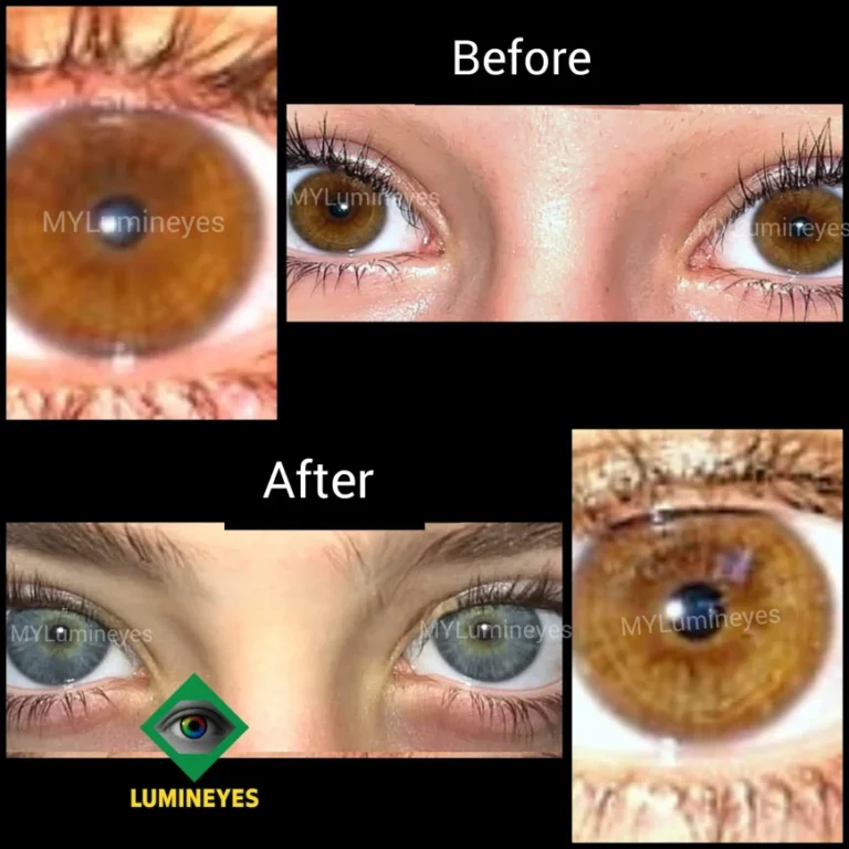
This statement confuses laser exposure with the final visual phenotype. Human iris color reflects the amount and optical distribution of melanin in the anterior iris, together with stromal architecture and light scattering. Histological work has shown that normal differences in iris color are related primarily to melanin content within a broadly similar population of stromal melanocytes rather than to a simple difference in melanocyte number.[7,8]
Within the Lumineyes™ interpretation, the laser exposure initiates a biological sequence rather than applying an artificial color. The visible appearance can evolve as pigment burden changes and as the residual stromal architecture contributes differently to light scattering. This is why pigment-clearance kinetics, baseline pigmentation, and the anatomy of the iris stroma are relevant to outcome interpretation.
Accordingly, “instant color change” is not a precise clinical endpoint. A more appropriate description is progressive visible phenotype evolution following pigment-modifying exposure.
“Any pigment release automatically means glaucoma.”
This statement merges three different phenomena: pigment liberation, intraocular-pressure (IOP) response, and glaucomatous optic neuropathy. They are related, but they are not interchangeable. Pigment dispersion syndromes demonstrate that liberated iris pigment can accumulate in the trabecular meshwork, reduce aqueous outflow, and contribute to ocular hypertension or pigmentary glaucoma in susceptible eyes.[9,10] That physiology provides an important reason to monitor pressure after pigment-altering procedures.
However, a transient postoperative pressure elevation is not, by definition, the same as chronic glaucoma. Glaucoma is defined by characteristic optic-nerve damage and functional loss, not by one pressure measurement alone. At the same time, transient does not mean trivial: severe pressure elevation after cosmetic iris laser procedures has been reported, including bilateral iatrogenic pigmentary glaucoma and more recent cases with markedly elevated IOP.[3,6]
The appropriate clinical position is therefore neither denial nor exaggeration. The relevant question is how pigment response, pressure kinetics, anterior-chamber findings, baseline anatomy, and recovery trajectory are monitored together. That logic is developed in the dedicated Early IOP Monitoring Framework.
“All cosmetic eye-color procedures have the same biological mechanism and the same risk logic.”
This is not a useful clinical assumption. Laser iris depigmentation modifies native iris pigment; keratopigmentation deposits exogenous pigment within the cornea; cosmetic iris implants introduce an intraocular device into the anterior segment. These interventions act on different tissues and create different surveillance priorities. Reviews of cosmetic eye-color surgery emphasize this procedural heterogeneity and also note that the long-term evidence base remains uneven across methods.[2,5]
A mechanistic difference does not prove that one procedure is universally safe or another universally unsafe. It means that outcomes and complications must be attributed to the tissue, material, device, technique, and follow-up system actually studied. For readers comparing procedures, see Eye Color Change Method Comparison, Keratopigmentation, and Eye Color Change Surgery.
“If postoperative IOP is normal, the eye has been proven structurally safe.”
IOP is an important functional variable, but it is not a histological assay of the trabecular meshwork, an endothelial cell count, an OCT map of the anterior segment, or a complete assessment of visual function. A normal reading can be reassuring for that time point while still leaving other questions unanswered.
The Lumineyes™ framework therefore treats pressure as part of a multidomain recovery assessment. The direction of change—resolving, persistent, or progressing—may be more informative than a single isolated value. This concept also connects to L-SAFE, a proposed closed-loop safety framework that separates response severity from response kinetics rather than treating every finding at the same nominal stage as biologically identical.
“A satisfactory final eye color is enough to define clinical success.”
Aesthetic appearance is an important patient-reported outcome, but it cannot stand alone as a clinical endpoint. A complete evaluation should distinguish visible phenotype from pressure behavior, inflammation, anterior-segment structure, corneal status, pupil function, visual symptoms, adverse events, and long-term stability.
This is also why guaranteed shade promises are scientifically weak. The final visible appearance depends not only on how much pigment is reduced but also on the native architecture of the iris and the way light interacts with the remaining stromal structure. The relationship between visible appearance, geometry, and optical signal is one of the questions addressed by the proposed Lumineyes Optical Architectural Typology (L-OAT) research framework described below.
3. The Lumineyes™ Conceptual Architecture
The Lumineyes™ research program uses three linked terms to distinguish the target, the response, and the next decision. These terms are intentionally narrower than the generic phrase laser iris depigmentation. They do not claim universal acceptance; their purpose is to make the protocol’s logic explicit enough to be examined, criticized, and prospectively tested.
Selective Stromal Melanin Modulation
SSMM describes the intended preferential interaction with the optically relevant melanin-bearing anterior stromal compartment, with progressive pigment reduction rather than intentional tissue ablation as the procedural objective.
Controlled Treatment-Related Pigment Response
The biological response to exposure—including pigment liberation, inflammatory activity, pressure behavior, and recovery—is treated as clinically meaningful information rather than background noise.
Response-Guided Staged Treatment
The next exposure is not defined solely by a fixed session count or calendar. The previous exposure’s response is reassessed before continuation, modification, deferral, or cessation is considered.
The dedicated Selective Stromal Melanin Modulation article defines the target concept in greater detail. Its key evidence boundary is important here: SSMM is a proposed clinical and procedural framework, not proof that laser energy interacts exclusively with pigment or that complete microscopic tissue preservation has already been demonstrated.
| Term | Primary question | Clinical function | Evidence boundary |
|---|---|---|---|
| Selective stromal melanin modulation | What is the intended target? | Defines the chromophore-bearing anterior stromal compartment and the intention to reduce pigment without deliberate stromal ablation. | Does not prove absolute cellular selectivity. |
| Controlled treatment-related pigment response | What happened after exposure? | Frames pigment liberation, inflammation, pressure behavior, and recovery as interpretable biological variables. | Does not imply that every response is harmless or desirable. |
| Response-guided staged treatment | What should happen next? | Uses the previous response to inform continuation, modification, delay, or cessation. | Does not prove that staging eliminates risk. |
4. L-SAFE and L-OAT: Extending the Framework Beyond Session Counting
L-SAFE: safety as state + trajectory
L-SAFE is a proposed Lumineyes™ closed-loop safety framework developed to formalize the difference between a response that is resolving and one that is progressing. The clinical premise is that two eyes with similar absolute findings can have different trajectories and therefore different implications for treatment readiness.
Its role in this article is conceptual: it reinforces the principle that re-exposure should be governed by the status of the previous response rather than by scheduling alone. L-SAFE is under development and should not be presented as an independently validated risk score.
L-OAT: phenotype as measurable architecture
Lumineyes Optical Architectural Typology (L-OAT) is a proposed multimodal hypothesis for quantitative iris phenotyping. It is intended to move beyond subjective color labels by relating visible appearance to iris geometry and OCT-derived optical signal.
Within the present framework, L-OAT addresses a different problem from L-SAFE: not “is the eye ready for another exposure?” but “how can baseline and evolving iris phenotype be represented quantitatively?” L-OAT remains conceptual and is not a validated diagnostic or predictive instrument. See the MyLumineyes™ Research Library for research-status documentation.
| Framework | Domain | Primary output | Current role |
|---|---|---|---|
| SSMM | Biophysical / anatomical target | Explicit definition of the intended melanin-bearing stromal target | Conceptual and procedural terminology |
| Controlled treatment-related pigment response | Biological response | Structured interpretation of pigment, inflammation, pressure and recovery | Clinical documentation framework |
| Response-guided staged treatment | Temporal decision-making | Continue / modify / defer / stop logic after reassessment | Clinical methodology |
| L-SAFE | Safety-control architecture | Severity state interpreted together with resolving vs progressing kinetics | Proposed closed-loop framework; validation required |
| L-OAT | Optical / structural phenotyping | Quantitative representation of visible phenotype, geometry and OCT-derived signal | Conceptual hypothesis; validation required |
5. Procedure-Specific Risk Interpretation
A clinically useful comparison begins with where the intervention occurs and what material or tissue is changed. It should not begin with a global label such as “eye-color surgery.” The table below therefore describes mechanism and surveillance domains rather than declaring an overall winner.
| Dimension | Laser iris depigmentation | Cosmetic keratopigmentation | Cosmetic iris implants |
|---|---|---|---|
| Primary tissue / location | Native iris, particularly the anterior pigment-bearing stromal compartment | Corneal stroma | Anterior segment with an intraocular implant positioned in front of the native iris |
| Material strategy | Reduces or modifies native pigment; no exogenous color pigment is added to the iris | Introduces exogenous pigment into corneal tissue | Introduces a synthetic intraocular device |
| Primary optical strategy | Changes the visible expression of native iris pigmentation | Changes apparent eye color by corneal pigment deposition | Changes apparent color by covering / altering the visible iris appearance |
| Key surveillance domains | Pigment response, inflammation, IOP / outflow, iris structure, pupil, cornea, retinal laser-related events | Corneal integrity, pigment distribution, infection / inflammation, endothelial status, visual quality | Endothelium, angle, inflammation, cataract, glaucoma, implant position and chronic intraocular effects |
| Evidence interpretation | Published evidence remains limited and protocol-heterogeneous; serious complications are documented in case reports | Published clinical literature is expanding, but technique and follow-up remain important to interpretation | Reviews document major complication concerns; regulatory and clinical context must be interpreted by jurisdiction and indication |
For a deeper biological comparison of deposited corneal pigment versus altered native iris pigment, see Biological Fate of Pigment in Cosmetic Iris Color Modification.
6. Patient Selection Is Foundational—but It Is Not the Whole Safety System
Online discussions often treat the laser platform itself as if it were the entire procedure. In reality, patient selection is a foundational safety prerequisite, but it does not replace appropriate energy delivery, staged exposure, postoperative surveillance, management of treatment-related responses, or long-term follow-up.
Within the Lumineyes™ framework, baseline assessment considers anterior-segment anatomy, pressure profile, iris phenotype, relevant ocular history, recovery behavior, and the ability to comply with follow-up. The exact eligibility and contraindication architecture is a separate methodological subject and should not be reduced to a few website checkboxes. For patient-facing context, see Who Is Not a Candidate for Eye Color Change? and Eye Color Change Safety.
Scope boundary: this article does not provide individualized candidacy rules or laser settings. Its purpose is evidence interpretation. Patient-specific suitability requires direct ophthalmic examination and clinical judgment.
7. Optical Reality: Color Is a Phenotype, Not a Paint Code
The visible iris phenotype is produced by more than pigment quantity alone. Human iris color reflects the interaction of melanin with stromal microstructure and light scattering.[7,8,11] As anterior pigment burden changes, the contribution of the underlying stromal optical architecture may become more apparent. This helps explain why nominally similar degrees of pigment reduction do not necessarily yield identical visible shades.
This point has practical and ethical implications. Guaranteed final-color promises imply a level of deterministic control that current tissue biology does not support. A more rigorous approach is to describe a range of plausible phenotype evolution while separating what can be measured from what remains uncertain. L-OAT was conceived precisely around that unresolved measurement problem: how to move from subjective labels such as “hazel,” “green,” or “light brown” toward a multimodal description of visible appearance, geometry, and optical signal.
8. What the Published Literature Actually Shows
| Source | What it contributes | What should not be inferred |
|---|---|---|
| Ruiz, 2021 — 1,176 eyes[1] | Prospective single-center clinical experience using a defined 532-nm Q-switched Nd:YAG protocol, with reported efficacy, predictability, satisfaction, follow-up, and complications. | It does not create a universal outcome or safety rate for all LID methods, including Lumineyes™. |
| D'Oria et al., 2022[2] | Review of cosmetic iris implants, laser iris depigmentation, and keratopigmentation, emphasizing different mechanisms, outcomes and complications. | A review of heterogeneous literature cannot substitute for protocol-specific prospective Lumineyes™ data. |
| Liu et al., 2023[3] | Documents severe bilateral iatrogenic pigmentary glaucoma and retinal vascular / macular injury after cosmetic iris laser treatment. | A case report does not establish incidence across all protocols; it establishes that severe complications are clinically possible. |
| Flores-Márquez et al., 2023[4] | Documents laser-induced maculopathy after cosmetic iris depigmentation. | It cannot quantify overall retinal complication risk, but it expands the relevant surveillance domain beyond the anterior segment. |
| D'Oria & Alió, 2025[5] | Updated narrative comparison of cosmetic eye-color techniques and continued emphasis on limited long-term LID evidence. | Its cross-procedure conclusions do not independently validate any proprietary LID protocol. |
| Jaber et al., 2026[6] | Reports markedly elevated IOP after multiple LID sessions, with additional corneal complications in one case. | Two cases cannot define a protocol-wide incidence rate; they reinforce the need for postoperative surveillance and protocol attribution. |
Safety message: laser iris depigmentation is not risk-free. Published literature includes pigment-related pressure elevation, pigmentary glaucoma, inflammation, macular injury, and corneal complications. “Staged,” “selective,” or “response-guided” terminology should never be used to imply that these risks disappear. The scientific question is whether a defined protocol can characterize, detect, manage, and ultimately quantify its own risk profile.
9. Evidence Boundaries for Lumineyes™
The strongest version of the Lumineyes™ research position is not an unqualified claim of superiority. It is a transparent separation between what is already established externally, what has been reported within the clinic, and what is still a research hypothesis.
| Statement | Status | Responsible interpretation |
|---|---|---|
| Anterior iris melanin and stromal optics are major determinants of visible iris color. | Established biology | Supported by human iris morphology and pigmentation literature. |
| Pigment liberation can interact with aqueous-outflow physiology and may be associated with IOP elevation. | Established principle | Supported by pigment dispersion / pigmentary glaucoma literature and relevant to LID monitoring, without implying biological equivalence. |
| LID can produce both desired cosmetic change and significant complications. | Published evidence | Supported by a clinical series, reviews and case reports; event frequency remains protocol-dependent and incompletely characterized. |
| SSMM accurately describes the intended Lumineyes™ treatment target. | Framework terminology | Useful as an operational definition; degree of microscopic selectivity requires prospective / experimental validation. |
| Response-guided staging is preferable to automatic fixed re-exposure for the Lumineyes™ protocol. | Clinical methodology | Mechanistically coherent and used as a clinical decision framework; comparative risk reduction still requires prospective evidence. |
| L-SAFE can classify safety state and kinetics. | Proposed framework | Conceptual closed-loop model under development; not yet a validated prediction score. |
| L-OAT can quantitatively phenotype the iris. | Conceptual hypothesis | Defines a multimodal measurement hypothesis; classification performance and reproducibility require validation. |
| Lumineyes™ has independently proven superior safety or efficacy to other eye-color procedures. | Not established | Should not be claimed without appropriately designed comparative evidence. |
10. From Clinical Archive to Testable Research
A research archive is most useful when it does more than repeat clinical claims. It should show how those claims could be tested. The MyLumineyes™ Research program is therefore organized around a progression from first-hand documentation to explicit definitions and, ultimately, protocol-specific analysis.
Document the protocol
Preserve treatment evolution, baseline assessment, response variables, follow-up, adverse events, and reasons for continuation or cessation.
Define the phenotype
Use structured iris classification and quantitative imaging concepts rather than relying only on subjective color names.
Define safety states
Separate absolute severity from response kinetics and make re-exposure decisions auditable.
Validate prospectively
Use prespecified endpoints, standardized imaging, IOP measurement, endothelial assessment, and complete adverse-event reporting.
Analyse longitudinally
Distinguish patients, eyes, sessions and follow-up periods; report missing data and uncertainty transparently.
Invite external scrutiny
Peer review, reproducible definitions, external replication and independent analysis address different layers of scientific confidence.
The How Clinical Evidence Is Evaluated in Laser Eye Color Change article explains this evidence philosophy in greater detail. The MyLumineyes™ Clinical Data page should be read as reported longitudinal clinical experience and data summaries unless and until individual analyses are formally defined, audited, and published under their stated research design.
11. Digital Misinformation Is a Communication Problem, Not the Scientific Center of the Article
The internet tends to reward certainty, novelty, visual transformation, and emotionally simple narratives. Medicine rarely behaves that way. Promotional content may compress a biological process into an “instant result,” while alarmist content may convert every temporary finding into a guaranteed chronic disease outcome. Both forms of simplification can erase the distinctions that matter clinically.
The appropriate response is not to replace negative slogans with positive slogans. It is to make the evidence category, mechanism, protocol, population, time frame, and uncertainty visible. That is the purpose of a research archive.
For a shorter public-facing version of this issue, see the external article Laser Eye Color Change: Clinical Reality vs Internet Myths. The present page is intentionally more technical and evidence-structured.
12. How to Read a Claim About Laser Eye Color Change
| Question | Why it matters |
|---|---|
| What exact procedure and protocol are being discussed? | “Laser eye color change” may conceal important differences in laser platform, wavelength, pulse characteristics, treatment density, interval and postoperative management. |
| Is the statement about possibility, incidence, or comparative risk? | A case report can establish possibility but cannot calculate incidence; a series can estimate outcomes within its own design but may not support cross-protocol comparison. |
| Are patients, eyes, and treatment sessions distinguished? | These denominators are not interchangeable and can materially change interpretation. |
| What is the follow-up period? | Early recovery, medium-term stability and long-term safety are different endpoints. |
| Are non-completers and adverse responses retained in the analysis? | Excluding patients who stop treatment can make a protocol appear more favorable than the complete clinical experience. |
| Is the outcome cosmetic, structural, physiological, or functional? | Color satisfaction cannot substitute for IOP, anatomy, endothelial health, visual function or adverse-event assessment. |
| Is a mechanism being presented as proof? | Biological plausibility can justify a hypothesis but does not by itself demonstrate clinical safety or efficacy. |
13. Limitations of the Present Framework
This article is a clinical commentary and evidence-interpretation framework. It does not report a new prospective cohort, randomized comparison, or independently audited Lumineyes™ dataset. Lumineyes™-specific concepts—including SSMM, controlled treatment-related pigment response, response-guided staged treatment, L-SAFE, and L-OAT—are presented with their current conceptual or clinical status and should not be interpreted as independently validated classifications unless supported by appropriate future evidence.
The broader LID literature also has important limitations. Published studies use different terminology, treatment parameters, follow-up periods and outcome definitions, and case reports are disproportionately useful for identifying severe events rather than estimating population incidence. Consequently, absence of a reported complication in one source is not proof that the event cannot occur, while occurrence in an individual case does not establish the frequency of that event under every protocol.
14. Conclusion
The most clinically useful question in laser eye color change is not whether an online claim is “positive” or “negative.” It is whether the claim is mechanistically coherent, supported by the correct type of evidence, attributable to the actual protocol studied, and expressed with appropriate uncertainty.
The Lumineyes™ research framework is intended to make those distinctions explicit. It separates the intended pigment target from the biological response, separates the biological response from the decision to re-expose, separates IOP from the broader concept of ocular safety, and separates visible phenotype from the structural and optical architecture that produces it.
That approach does not remove uncertainty. It makes uncertainty measurable. For a developing area of aesthetic ophthalmology, that is a more scientifically useful starting point than either promotional certainty or undifferentiated fear.
Research FAQ
Is Lumineyes™ presented here as proven safer than every other laser iris depigmentation protocol?
No. The article describes a protocol-specific clinical and research framework. Independent comparative superiority requires appropriately designed comparative data.
Does pigment release automatically mean pigmentary glaucoma?
No. Pigment liberation, transient IOP elevation, ocular hypertension, and glaucomatous optic neuropathy are distinct clinical states. Pigment-related outflow stress is clinically relevant, however, and significant IOP elevation after cosmetic iris laser treatment has been reported.
What is selective stromal melanin modulation?
SSMM is the Lumineyes™ operational term for intended preferential interaction with the optically relevant melanin-bearing anterior stromal compartment, with progressive pigment reduction rather than intentional tissue ablation as the objective. It remains a framework requiring prospective validation of its degree of selectivity.
What is L-SAFE?
L-SAFE is a proposed closed-loop safety framework that distinguishes response severity from response kinetics, including whether a treatment-related finding is resolving or progressing. It is not presented as an independently validated prediction score.
What is L-OAT?
L-OAT, or Lumineyes Optical Architectural Typology, is a proposed multimodal framework for quantitative iris phenotyping using visible appearance, iris geometry and OCT-derived optical signal. It is conceptual and not yet a validated diagnostic or predictive instrument.
Why are keratopigmentation, iris implants and laser iris depigmentation not treated as one evidence category?
Because they act on different tissues and use different mechanisms: native iris pigment modification, corneal pigment deposition, and intraocular implantation. Their outcome measures and complication pathways therefore require procedure-specific interpretation.
References
- Ruiz PG. Photoablative cosmetic iridoplasty: effective, safe, and predictable—eye color change in 1176 eyes. Int Ophthalmol. 2021;41(4):1381-1393. doi:10.1007/s10792-021-01693-5. PMID: 33484383.
- D'Oria F, Abu-Mustafa SK, Alio JL. Cosmetic Change of the Apparent Color of the Eye: A Review on Surgical Alternatives, Outcomes and Complications. Ophthalmol Ther. 2022;11(2):465-477. doi:10.1007/s40123-022-00458-2. PMID: 35061240.
- Liu J, Korban S, Moster MR, Rhéaume MA, Wang Q. Bilateral severe iatrogenic pigmentary glaucoma following laser treatment for cosmetic iris color change. Am J Ophthalmol Case Rep. 2023;32:101927. doi:10.1016/j.ajoc.2023.101927. PMID: 37771388.
- Flores-Márquez A, Moreno-Gutiérrez JA, Chinchurreta-Capote A, García-Martín F, Rocha-de-Lossada C. Laser-induced maculopathy after iris depigmentation cosmetic treatment. Can J Ophthalmol. 2023;58(1):e29-e31. doi:10.1016/j.jcjo.2022.05.012. PMID: 35809624.
- D'Oria F, Alio JL. Surgical Techniques for Cosmetic Eye Color Change: A Narrative Review. Ophthalmol Ther. 2025;14(8):1685-1694. doi:10.1007/s40123-025-01177-0. PMID: 40483374.
- Jaber W, Nemet M, Waisbourd M. Laser Iris Depigmentation Resulting in Markedly Elevated Intraocular Pressure and Herpes Simplex Keratitis with Corneal Scarring: Case Report. Case Rep Ophthalmol. 2026;17(1):312-317. doi:10.1159/000551226.
- Imesch PD, Wallow IHL, Albert DM. The color of the human eye: a review of morphologic correlates and of some conditions that affect iridial pigmentation. Surv Ophthalmol. 1997;41 Suppl 2:S117-S123. doi:10.1016/S0039-6257(97)80018-5. PMID: 9154287.
- Wilkerson CL, Syed NA, Fisher MR, Robinson NL, Wallow IHL, Albert DM. Melanocytes and iris color: light microscopic findings. Arch Ophthalmol. 1996;114(4):437-442. doi:10.1001/archopht.1996.01100130433014. PMID: 8602782.
- Scuderi G, Contestabile MT, Scuderi L, Librando A, Fenicia V, Rahimi S. Pigment dispersion syndrome and pigmentary glaucoma: a review and update. Int Ophthalmol. 2019;39(7):1651-1662. doi:10.1007/s10792-018-0938-7. PMID: 29721842.
- Niyadurupola N, Broadway DC. Pigment dispersion syndrome and pigmentary glaucoma—a major review. Clin Exp Ophthalmol. 2008;36(9):868-882. doi:10.1111/j.1442-9071.2009.01920.x. PMID: 19278484.
- Sturm RA, Larsson M. Genetics of human iris colour and patterns. Pigment Cell Melanoma Res. 2009;22(5):544-562. doi:10.1111/j.1755-148X.2009.00606.x. PMID: 19619260.
- Anderson RR, Parrish JA. Selective photothermolysis: precise microsurgery by selective absorption of pulsed radiation. Science. 1983;220(4596):524-527. doi:10.1126/science.6836297. PMID: 6836297.
- Hu DN, Simon JD, Sarna T. Role of ocular melanin in ophthalmic physiology and pathology. Photochem Photobiol. 2008;84(3):639-644. doi:10.1111/j.1751-1097.2008.00316.x. PMID: 18346089.
- Mete M. Cosmetic Eye Colour Change Requires Procedure-Specific Evidence: A Commentary on Laser Iris Depigmentation and Keratopigmentation. Zenodo. 2026. Version 1.0. doi:10.5281/zenodo.23014030.
Methodological and conflict-of-interest disclosure. Mustafa Mete, MD, is the developer of the Lumineyes™ methodology and of the Lumineyes™-specific terminology and conceptual frameworks discussed on this page. Lumineyes™ clinical descriptions are therefore first-hand and should not be interpreted as independent validation. External literature is cited separately so that established biology, published LID evidence, clinic-based observation, and proposed frameworks remain distinguishable.
Medical information notice. This article is intended for scientific and educational communication. It does not provide individualized diagnosis, treatment instructions, laser settings, or candidacy decisions.
About the author. Mustafa Mete, MD, is an ophthalmologist and developer of the Lumineyes™ methodology. His research interests include laser–tissue interaction, iris pigmentation biology, anterior-segment response, IOP kinetics, optical phenotyping, and response-guided treatment frameworks. Additional project documentation is available through the MyLumineyes™ Research Hub and Research Library.