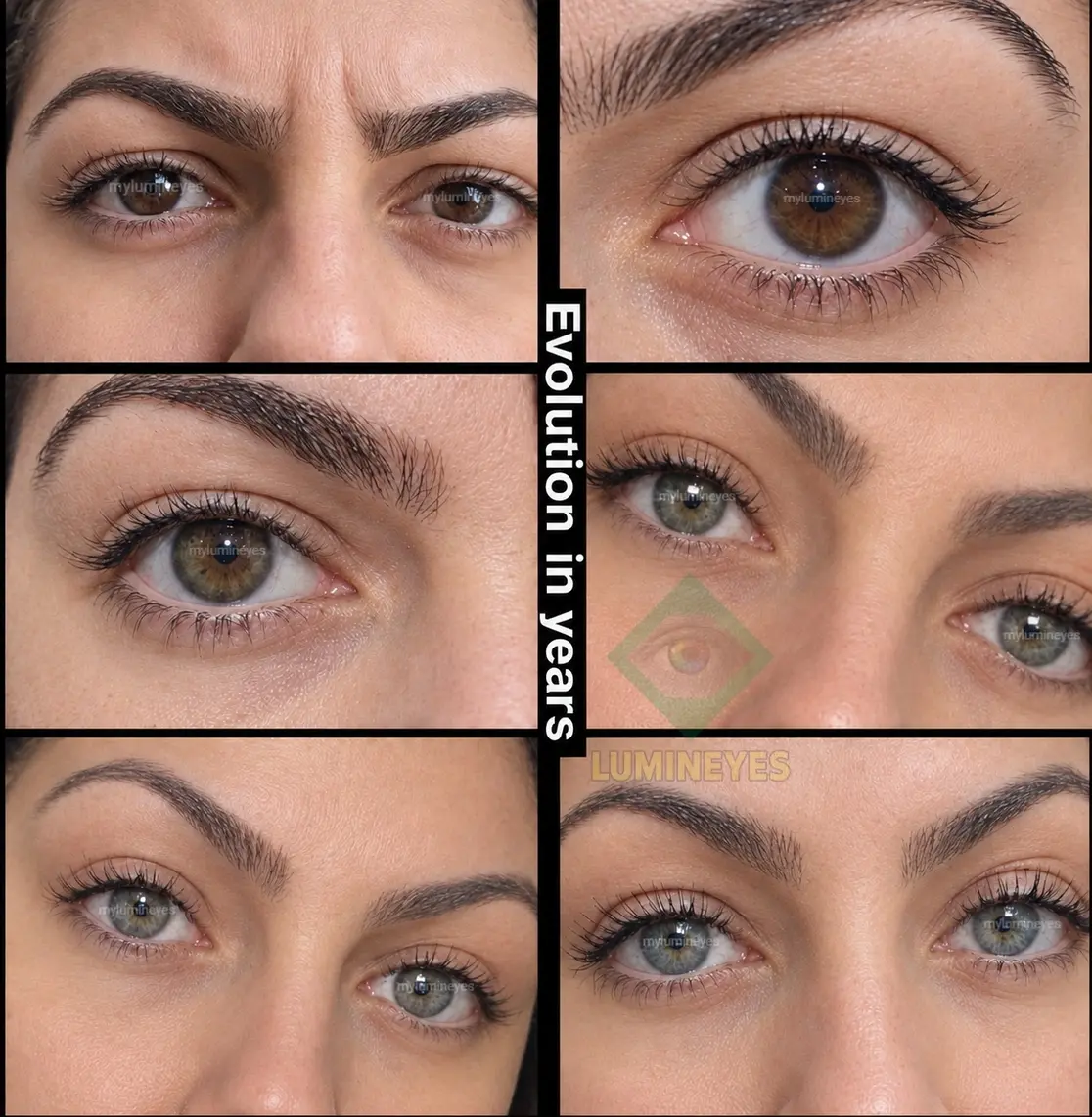
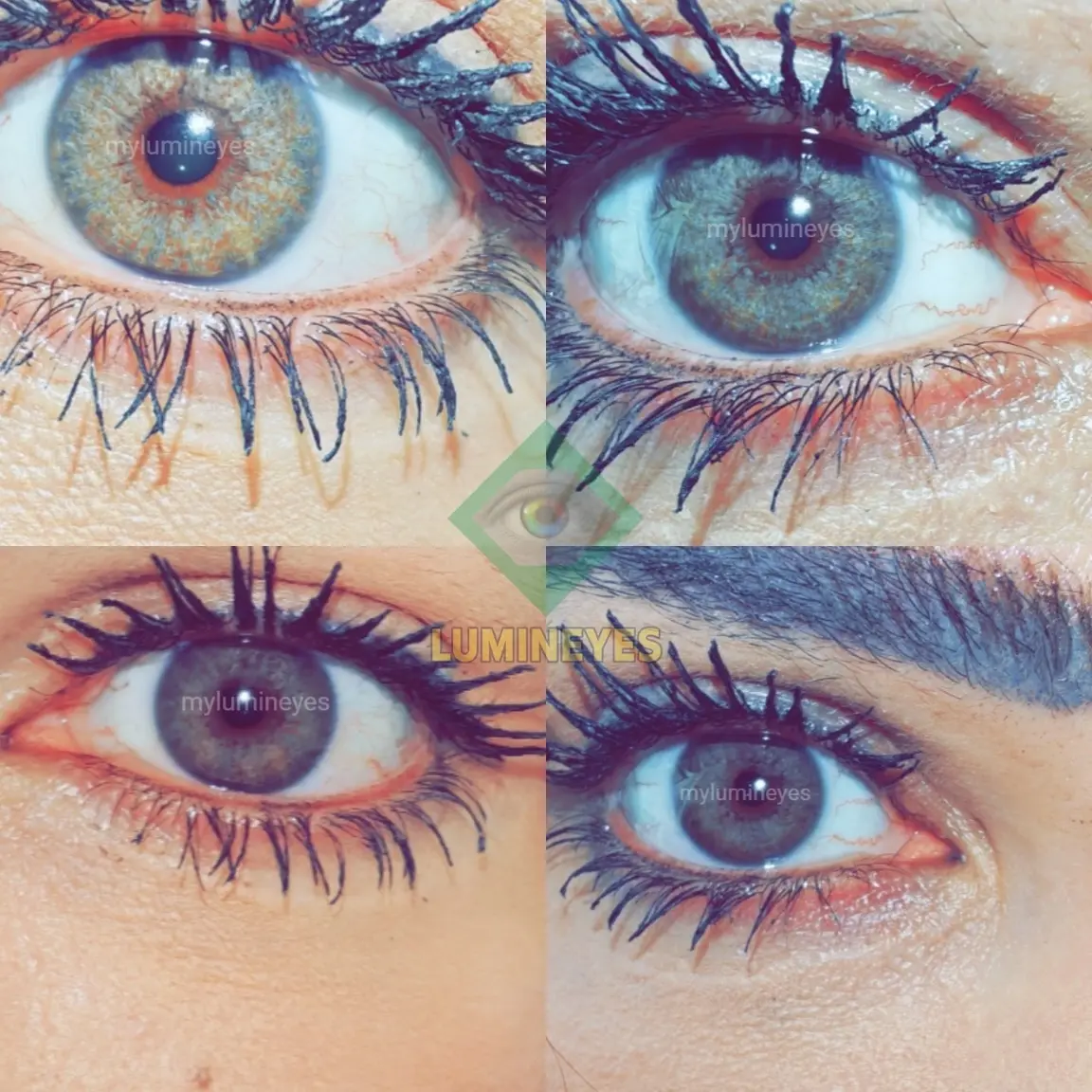
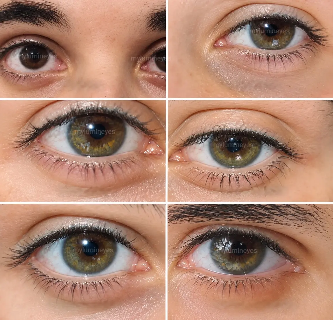
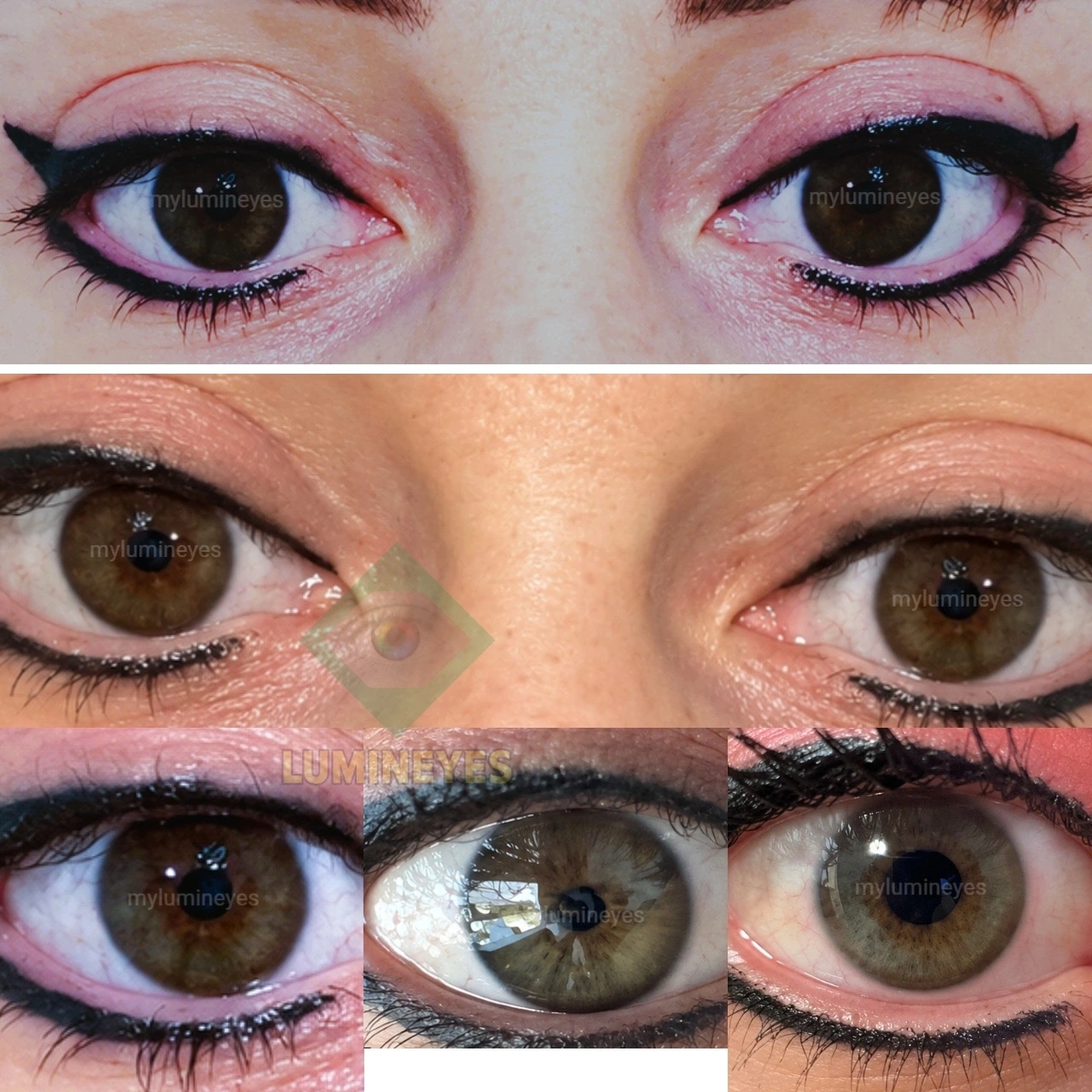
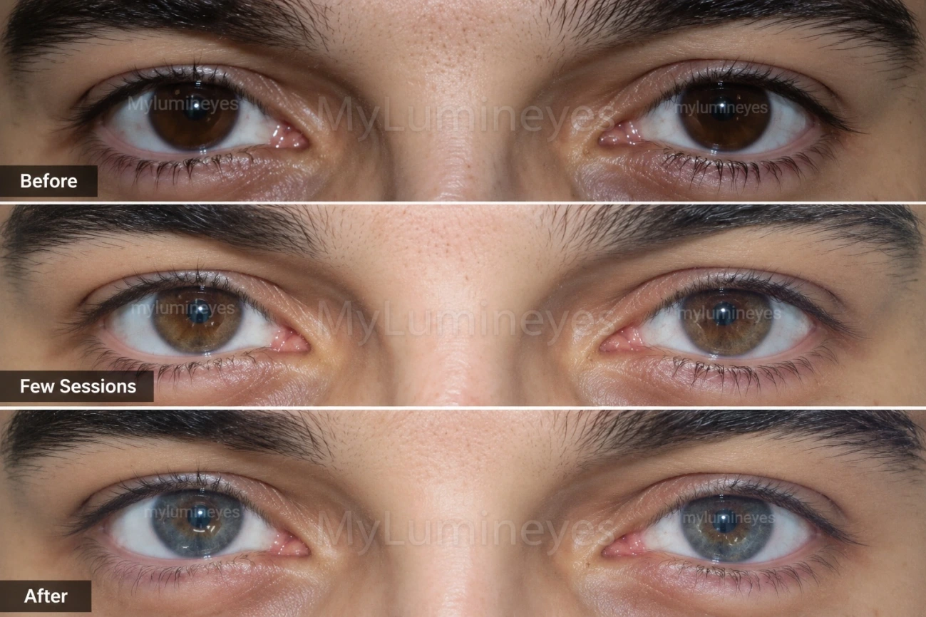
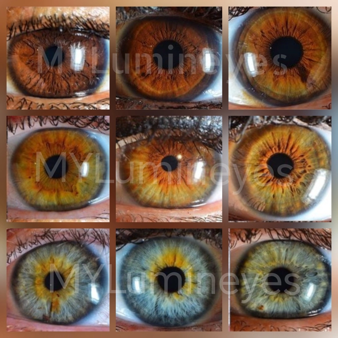
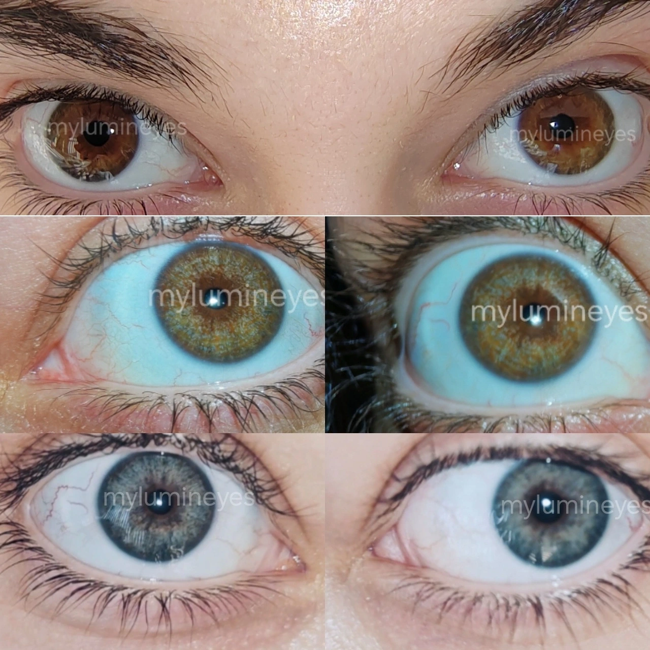
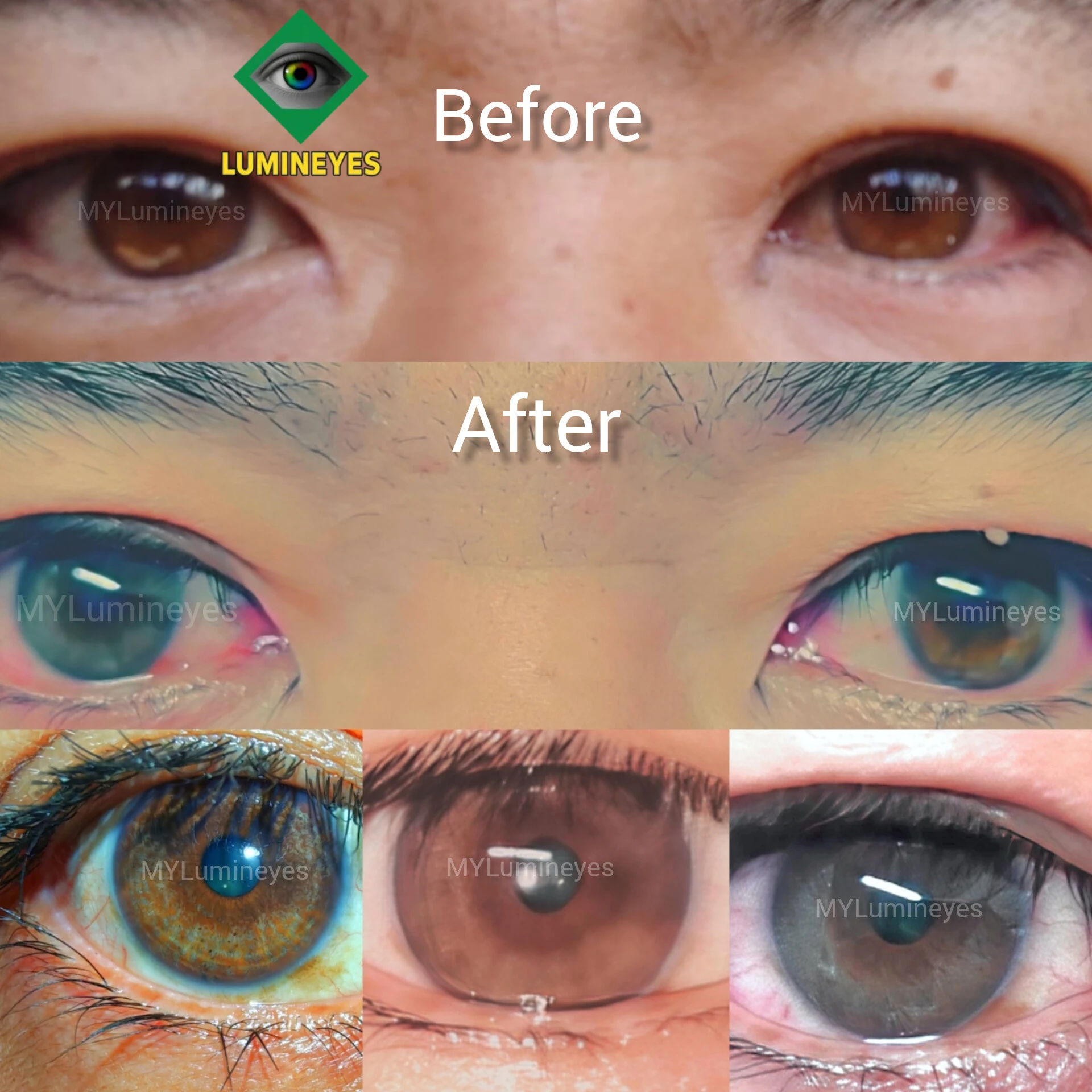
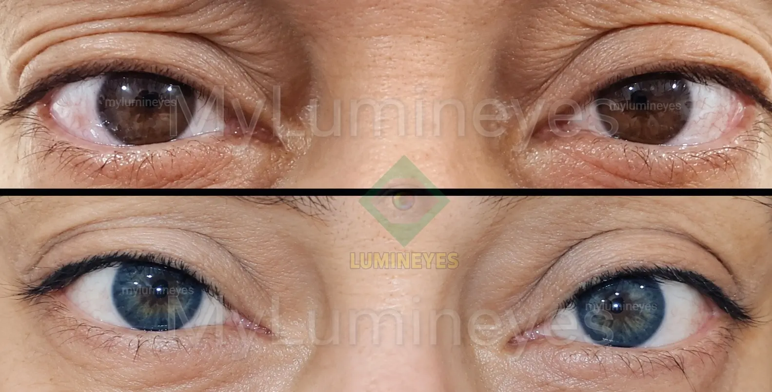
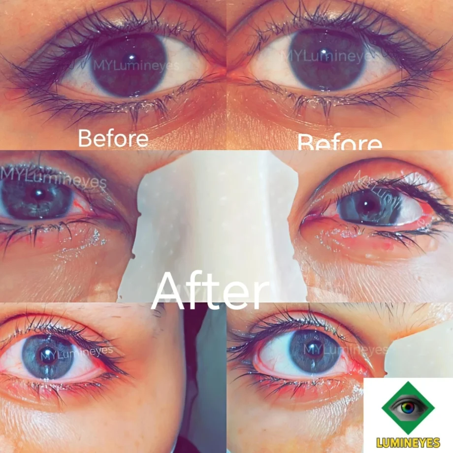
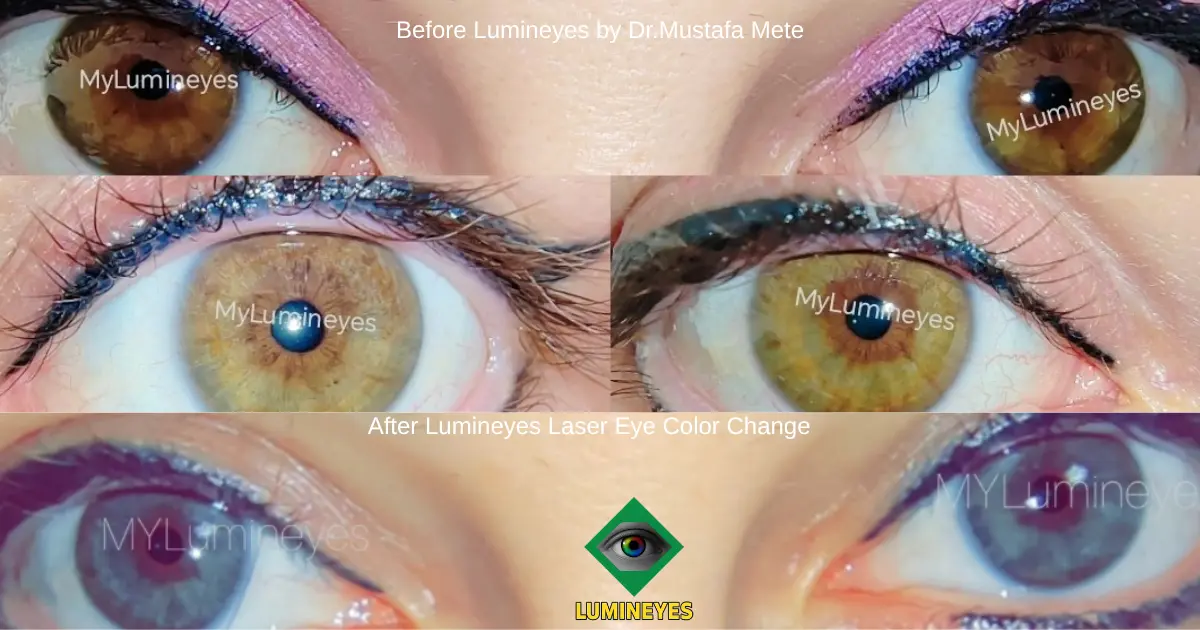
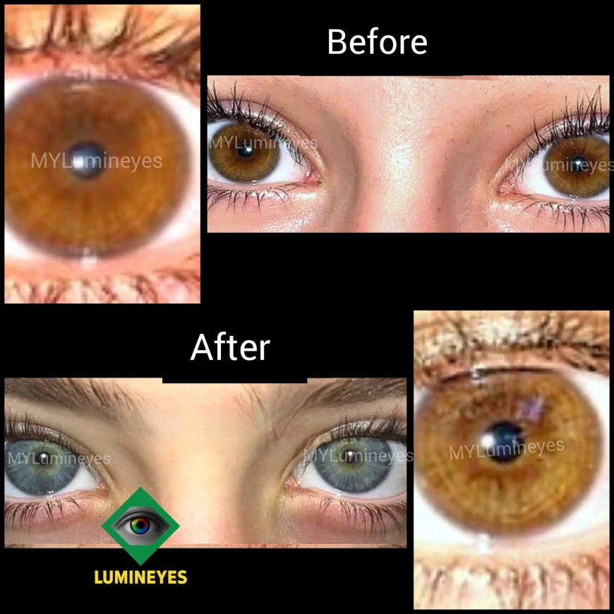
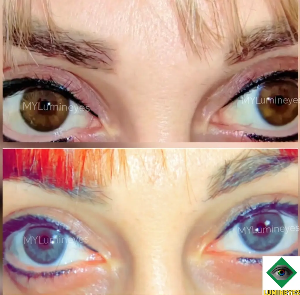
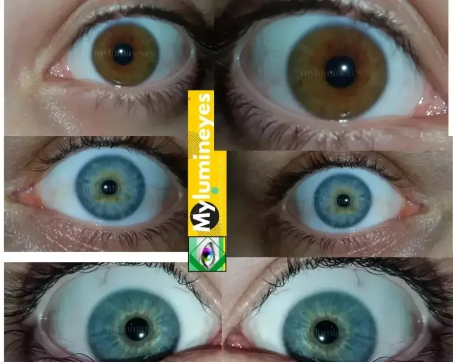
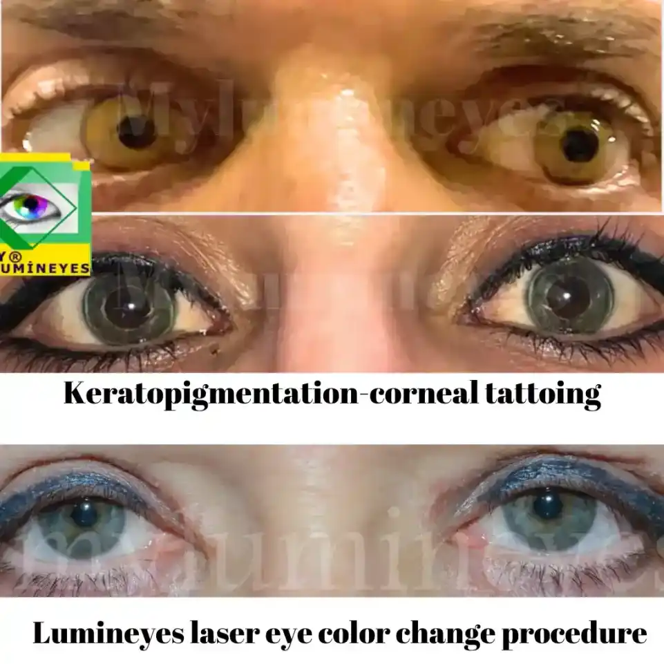
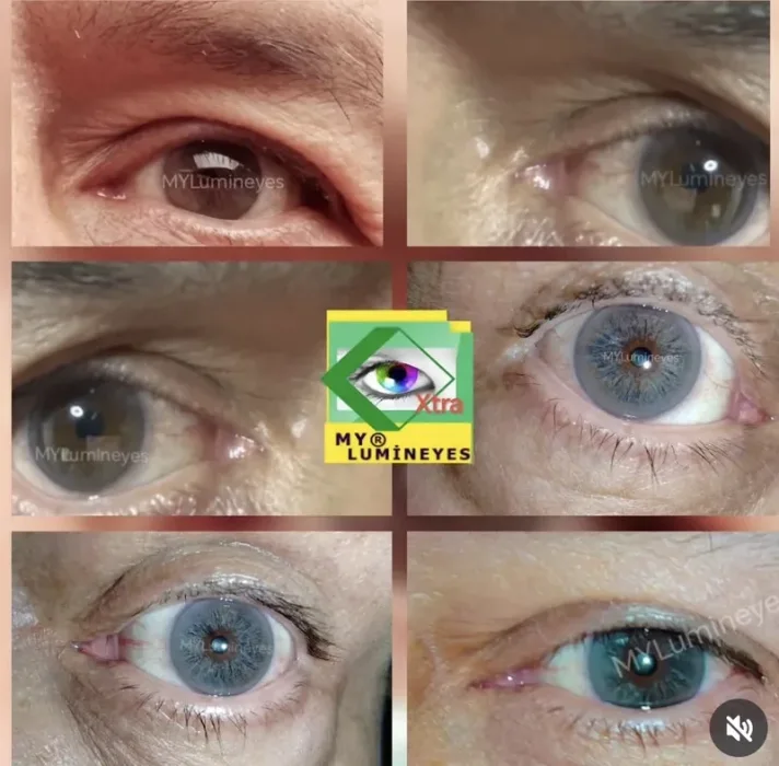
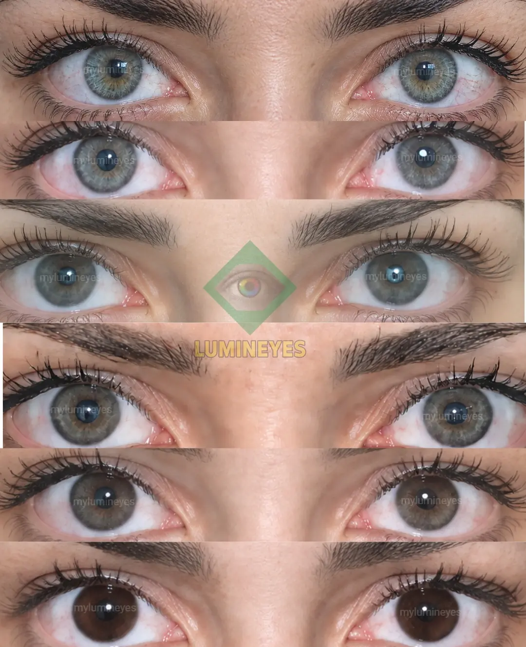
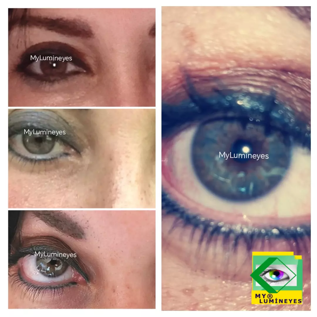
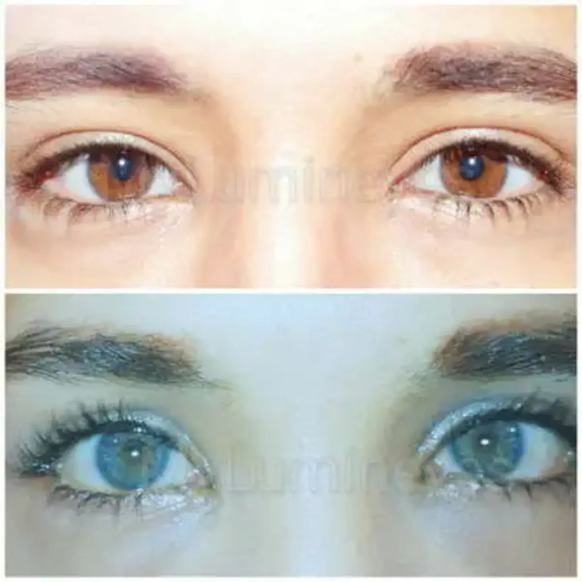
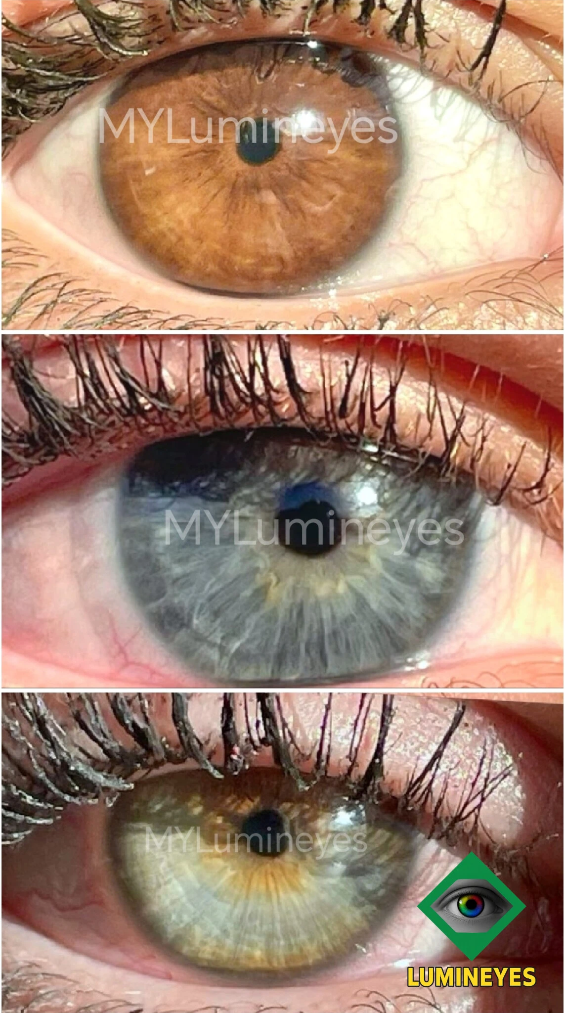
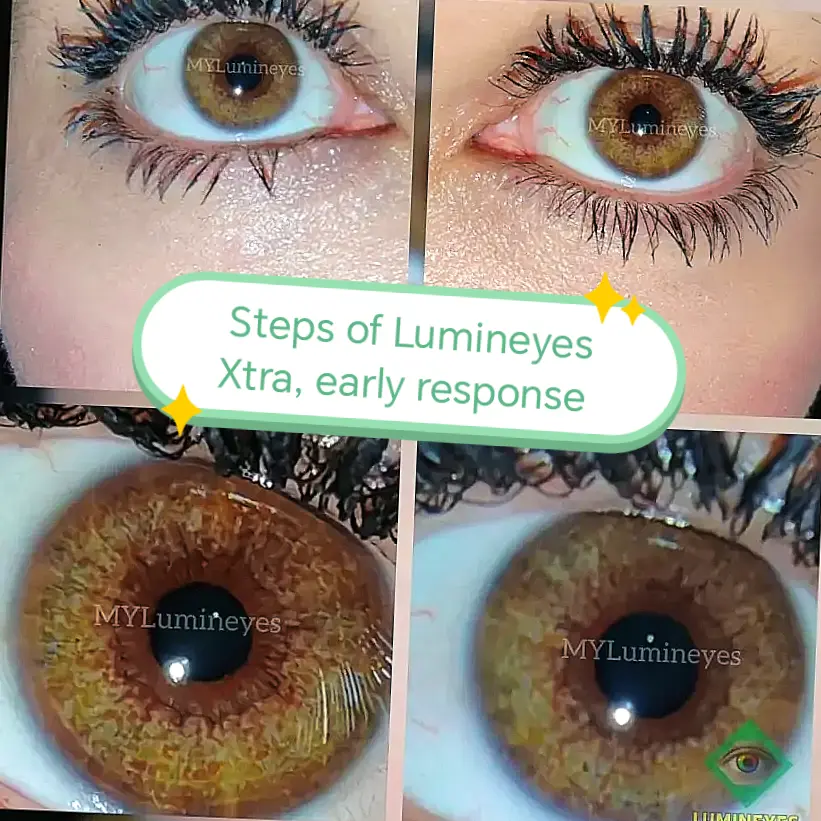
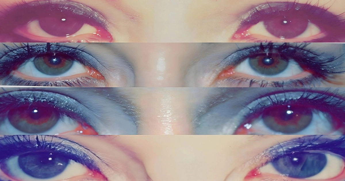
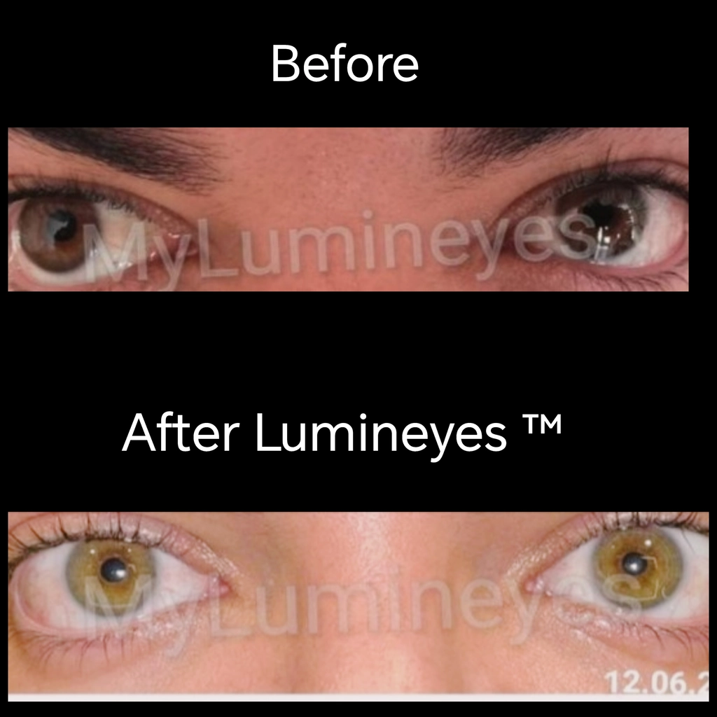
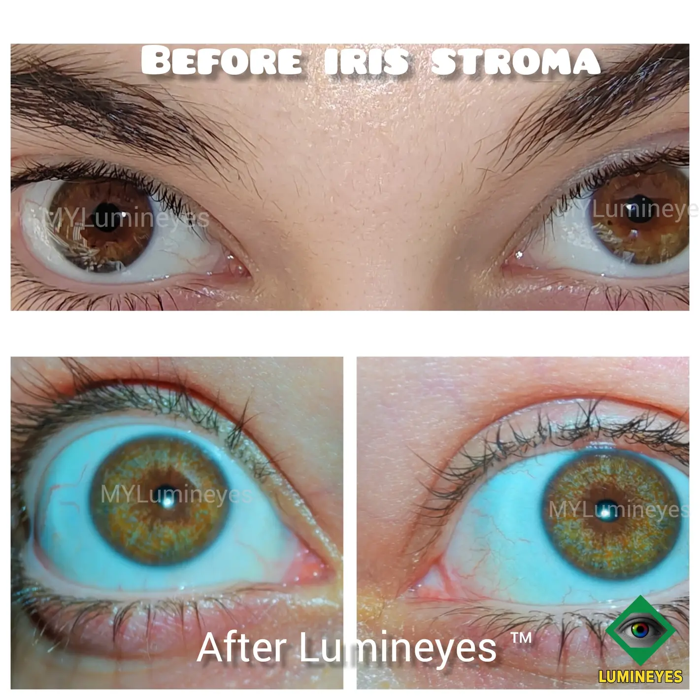
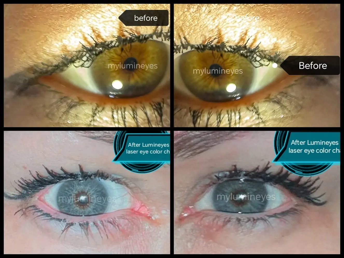
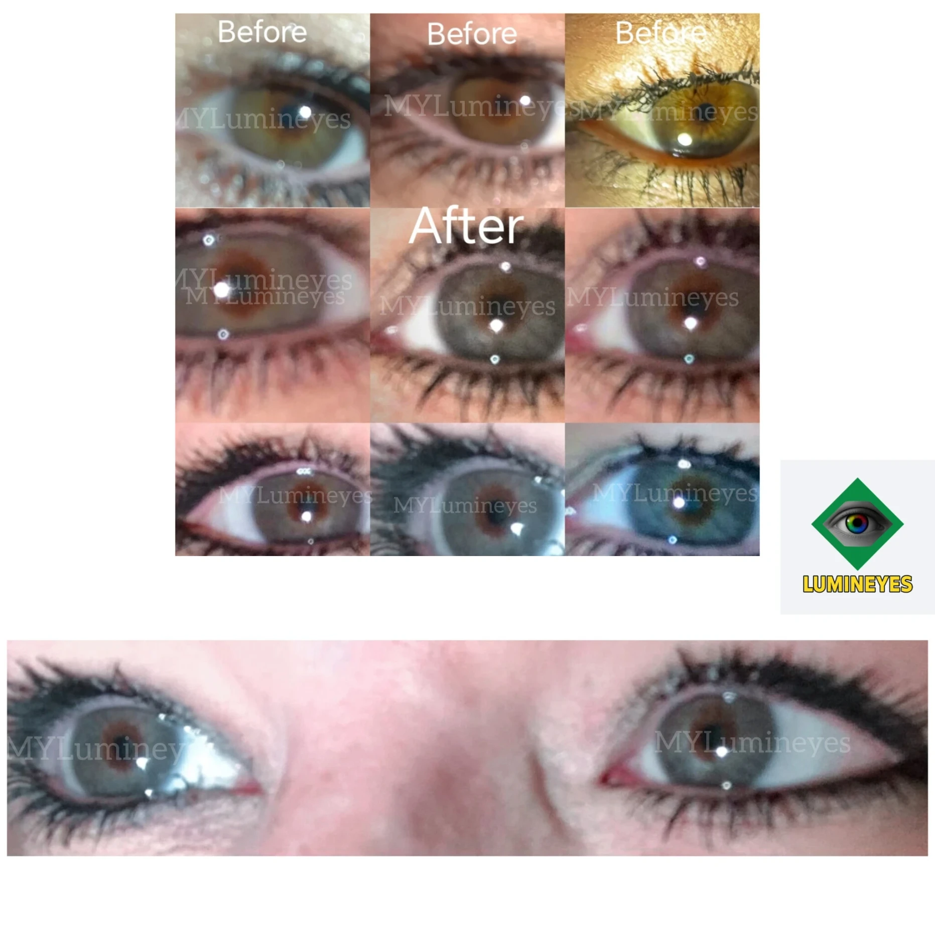
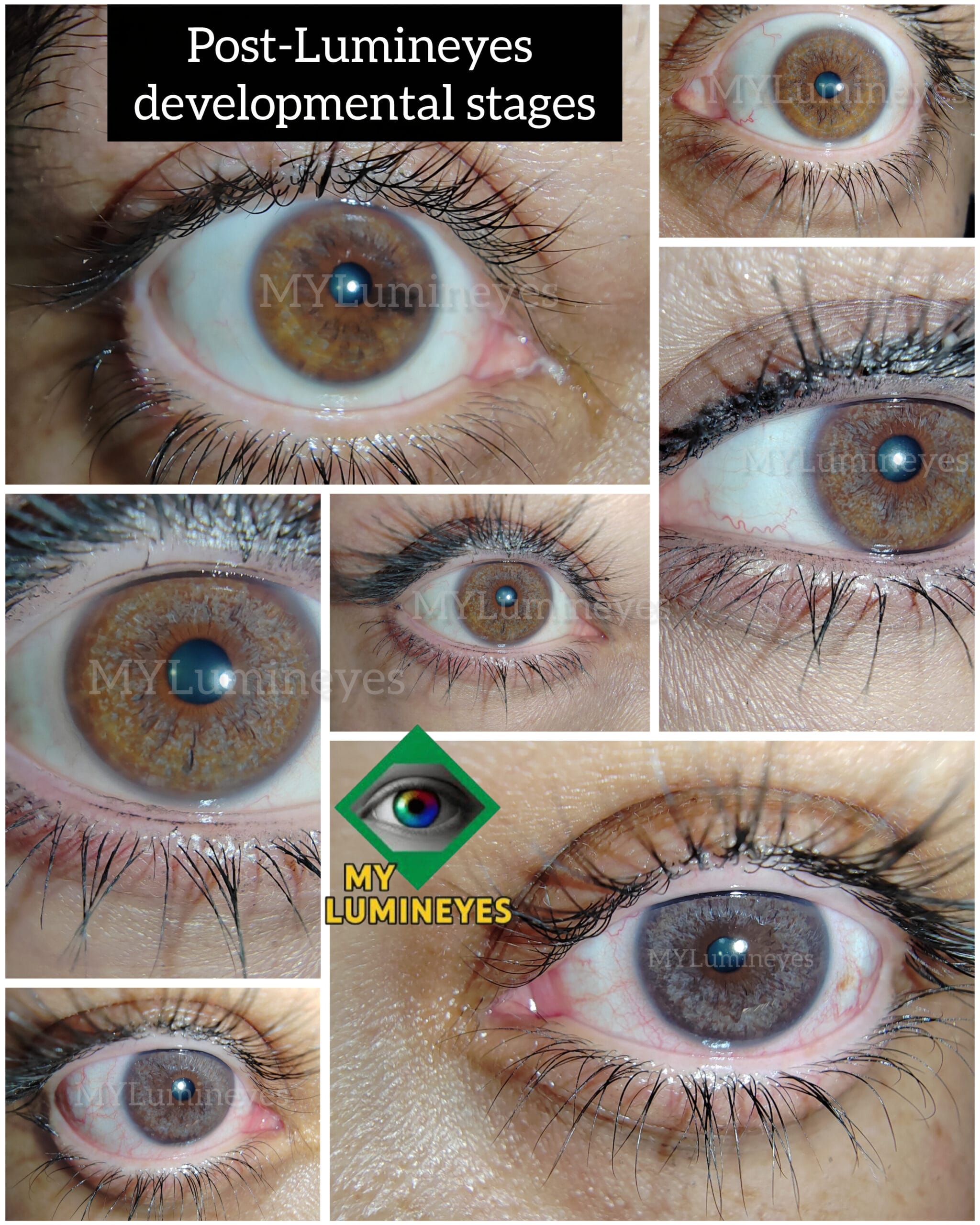
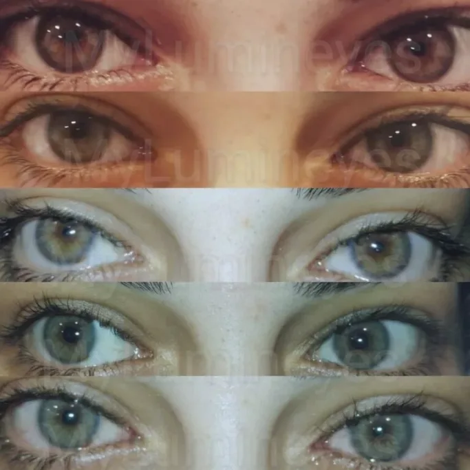
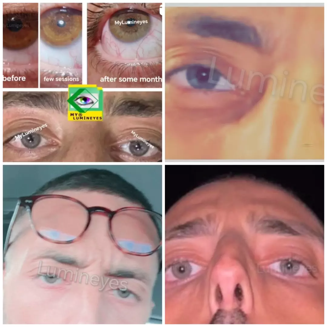
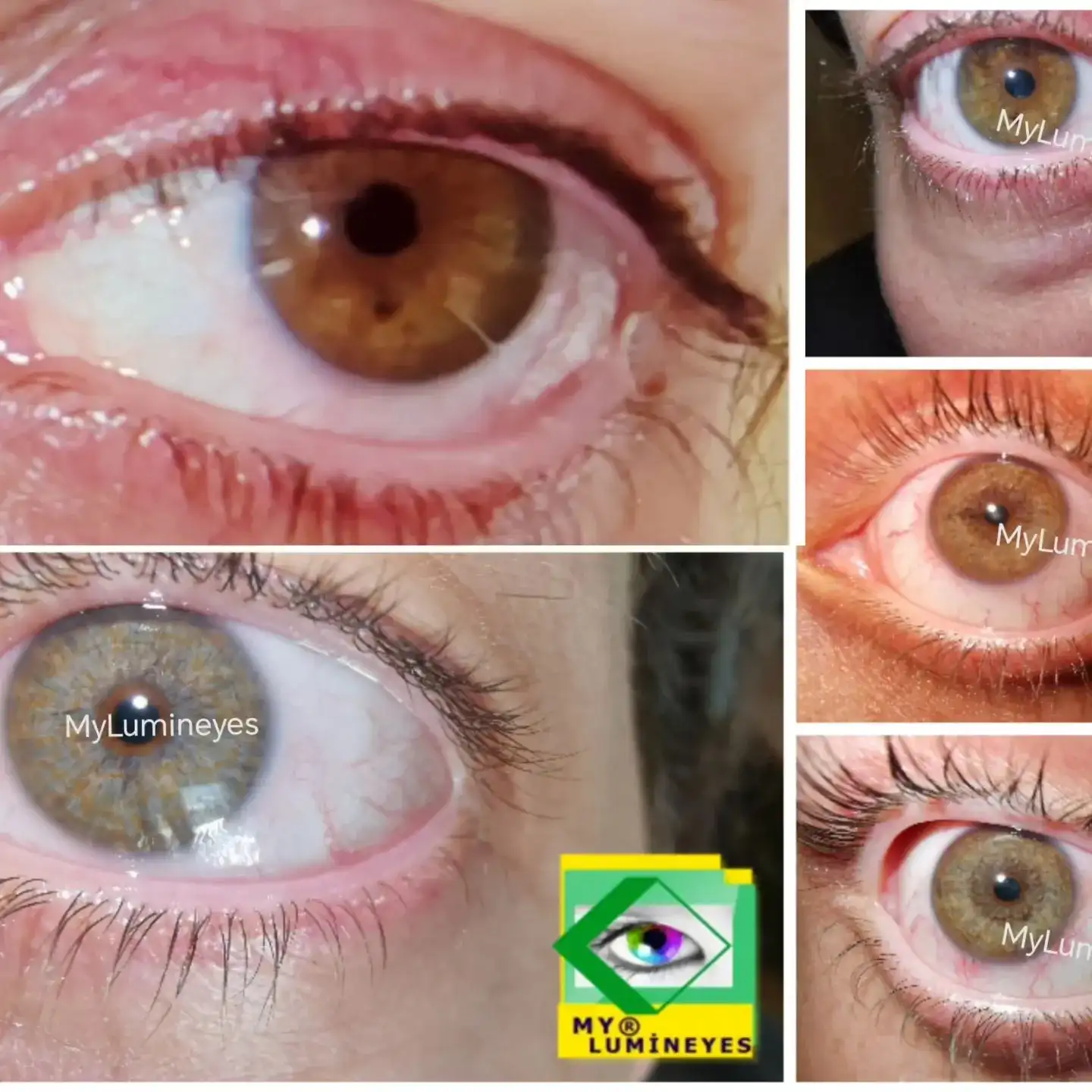
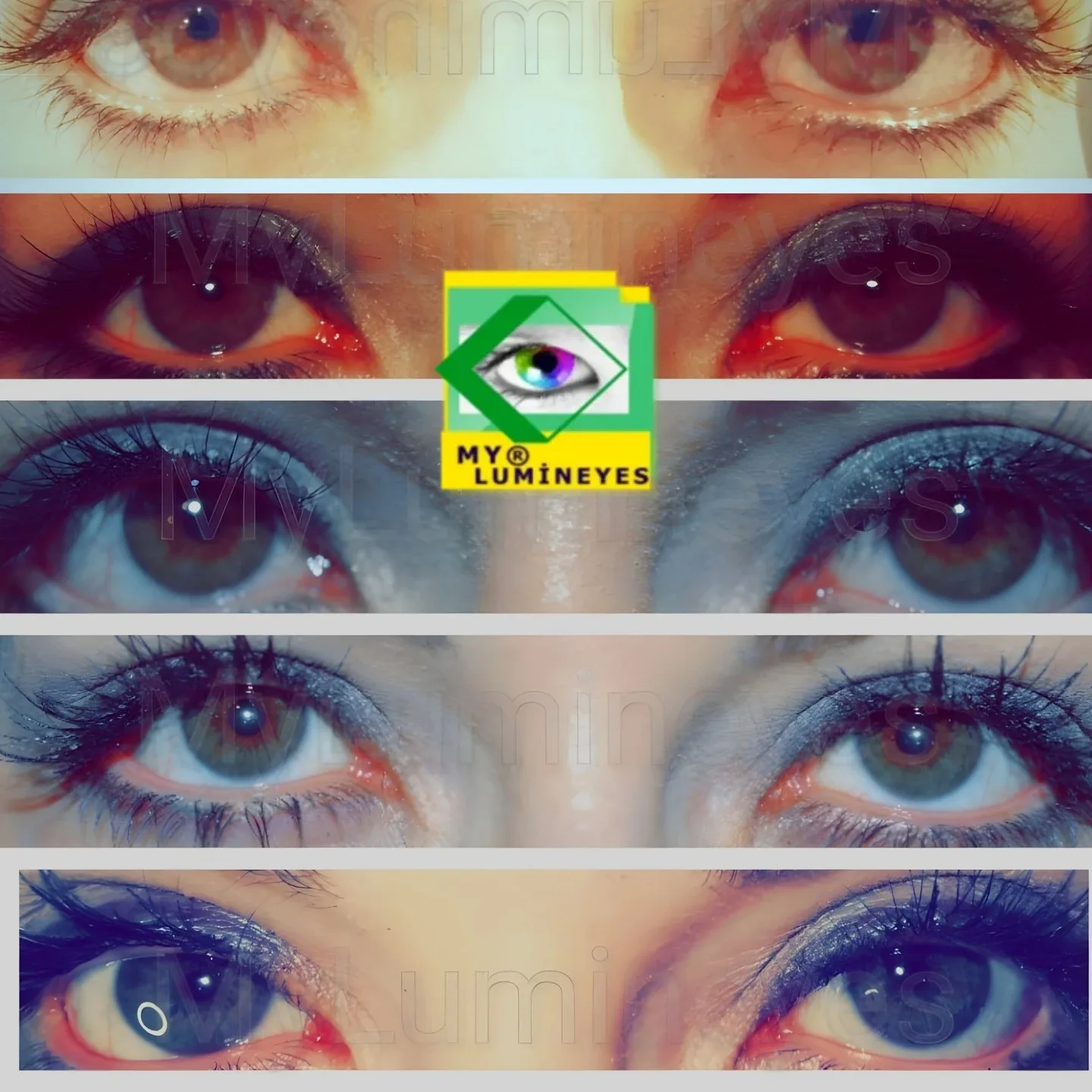
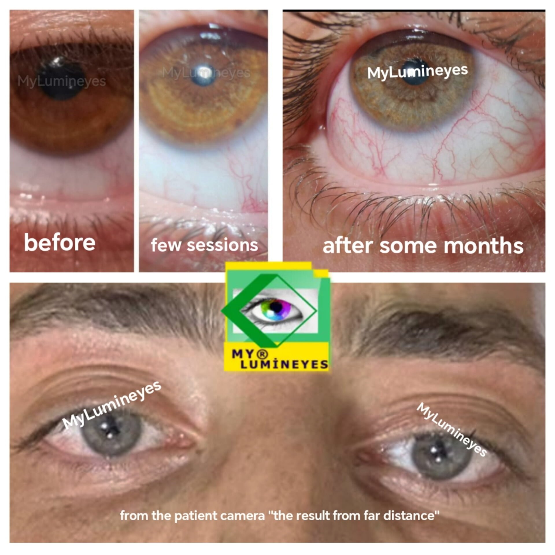
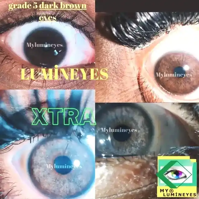
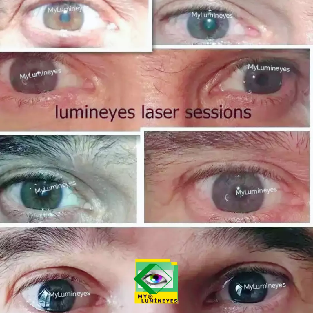
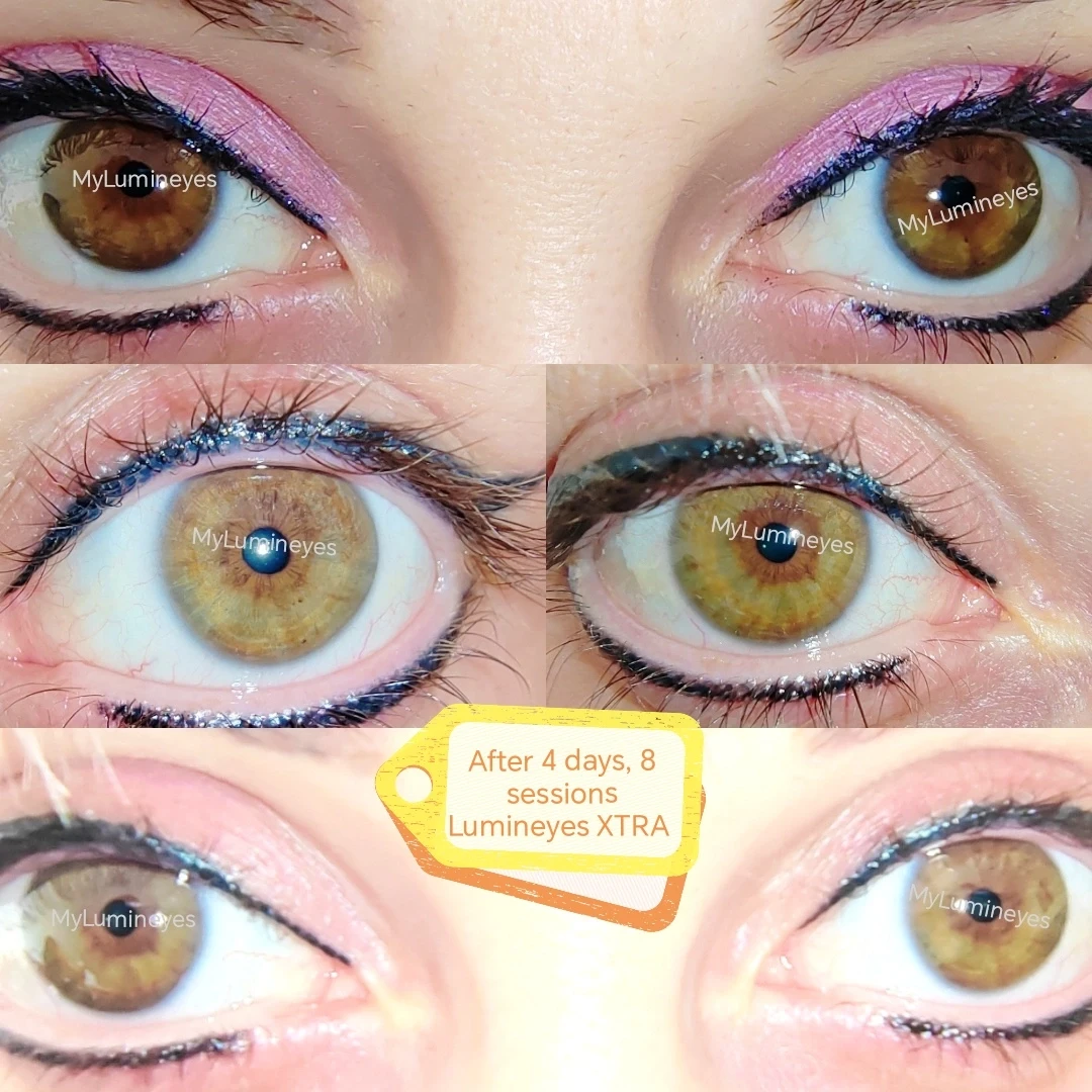
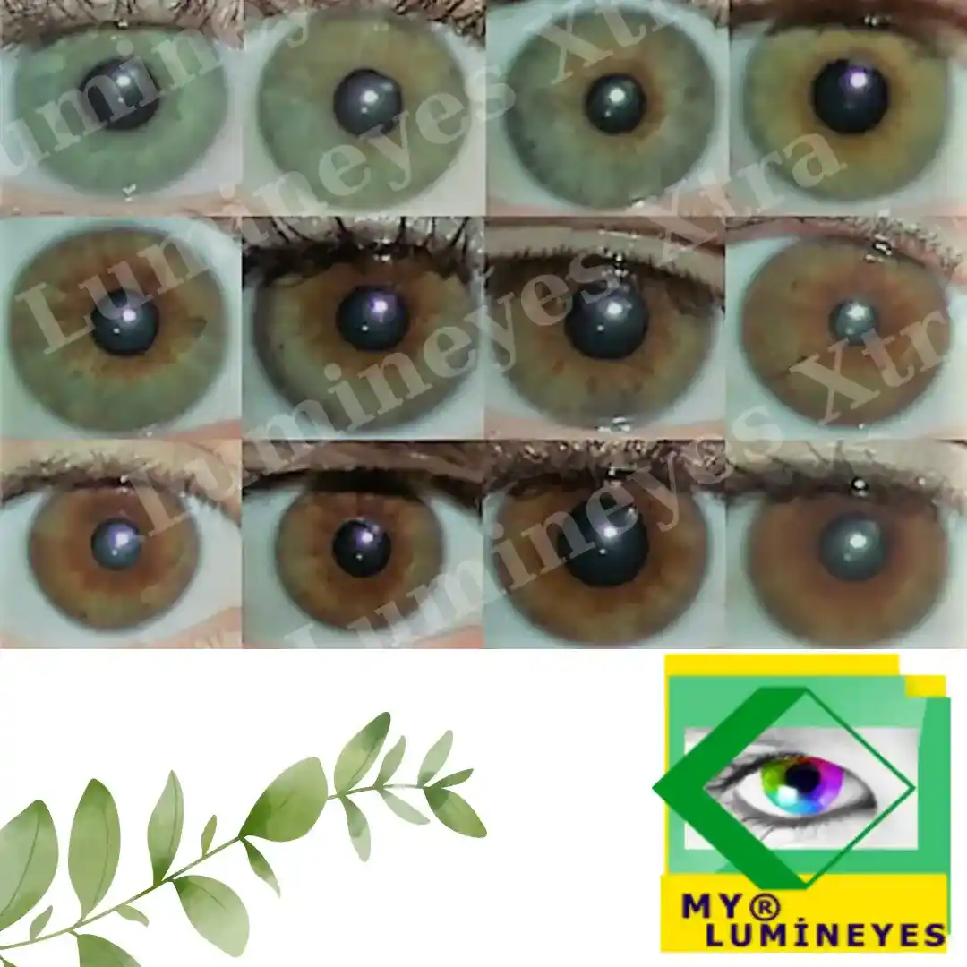
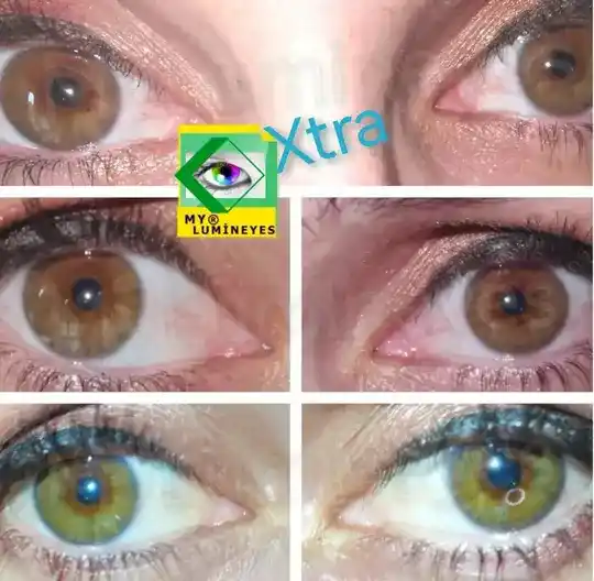
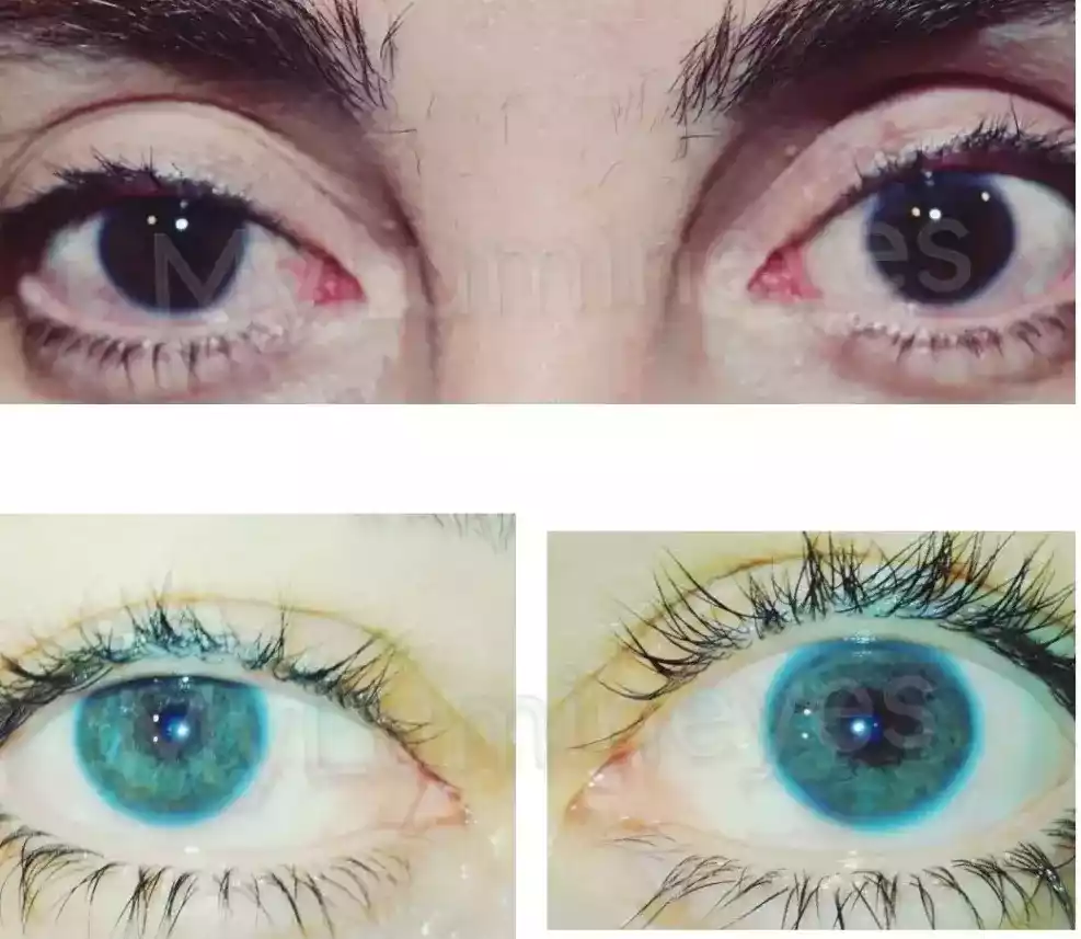
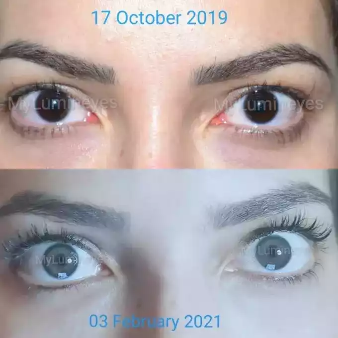
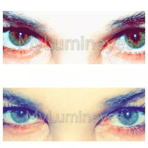
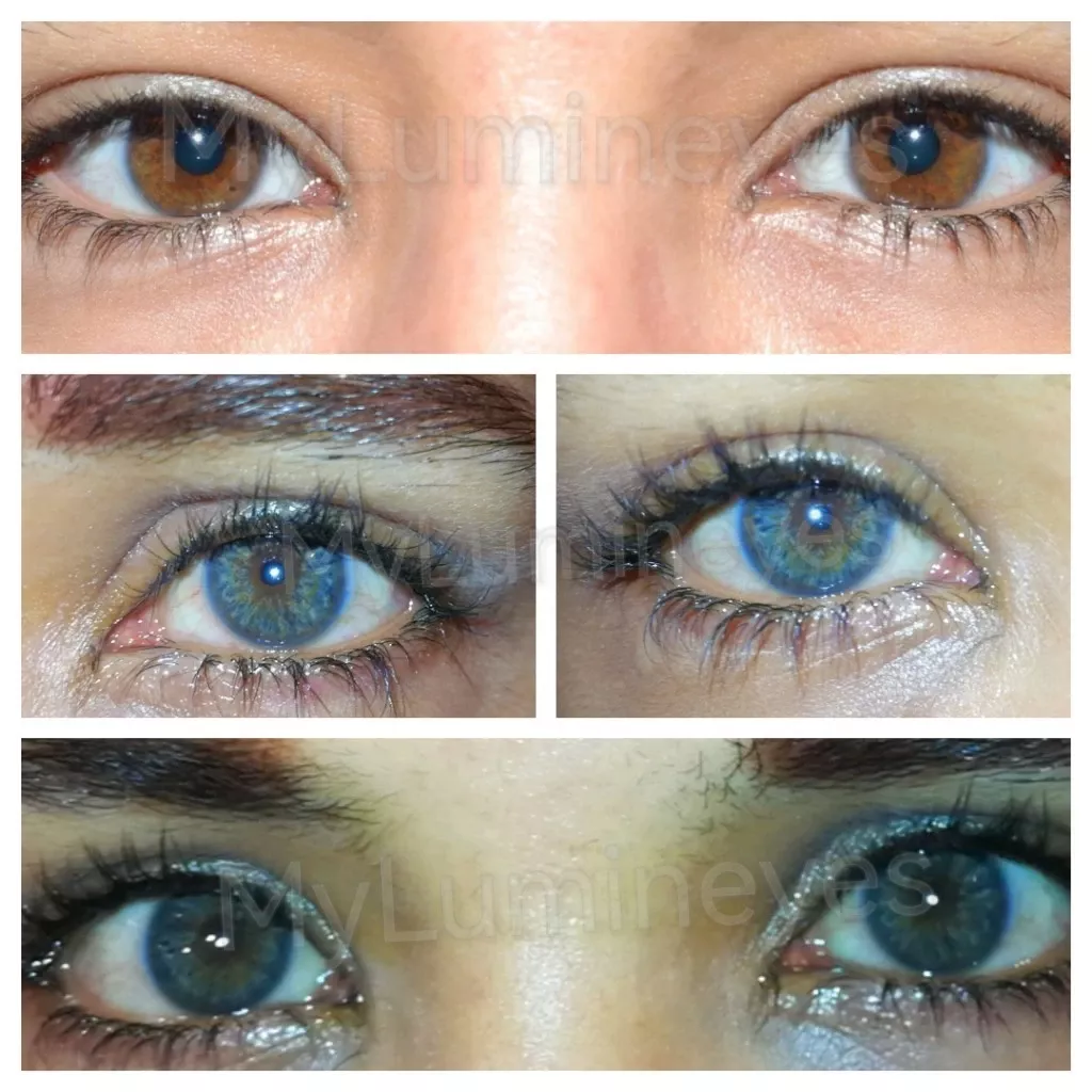
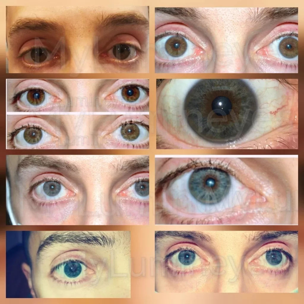
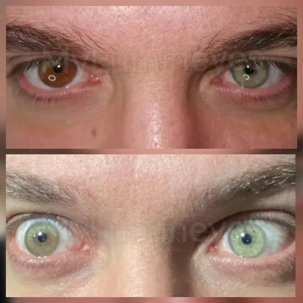
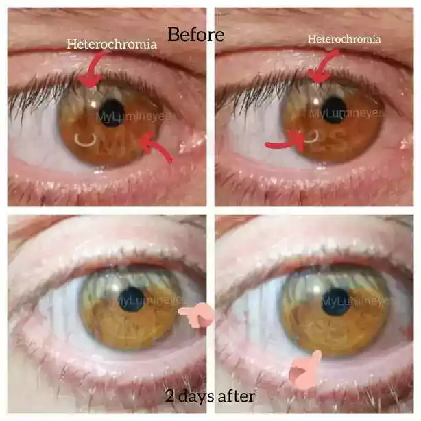
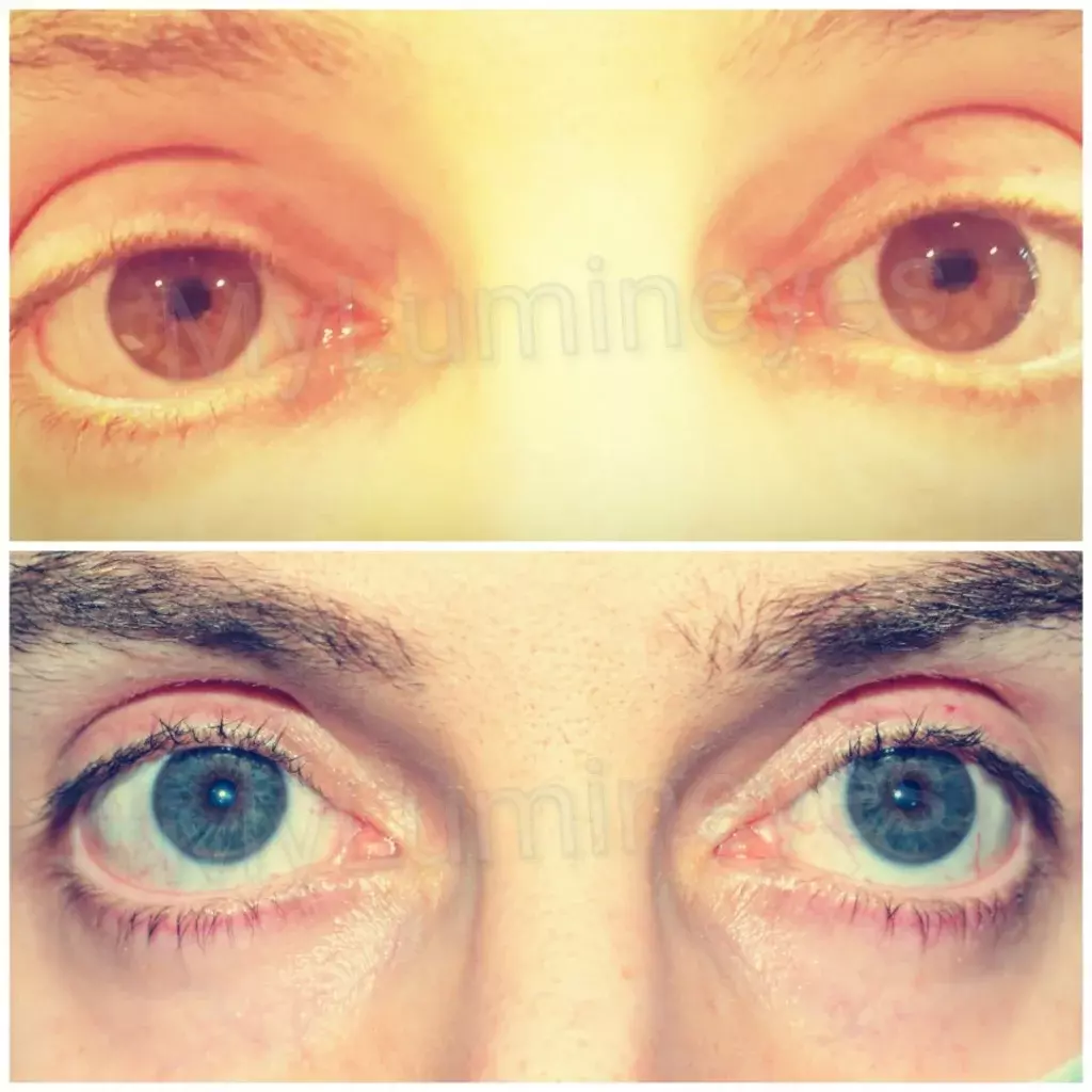
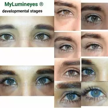
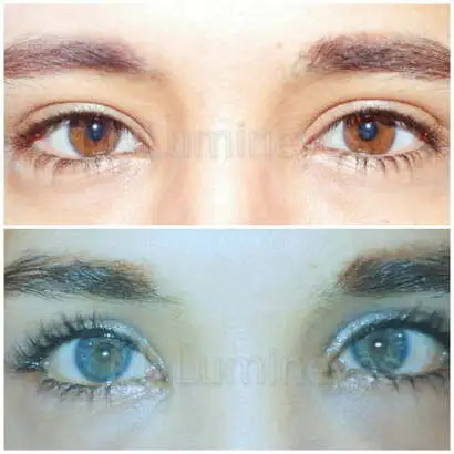
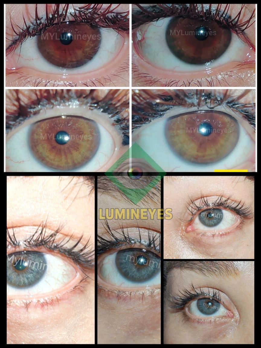
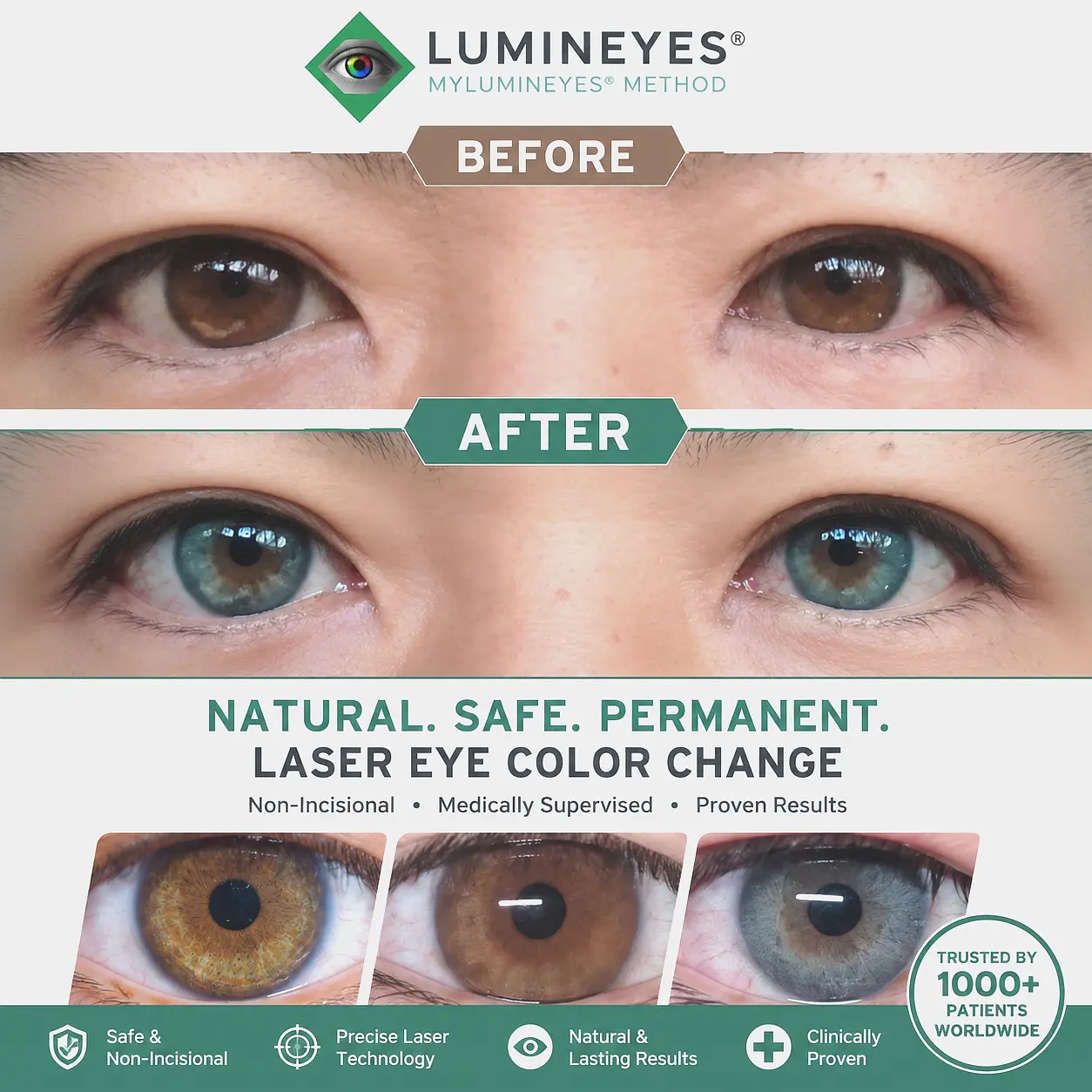
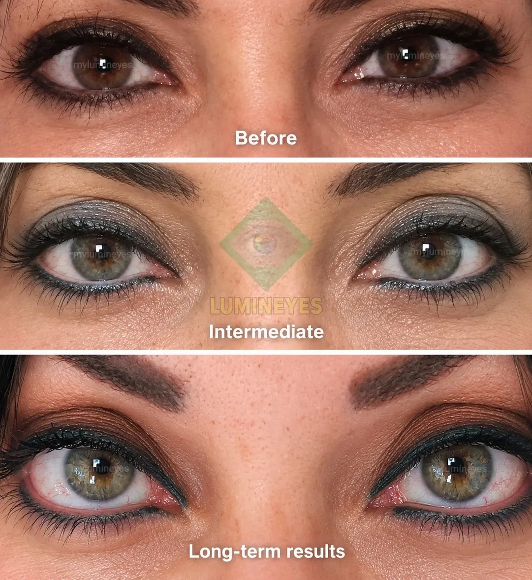
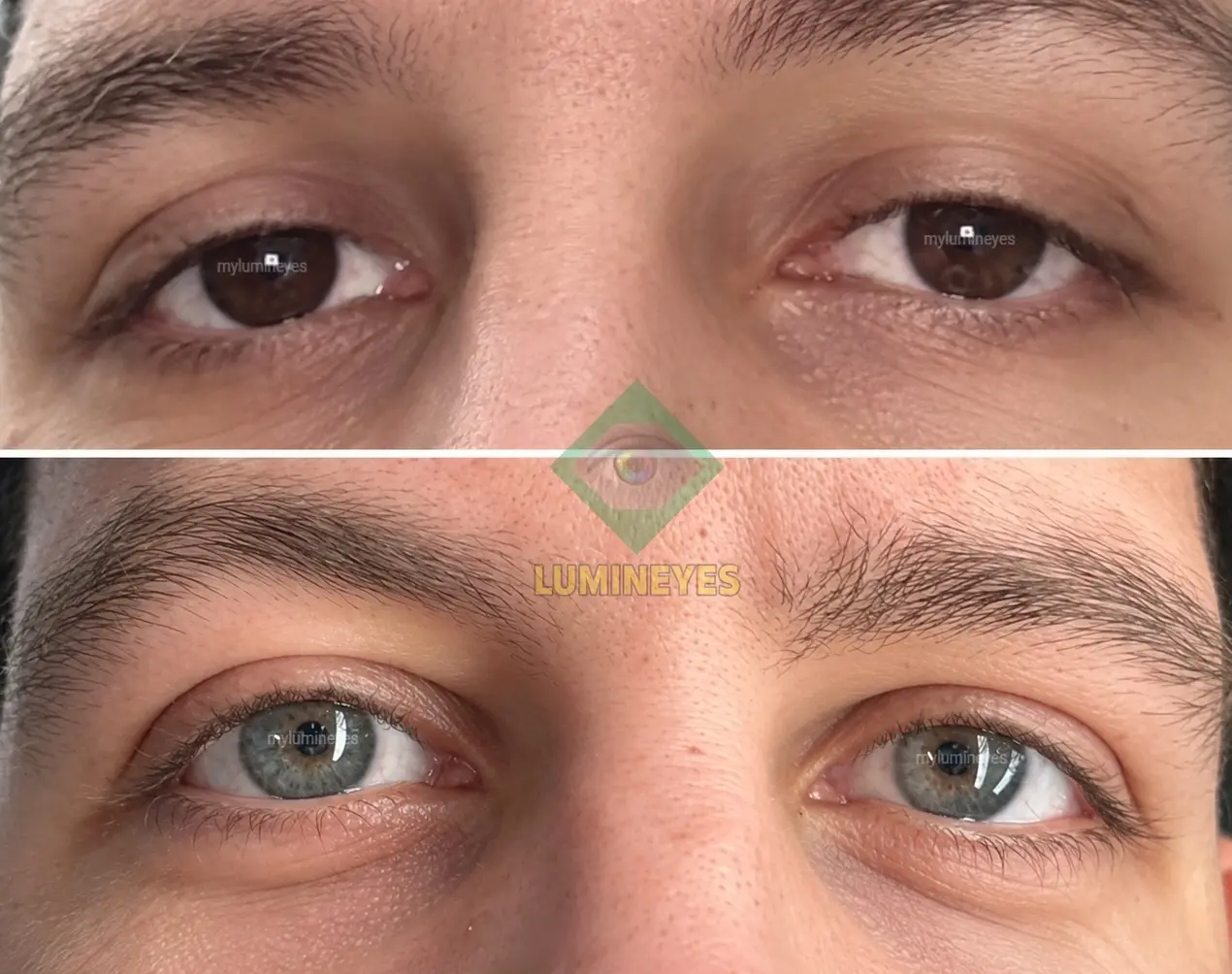
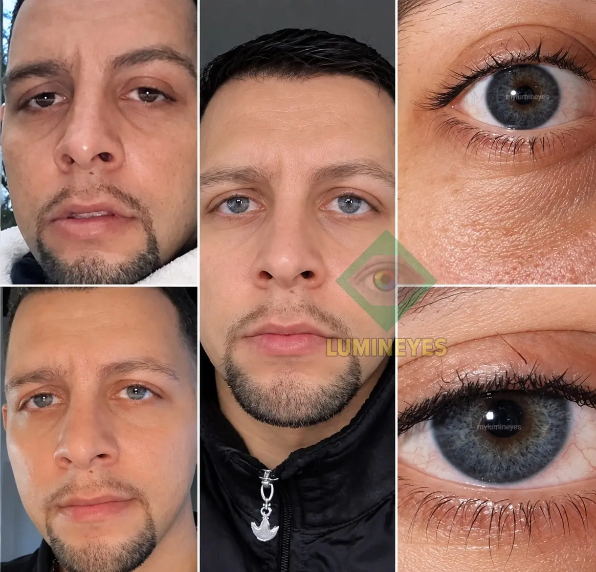
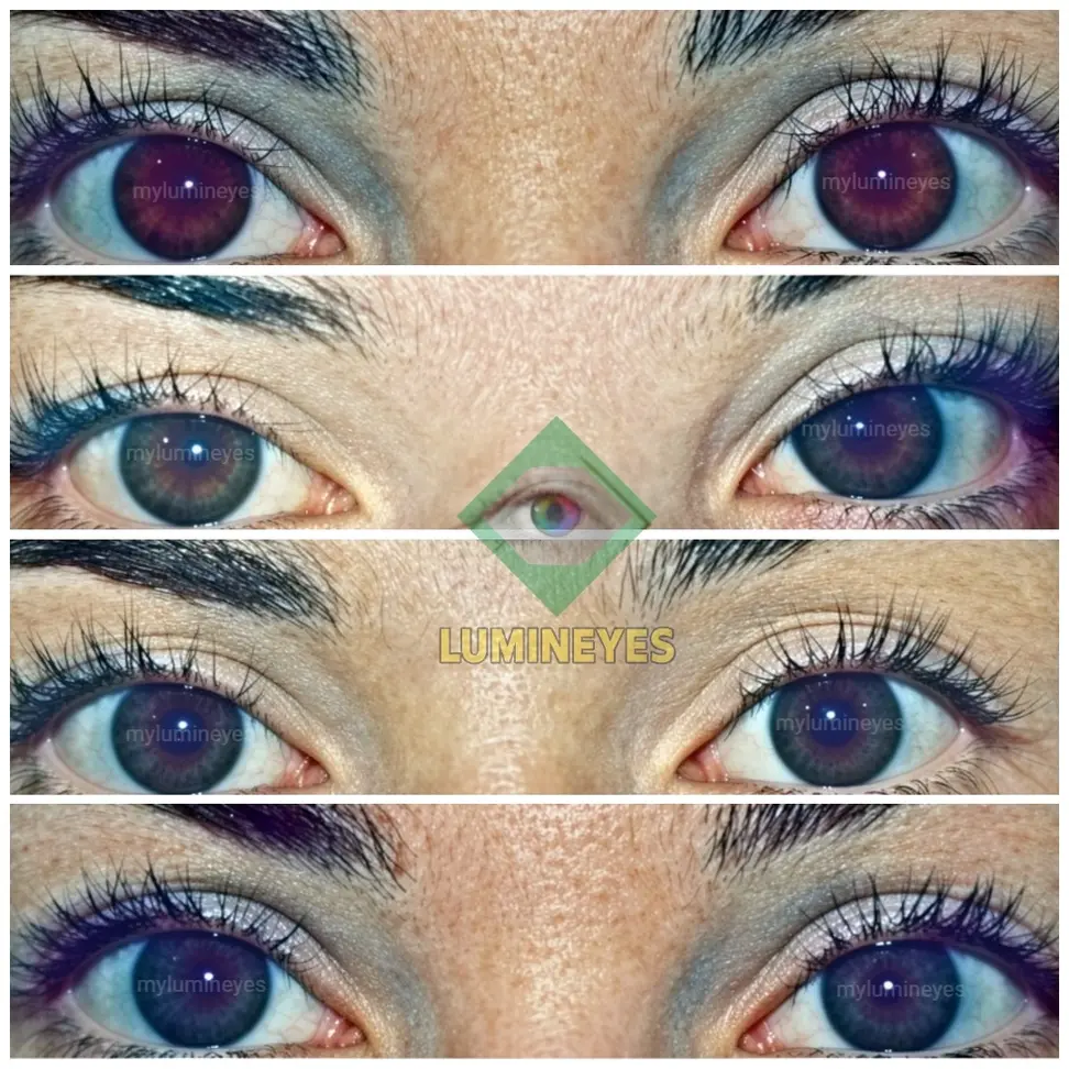
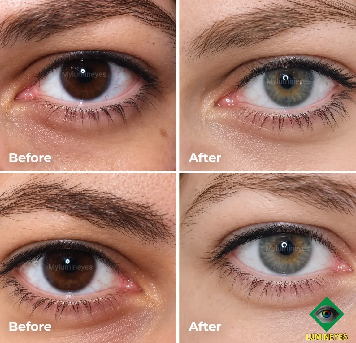
Authentic before-and-after clinical photographs provide real insight into how visible iris lightening develops through the Lumineyes® laser method. All documented transformations represent genuine patient journeys evaluated under medical supervision by Op. Dr. Mustafa Mete. No cosmetic filters, digital manipulation, or artificial color enhancements are used.
Because laser iris depigmentation operates through gradual, response-guided pigment reduction rather than a single cosmetic alteration, visible change unfolds across planned treatment stages. Baseline melanin density, iris structural features, and biological clearance kinetics determine the required treatment pathway for each individual.
For a detailed breakdown of the biological mechanism and ophthalmic safety protocols behind these results, explore our primary Laser Eye Color Change Procedure pillar guide and Laser Iris Depigmentation technical resource.
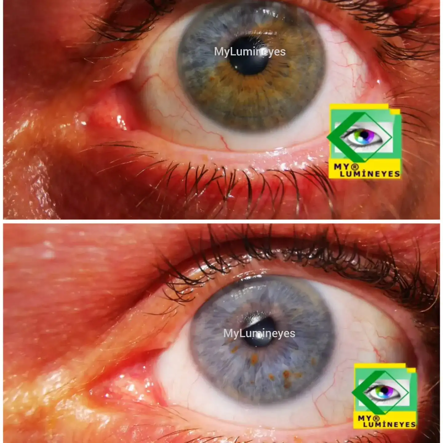
The procedure applies selective stromal melanin modulation to visible anterior iris pigment. As targeted melanin granules undergo controlled laser interaction, natural ocular cellular pathways clear dispersed pigment progressively over subsequent weeks.
Because the laser targets only existing superficial melanin, it does not introduce synthetic dye or artificial colors. Instead, gradual pigment reduction reveals the underlying, natural stromal optical tones unique to each patient's iris structure. Treatment progression is monitored continuously by the treating physician between stages to ensure ocular safety and healthy clearance.
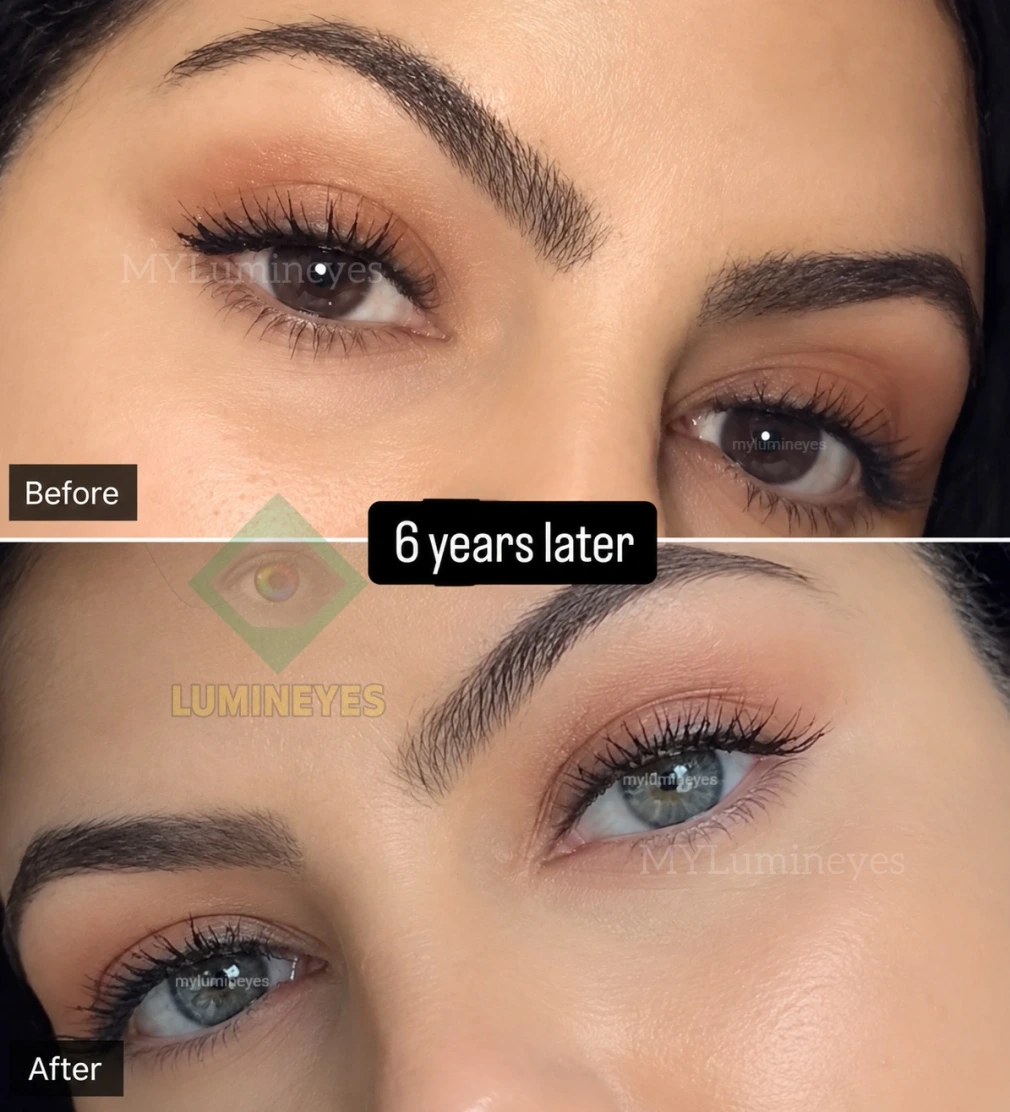
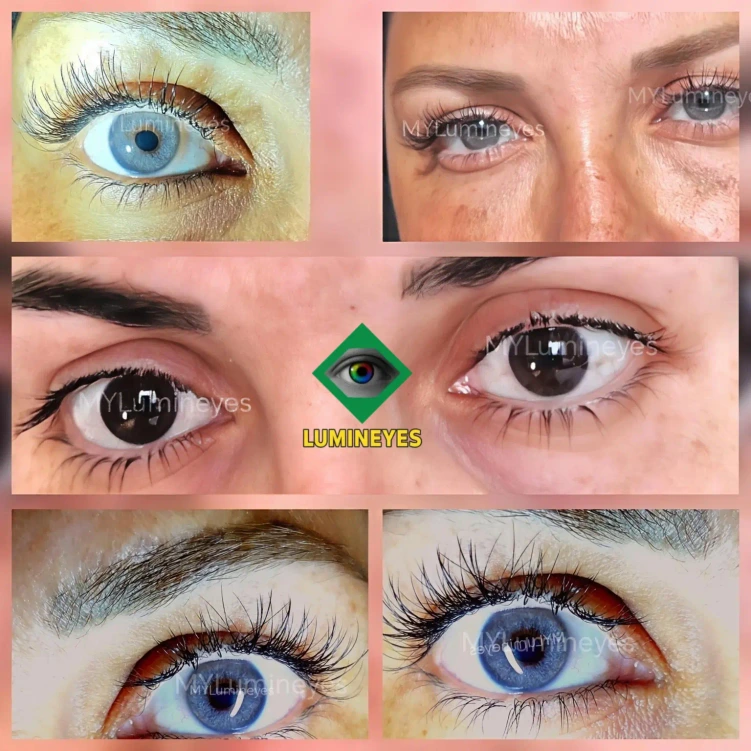
Longitudinal follow-up photograph documenting stable iris appearance six years following completed Lumineyes® laser iris depigmentation. Results stabilize once the eye reaches a healthy clinical plateau phase.
No clinic can predict or guarantee an exact cosmetic shade because final iris appearance is governed by seven distinct biological and clinical factors:
To learn more about natural iris pigmentation grading, review the Lumineyes® Iris Pigmentation Scale (LIPS) research framework.
 Lumineyes Assistant
×
Lumineyes Assistant
×