Keratopigmentation for Eye Color Change
Keratopigmentation is a corneal pigment-based procedure used to change the visible appearance of eye color by placing pigment within the corneal tissue. Because the cornea is an optical structure rather than the natural pigment-bearing tissue of the iris, the resulting color is a visual simulation rather than a change in the natural iris itself.
The appearance, stability, and optical characteristics of keratopigmentation can vary between individuals. This clinical guide explains how the procedure works, what tissue it changes, its cosmetic characteristics, potential medical and optical considerations, long-term stability, reversibility, and how it differs from approaches that target the iris itself.
Keratopigmentation is one of the surgical approaches discussed in our broader eye color change surgery guide , where different methods are considered from anatomical and clinical perspectives.
What Is Keratopigmentation?
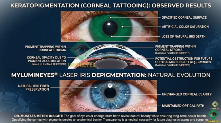
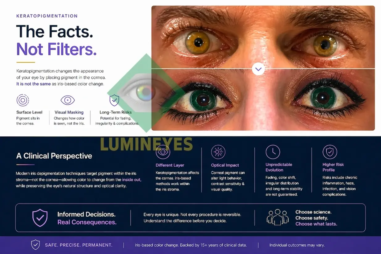
Keratopigmentation, also called corneal tattooing, changes the visible color of the eye by introducing pigment into the cornea. The natural iris remains underneath the cornea; the cosmetic effect is produced by the pigment positioned within the corneal tissue.
This anatomical distinction is central to understanding the procedure. The cornea is normally transparent and forms an important part of the eye's optical system, while the iris is the natural colored structure surrounding the pupil.
The procedure therefore does not simply "change the iris color." Instead, it creates a new visible color effect through pigment placed in the cornea.
How Keratopigmentation Changes the Appearance of the Eye
Keratopigmentation creates the cosmetic effect by introducing pigment into the corneal tissue. The resulting pigment pattern is viewed through the anterior corneal surface and changes the apparent color of the eye.
- Corneal pigment placement: the visible color is produced within the cornea rather than by directly altering natural iris pigmentation.
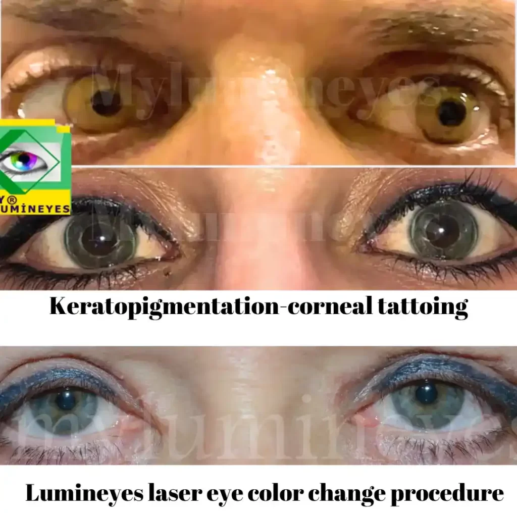
- Simulated iris appearance: the cosmetic pattern may not reproduce the complete radial texture, depth, and biological variation of a natural iris.
- Lighting-dependent appearance: perceived color can vary according to illumination, viewing angle, pupil size, and optical characteristics.
- Individual variation: corneal anatomy, pigment distribution, healing, and the technique used can influence the final appearance.
These differences are particularly relevant when considering the biological limits of eye color change and the distinction between changing the appearance of an eye and changing its natural anatomical pigmentation.
Keratopigmentation Overview
| Feature | Keratopigmentation |
|---|---|
| Type of procedure | Corneal pigment-based intervention, commonly described as corneal tattooing. |
| What changes? | Pigment is introduced into the cornea; the natural iris remains anatomically unchanged. |
| Visible appearance | Creates a simulated eye-color effect that may differ from the texture and optical depth of a natural iris. |
| Optical considerations | Because the cornea is part of the optical system, transparency, regularity, and visual quality are relevant clinical considerations. |
| Long-term stability | Cosmetic appearance may evolve with healing, tissue remodeling, pigment distribution, or fading. |
| Reversibility | Pigment removal can be difficult and may require additional corneal intervention. |
Cosmetic and Optical Considerations
- Color uniformity: pigment distribution can vary between areas and may influence consistency of the cosmetic result.
- Natural iris detail: a corneal pigment pattern does not reproduce the complete biological architecture of the natural iris.
- Color accuracy: achievable shades depend on pigment characteristics, placement, corneal anatomy, and lighting conditions.
- Light interaction: pigment within a normally transparent optical structure can influence how light is transmitted or scattered.
Corneal Pigment Placement vs. Iris-Based Eye Color Change
The central anatomical distinction is straightforward: keratopigmentation changes the cornea, whereas iris-based approaches target pigmentation within the iris.
This distinction matters because the cornea is a transparent, highly specialized optical structure that forms the anterior refractive surface of the eye. The iris, by contrast, is the pigmented structure surrounding the pupil and is the anatomical tissue responsible for the natural coloration of the eye.
The broader differences between available approaches are discussed in our clinical comparison of eye color change methods , including the anatomical target and the way each approach produces its visible effect.
Who May Not Be an Ideal Candidate?
Suitability for any corneal procedure requires an individual ophthalmic assessment. Particular caution may be appropriate in people with:
- Corneal disease or previous corneal surgery.
- Thin or irregular corneas.
- Chronic dry eye or recurrent ocular inflammation.
- Glaucoma or suspected glaucoma requiring ongoing monitoring.
- Previous complex ocular surgery.
- Conditions that may affect corneal healing or ocular stability.
A complete ophthalmic examination is essential because suitability cannot be determined from eye color alone.
Medical and Optical Considerations
Because keratopigmentation is an invasive corneal surgical procedure, its consequences extend well beyond the cosmetic appearance of the eye. Pigment is deliberately introduced into normally transparent corneal tissue, making corneal structure, transparency, regularity, healing, and long-term optical function clinically relevant.
The nature and severity of complications depend on the technique, instrument and surgical approach, pigment characteristics, location and depth of pigment placement, the amount of tissue manipulated, individual corneal anatomy, and the patient's healing and inflammatory response. These variables do not make the underlying issue less important: the cornea is being surgically altered for a cosmetic indication.
1. Short-Term and Perioperative Considerations
- Pain and photophobia: corneal manipulation can produce significant discomfort, foreign-body sensation, tearing, and light sensitivity during the early postoperative period.
- Inflammatory response: tissue manipulation and pigment placement can trigger postoperative inflammation that may affect comfort, corneal clarity, and visual quality.
- Epithelial disturbance and variable healing: recovery is not identical in every patient, and delayed or abnormal healing can increase the clinical burden of the procedure.
2. Corneal and Optical Complications
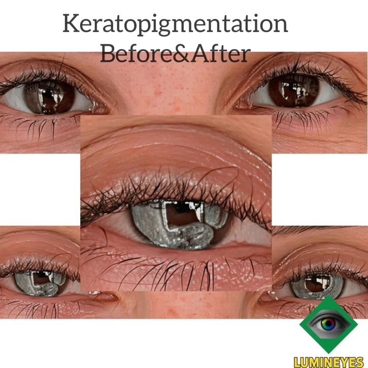
- Induced or irregular astigmatism: altering corneal tissue can disturb the regularity of the corneal optical surface and may produce irregular optical effects that are not adequately described by simple refractive-error measurements.
- Reduced optical quality: changes in transparency, pigment distribution, light transmission, or light scattering may contribute to glare, halos, reduced contrast, dysphotopsia, or other visual disturbances.
- Stromal damage and structural change: corneal manipulation may be associated with stromal injury, thinning, scarring, or other structural changes depending on the technique and individual response.
- Neovascularization and chronic corneal reaction: abnormal healing or persistent tissue reaction may result in vascular or inflammatory changes that can compromise the cornea.
- Pigment instability and migration: pigment distribution and the cosmetic appearance may change with healing, remodeling, tissue reaction, or pigment displacement over time.
3. Infectious Complications
An invasive procedure that disrupts the corneal tissue also introduces an infectious risk. Infectious keratitis can progress to severe corneal ulceration, stromal destruction, corneal melting, perforation, or other sight-threatening complications.
In a severe uncontrolled ocular infection, infection may extend beyond the cornea into deeper ocular structures, potentially resulting in intraocular infection including endoftalmi. Such progression is not an expected outcome of keratopigmentation, but it illustrates why a cosmetic corneal procedure cannot be regarded as equivalent to a superficial or risk-free cosmetic treatment.
4. Long-Term Consequences and Future Eye Surgery
The implications of keratopigmentation do not necessarily end when the cosmetic result has stabilized. Permanent corneal pigmentation and the structural changes associated with the procedure become part of the patient's long-term ophthalmic history.
If the patient subsequently requires refractive surgery, cataract surgery, corneal surgery, retinal examination or retinal treatment, or other ophthalmic intervention, the previous keratopigmentation may need to be taken into account during examination, optical assessment, imaging, surgical planning, visualization, and postoperative follow-up.
This does not mean that every future ophthalmic procedure will necessarily be prevented. The practical implications depend on the location, depth, density, and stability of the pigment and on the condition of the cornea at the time of the later procedure. Nevertheless, the patient has undergone a permanent alteration of an important optical tissue, and that alteration can remain clinically relevant for the rest of the eye's subsequent care.
Corneal Transparency and Future Ocular Care
The cornea must remain sufficiently transparent and optically regular for normal vision and for reliable clinical examination. Permanent pigment within the corneal stroma therefore creates a fundamentally different clinical situation from an eye in which the cornea has not been used as the site of cosmetic pigmentation.
Previous keratopigmentation should remain an important part of the patient's ophthalmic history. The significance for future management depends on the individual patient, the location and characteristics of the pigment, the condition of the cornea, and the type of subsequent ophthalmic treatment required.
Reversibility and Long-Term Stability
One of the most important questions before keratopigmentation is what happens if a patient later wants a different color or no longer wants the pigment. Unlike a temporary cosmetic surface treatment, intrastromal pigment is placed within corneal tissue.
Pigment removal can therefore be substantially more complicated than pigment placement. The feasibility of reversal depends on factors including the pigment used, its depth and distribution, the amount of material, the original technique, and the condition of the cornea.
Keratopigmentation should consequently be considered a significant corneal intervention rather than a simple cosmetic treatment that can automatically be reversed.
Iris-Based Approaches and Anatomical Target
Another approach to changing eye color is to target pigmentation within the iris rather than placing cosmetic pigment within the cornea. The Lumineyes laser eye color change procedure is based on this different anatomical approach.
The distinction is anatomical: keratopigmentation produces a visible color change through pigment placed within the cornea, whereas an iris-based approach is intended to modify pigmentation within the iris itself without using the cornea as the site of cosmetic pigment placement.
The clinical rationale and available documentation for the iris-based approach are presented in the Lumineyes clinical safety and research documentation .

Conclusion
Keratopigmentation can create a visible change in eye color by placing pigment within the cornea. The key anatomical distinction is that the procedure uses the cornea as the site of pigment placement, whereas the natural coloration of the eye originates from pigmentation within the iris.
Understanding the cosmetic effect, corneal involvement, optical considerations, long-term stability, and limitations of pigment removal is essential before considering keratopigmentation for cosmetic purposes.
The American Academy of Ophthalmology has also discussed concerns surrounding cosmetic eye-color-changing procedures, including procedures involving pigment placement in the cornea. Patients should receive an appropriate ophthalmic assessment and understand the potential consequences before treatment.
Frequently Asked Questions About Keratopigmentation
Is keratopigmentation safe for cosmetic eye color change?
Keratopigmentation is a corneal procedure, so its safety cannot be assessed solely by the cosmetic result. The technique involves manipulating corneal tissue and introducing pigment into the stroma. Potential effects on healing, corneal transparency, optical quality, pigment stability, and future ocular care should be considered on an individual basis.
How does keratopigmentation change eye color?
Keratopigmentation changes the visible appearance of the eye by placing pigment within the cornea. The pigment is seen in front of the natural iris and therefore creates a simulated eye-color effect rather than changing the iris's natural pigmentation.
Does keratopigmentation change the natural iris?
No. Keratopigmentation does not directly change the natural pigmentation of the iris. The iris remains anatomically present underneath the cornea, while pigment introduced into the corneal tissue creates the visible color effect.
Does keratopigmentation look natural?
The cosmetic appearance varies between patients and techniques. Because the pigment is positioned within the cornea rather than being part of the biological iris, it may not reproduce the full radial fiber pattern, depth, and light behavior of a natural iris. The final appearance can also depend on lighting, pigment distribution, healing, and individual anatomy.
Can keratopigmentation be fully reversed?
Reversal can be difficult. Once pigment has been introduced into the corneal stroma, removing it may require additional corneal intervention, and complete removal cannot be assumed in every patient. The feasibility of pigment removal depends on factors including the type, depth, distribution, and amount of pigment and the condition of the cornea.
How is keratopigmentation different from iris-based laser eye color change?
The main difference is the anatomical target. Keratopigmentation places pigment into the cornea to create a new visible color, while iris-based laser approaches are designed to act on pigmentation within the iris. The distinction is clinically important because the cornea is a transparent optical structure, whereas the iris is the natural tissue responsible for the pigmentation of the eye.
More information about the iris-based approach is provided in the clinical guide to laser eye color change .