Lumineyes™ Clinical Data: Nine Years of Longitudinal Clinical Observations
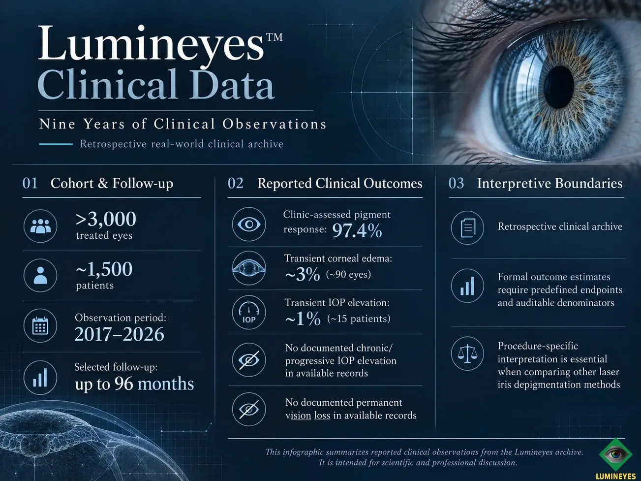
A physician-developed laser iris depigmentation protocol documented across approximately nine years of clinical practice: more than 3,000 treated eyes, approximately 1,500 patients, and selected follow-up extending to eight years
Quick Answer
This page presents the single-practitioner retrospective clinical archive associated with the contemporary Lumineyes™ protocol, developed and applied by Mustafa Mete, MD. Across approximately nine years of clinical practice, the archive encompasses more than 3,000 treated eyes in approximately 1,500 patients, with selected longitudinal observations extending to 96 months. The patient, eye, treatment-session, and follow-up units are distinguished throughout this report.
The available clinical records report no documented chronic or progressive IOP elevation and no documented permanent vision loss, alongside medically managed transient IOP elevations, transient corneal edema, mild iridocyclitis, and other generally short-lived anterior-segment responses. These are the practice’s reported observations, not pooled complication estimates from heterogeneous laser iris depigmentation methods. Exact endpoint denominators, follow-up completeness, and independent adjudication remain distinct requirements for formal statistical publication.
This Research Archive version deliberately separates reported clinical observations from mechanistic interpretation. The dedicated owner pages for selective stromal melanin modulation (SSMM), early IOP kinetics, L-SAFE, and L-OAT address those domains in greater depth.
Structured Abstract
Development-history clarification. The Lumineyes™ development history predates the approximately nine-year contemporary clinical observation period reported here. Duration of methodological development, years of clinical application, and the maximum follow-up of an individual eye are three separate measures; selected 96-month observations must not be presented as cohort-wide follow-up.
1. Dataset Provenance and Current Evidence Status
The clinical archive summarized here was accumulated during routine ophthalmic practice rather than under a prospectively registered observational protocol. That distinction is fundamental. A clinical archive can be large, longitudinal, and scientifically valuable while still requiring source verification, data cleaning, endpoint harmonization, and formal statistical design before it becomes a research-ready cohort.
| Parameter | Current record | Research interpretation |
|---|---|---|
| Approximate treated eyes | 3,000+ | Large single-practice clinical archive; exact eligibility and analysis denominators should be locked before formal analysis. |
| Approximate patients | ~1,500 | Participant-level denominators must remain distinct from eye-level and session-level denominators. |
| Clinical application period | Approximately nine years | Duration of the contemporary clinical observation archive, distinct from maximum individual follow-up. |
| Maximum reported follow-up | Up to 96 months in selected cases | Maximum follow-up is not equivalent to cohort-wide follow-up. Follow-up distribution and completeness must be reported separately. |
| Design | Retrospective longitudinal clinical observation | Not randomized; not prospectively registered; no untreated or alternative-treatment comparator. |
| Setting | Private clinical practice / single practitioner | Potential selection, referral, documentation, operator, and follow-up biases require explicit discussion. |
| External validation | Not yet performed | Independent replication and/or endpoint adjudication remain future research requirements. |
| Current analytical status | Descriptive Research Archive summary | Not yet a locked statistical dataset or peer-reviewed cohort analysis. |
The present page reports retrospective real-world clinical observations. It should be read as a transparent description of the available archive—not as an independently adjudicated registry, a prospectively derived incidence study, or a comparative safety trial.
2. Data Architecture: Participants, Eyes, Exposures, Visits, and “Cases” Are Different Units
A future peer-reviewed analysis cannot treat “patients,” “eyes,” “cases,” and “treatment sessions” as interchangeable denominators. Bilateral eyes are correlated within a participant, repeated treatment exposures are correlated within an eye, and duration of follow-up varies across individuals.
| Unit | What it represents | Why it matters |
|---|---|---|
| Participant | One individual receiving care | Appropriate denominator for participant-level adverse events, demographic variables, and participant-reported outcomes. |
| Eye | One treated or observed eye | Appropriate for eye-specific IOP, corneal, inflammatory, structural, and phenotype outcomes; paired-eye correlation must be addressed. |
| Exposure / session | One laser treatment event | Relevant when response or event probability may depend on cumulative exposure, treatment density, or interval. |
| Checkpoint / visit | One follow-up assessment | Relevant for longitudinal trajectories and repeated-measures analysis. |
| “Case” | Legacy clinical-record label | Must be mapped to a precise analytic unit before manuscript submission. |
This distinction is particularly important because several values in the current clinical summary are expressed using different units. Rather than hiding that inconsistency, the Research Archive should preserve it as an explicit pre-publication data-audit requirement.
3. Reported Clinical Outcomes: Observation Versus Statistical Readiness
The table below preserves the main values reported in the existing clinical page while separating the clinical observation from the statistical status of the endpoint. This prevents descriptive numbers from being mistaken for independently validated incidence estimates.
| Endpoint | Current reported observation | Current analytic unit | Before peer-reviewed inferential use |
|---|---|---|---|
| Clinic-assessed pigment response | 97.4% | Reported as “cases” | Audit required Define the endpoint operationally; provide numerator/denominator; specify image/clinical criteria; distinguish pigment response from final visible phenotype; define treatment-course completeness. |
| Chronic/progressive IOP elevation | 0 documented cases | Not yet specified as a locked denominator on this page | Audit required Prespecify definition, time window, denominator, medication context, follow-up completeness, and uncertainty interval. |
| Permanent vision loss | 0 documented cases | Not yet specified as a locked denominator | Audit required Define the visual endpoint, attribution, follow-up availability, and whether best-corrected acuity and other functional outcomes were systematically recorded. |
| Transient corneal edema / Descemet folds | ~3% of treated eyes (approximate) | Eye level | Reported Confirm exact numerator/denominator, diagnostic definition, severity, timing, management, and resolution criteria. |
| Transient IOP elevation | ~1% (~15 patients) | Participant level in the current page | Audit required Determine whether event ascertainment and analysis should be participant- or eye-level; define threshold, timing, duration, medication use, and bilateral events. |
| Mild iridocyclitis | Infrequently observed; final rate under reconciliation | Legacy “case” unit | Audit required Map “case” to participant or eye; define inflammatory criteria and grading; document timing, treatment, and resolution. |
| Transient pupillary fibrin / membrane | Reported transient finding; exact count pending reconciliation | Not fully specified | Audit required Establish the eye-level and participant-level counts and episode duration. Analyze persistent pupil shape or symmetry changes as a separate endpoint rather than combining them with transient fibrin/membrane events. |
| Posterior synechiae | No cases documented to date | Not yet locked | Audit required Prespecify ascertainment method, denominator, and observation period before inferential use. |
| Long-term endothelial observations | Reported as stable in available follow-up | Subset / availability not yet quantified on this page | Not a formal endothelial study Require standardized specular microscopy, baseline/follow-up pairing, device consistency, image-quality rules, sample size, and missing-data reporting. |
Why this table is deliberately strict. The table preserves the reported clinical observations without inventing reconciled counts. Where earlier approximate tallies and reporting units have not yet been matched to source records, provisional frequencies are deliberately not promoted to exact event rates. This does not diminish the observations; it defines how they can be reported reproducibly.
4. Zero-Event Observations: “None Documented” Is Not “Zero Risk”
The archive reports no documented chronic/progressive IOP elevation and no documented permanent vision loss within the available records. These observations are relevant, particularly in a large clinical archive, but they must be expressed using language that preserves statistical uncertainty.
“No event was observed or documented within the available dataset and follow-up.”
This wording is more appropriate than “0% risk” because an observed count of zero does not make the true population risk mathematically zero. The inferential value of a zero-event observation depends on the exact denominator, follow-up completeness, duration, event definition, ascertainment method, and loss to follow-up.
Formal publication should therefore report an appropriate uncertainty interval once the endpoint denominator and follow-up population are locked.
5. Longitudinal Follow-Up: Maximum Duration Is Not Cohort Duration
The longest available observations are reported to extend to approximately 96 months in selected cases. This is clinically valuable because delayed or progressive findings may not be apparent in short-term follow-up. However, the correct interpretation is not that the entire cohort was observed continuously for eight years.
A formal longitudinal analysis should report the distribution of follow-up, not only its maximum. It should identify the number of participants or eyes contributing to defined time horizons and quantify loss to follow-up or unavailable observations.
| Metric | Why it is required |
|---|---|
| Median follow-up and interquartile range | Describes the typical follow-up experience rather than the longest case. |
| Minimum / maximum follow-up | Defines the observed temporal range. |
| n available at prespecified intervals | Shows how much of the cohort contributes to 6-, 12-, 24-, 36-, 60-, or 96-month conclusions. |
| Loss to follow-up | Allows assessment of attrition bias and whether missingness may be related to outcome. |
| Reason for missing follow-up | Separates administrative absence from outside follow-up, treatment discontinuation, or other known causes. |
| Time-to-event structure | Allows delayed adverse events to be analysed against the population actually under observation at each time point. |
6. Intraocular Pressure: Observation, Kinetics, and Outcome Definition
The present archive reports transient early IOP elevations in approximately 1% of patients, managed medically in the recorded episodes, and no documented chronic or progressive IOP elevation in the available longitudinal records. Early pressure spikes and late pressure measurements are separate outcomes: the same time point, treatment context, and denominator must not be assigned to both. Selected long-term examinations have included lower IOP measurements, which are retained as observations without attributing a proven causal mechanism or assuming a uniform effect across the cohort.
These observations should be interpreted against the broader ophthalmic context. Pigment dispersion can increase trabecular pigmentation and is associated with reduced aqueous outflow and pigmentary glaucoma in susceptible eyes.[8] Published reports following cosmetic iris laser treatment include refractory pigmentary glaucoma, severe bilateral pigmentary glaucoma, and markedly elevated IOP.[3–6] These reports document that pressure-related complications are possible after some cosmetic iris laser procedures. Where laser settings and clinical protocols are not disclosed, they cannot be used as if they described the contemporary Lumineyes™ technique or estimated its event rates.
Within the Lumineyes™ research architecture, detailed time-dependent interpretation is examined in the Early IOP Monitoring Framework, while pre-exposure assessment is addressed through L-SAFE. The Blood–Aqueous Barrier Dynamics review separately examines the timing and interpretation of anterior-chamber responses without treating them as measured outcomes of this cohort.
| Variable | Recommended analytical treatment |
|---|---|
| Baseline IOP | Record per eye with tonometry method and relevant medication context. |
| Early postoperative IOP | Use prespecified time windows rather than an undefined “postoperative” category. |
| Peak IOP | Report absolute value, change from baseline, timing, laterality, and management. |
| Duration of elevation | Separate brief, medically responsive events from persistent or progressive abnormality. |
| Medication exposure | Do not interpret normalized IOP under treatment as equivalent to untreated physiological stability. |
| Angle / pigment findings | Report where gonioscopic documentation is available; do not infer trabecular preservation from pressure alone. |
| Longitudinal trajectory | Use repeated-measures methods that account for eye-within-participant correlation where appropriate. |
7. Corneal and Endothelial Outcomes
The archive reports transient corneal edema / Descemet folds in approximately 3% of eyes, described as resolving during recovery. It also describes corneal transparency and endothelial findings as stable in available follow-up. Those observations are clinically relevant but should remain separate from the stronger claim of prospectively demonstrated long-term endothelial safety.
Specular microscopy is well suited to longitudinal endothelial assessment when acquisition quality, sampling, and analysis are standardized.[9] A future endothelial study should define endothelial cell density (ECD), coefficient of variation, hexagonality, image-quality criteria, device consistency, central corneal thickness where relevant, and paired baseline-to-follow-up change. The measurement principles and limitations are reviewed in Corneal Endothelial Assessment and Specular Microscopy.
Research boundary. “Stable endothelial observations in available follow-up” is an observational statement. It should not be converted into “no endothelial effect” unless a prospectively defined and adequately powered endothelial study supports that conclusion.
8. Retina, Optic Nerve, and Visual Function
The clinical archive reports no documented permanent vision loss in the available records and describes preserved retinal and optic-nerve structural findings in available long-term imaging. The scientifically appropriate wording is limited to what was clinically documented; it should not imply absence of microscopic or subclinical change.
Posterior-segment surveillance matters because published case literature includes laser-induced maculopathy and other posterior-segment injury after cosmetic iris laser procedures.[5,7] These reports concern patients treated through separate, incompletely characterized laser iris procedures and are not Lumineyes™ clinical cases. They establish a reason to examine posterior-segment outcomes where indicated, but neither define the event rate of the Lumineyes cohort nor establish technical equivalence between the procedures.
Future formal analysis should specify which imaging modalities were available, the proportion of the cohort imaged, image-quality criteria, segmentation procedures where relevant, and whether interpretation was masked or independently adjudicated.
9. Pigment Response: Interpretation of the 97.4% Clinic-Assessed Endpoint
The clinical archive records a 97.4% clinic-assessed pigment-response endpoint. It reflects the treating practice’s documented response assessment rather than a guaranteed final eye color or an independently validated image-derived measure. In a formal clinical manuscript, the endpoint should be defined with its original numerator, denominator, assessment time point, and criteria.
“Pigment discharge” must be distinguished from several different outcomes:
- immediate visible lightening;
- measurable reduction in visible pigmentation;
- pigment or particulate material observed in the anterior chamber;
- resolution of anterior-chamber pigment;
- residual angle pigmentation;
- completion of a treatment course;
- and final visible iris phenotype.
To translate the reported clinic endpoint into a reproducible research variable, the final analysis should specify how incomplete courses, repeated exposures, examiner judgement, and serial image findings were treated. The 97.4% observation remains part of the archive; the statistical definition determines what it can validly quantify.
The intended biological target concept is developed in Selective Stromal Melanin Modulation (SSMM). The separate P-CLEAR Framework provides a conceptual distinction between pigment release, biological processing, and clearance; neither framework is retroactively treated as a measured endpoint of historical records.
10. Mechanistic Context: Important, but Separate From the Dataset
The original Clinical Data page included an extended discussion of selective photothermolysis / photoacoustic pigment disruption, macrophage processing, aqueous outflow, and delayed pigment clearance. These mechanisms remain relevant to interpretation, but the retrospective archive did not prospectively measure laser–tissue energy deposition, macrophage recruitment, cytokine kinetics, pigment-particle transport, extracellular-matrix turnover, or a complete clearance pathway.
Selective photothermolysis provides a general framework for preferential laser interaction with an absorbing chromophore, but biological selectivity in the iris depends on wavelength, pulse duration, spot characteristics, pigment distribution, local anatomy, and the actual tissue response.[11] Ocular melanin also has complex physiological and photobiological roles that should not be reduced to a single “pigment removal” mechanism.[12]
Accordingly, mechanistic models provide context while this page remains the primary owner of the clinical observations. Beyond Melanin Clearance examines how visible pigment reduction, melanocyte function, stromal signalling, and extracellular-matrix responses represent different biological domains and should not be inferred from appearance alone.
Figure 1. Original Research Archive schematic. A large clinical archive becomes stronger scientific evidence through explicit data provenance, endpoint definition, statistical discipline, and independent evaluation—not through stronger wording alone.
11. Temporal Evolution of Visible Iris Phenotype
Serial clinical observation indicates that visible iris phenotype can continue evolving beyond the immediate treatment interval. The trajectory is relevant to response-guided practice; the archival observations do not, by themselves, quantify individual cell-transport or stromal-remodelling pathways.
Future objective phenotype work should use standardized photography, quantitative colorimetry, spatial mapping, and validated imaging methodology. The proposed Lumineyes Optical Architectural Typology (L-OAT) provides a separate conceptual framework for multimodal iris phenotyping; historical photographs should not be retroactively labelled L-OAT data unless they satisfy the acquisition, calibration, registration, and quality requirements of that framework.
12. Patient Selection and Clinical Risk Management
Patient selection and clinical judgement are integral parts of the Lumineyes™ treatment model. Their effects should be documented as part of the clinical protocol rather than assumed to explain the observed outcomes. Selection criteria, exclusion patterns, adherence, baseline ocular characteristics, and treatment discontinuation can all influence the composition of a real-world cohort.
A formal study should therefore describe eligibility criteria, reasons for exclusion, reasons for discontinuation, and baseline ocular features using prespecified categories. Patient selection should be analysed as part of the clinical system rather than invoked post hoc as proof of causation.
The broader methodology of baseline assessment, monitoring, selective advanced testing, and evidence interpretation is discussed in How Clinical Evidence Is Evaluated in Laser Eye Color Change.
13. External Literature: Distinguishing Laser Platforms, Methods, and Clinical Outcomes
Laser iris depigmentation (LID) is a procedural category, not one uniform intervention. Published approaches differ in the laser platform, wavelength, pulse characteristics, delivered exposure, anatomical targeting, cumulative treatment pattern, interval between courses, patient selection, adjunctive medication, and postoperative monitoring. The fact that two procedures both address iris pigment—or even use lasers within the same wavelength family—does not establish equivalent tissue interaction or clinical management.
The contemporary Lumineyes™ protocol is a physician-developed, non-incisional, selective stromal melanin modulation approach based on individualized examination, staged and response-guided exposure, defined monitoring, and clinical stop criteria. Its longitudinal outcomes are reported from its own clinical archive rather than calculated from unrelated LID methods. Distinct protocols also require distinction in the opposite direction: neither unfavorable case reports nor reassuring outcomes of other methods can be automatically transferred to Lumineyes™.
| Source | Published technique or clinical context | What the report establishes—and does not establish |
|---|---|---|
| Grimaldos Ruiz, 2021; 1,176 eyes[1] | Published prospective photoablative cosmetic iridoplasty (PCI) series using a 532-nm Crystal Q-switched Nd:YAG platform with 3–4 ns pulses and staged treatment phases. | A substantial different clinical protocol with reported outcomes. Shared wavelength characteristics do not imply identical spot geometry, energy distribution, tissue targeting, patient selection, or Lumineyes™ event rates. |
| D'Oria et al., 2022; D'Oria & Alió, 2025[2,10] | Reviews of heterogeneous techniques for cosmetic alteration of eye color, including laser approaches. | Provide comparative context and identify unanswered safety questions; they do not supply direct outcome data for this Lumineyes™ clinical archive. |
| Ong et al. and Swampillai et al.[3,4] | External case reports describing pigmentary glaucoma after cosmetic iris laser treatment, with incomplete protocol-level comparability to Lumineyes™. | Establish that serious events can occur in particular treated individuals; do not estimate population incidence or establish that the same protocol was used. |
| Liu et al., 2023[5] | External case after four bilateral laser sessions, with reported postoperative pilocarpine use, severe IOP elevation, pigmentary glaucoma, paracentral acute middle maculopathy, and intraretinal hemorrhages. The authors could not determine the original laser parameters. | The laser platform and exposure settings cannot be reconstructed reliably. This is not a Lumineyes™ case and must not be entered into, or used as a direct estimate for, the Lumineyes™ cohort. |
| Flores-Márquez et al., 2023[7] | Separate report of maculopathy following cosmetic iris depigmentation; detailed technical comparability cannot be established from the citation alone. | Signals a posterior-segment outcome requiring attention; does not measure Lumineyes™-specific frequency. |
| Jaber et al., 2026[6] | External case report of markedly elevated IOP and corneal complications following laser iris depigmentation; complete individualized treatment parameters are not supplied for direct reconstruction. | Documents a clinically important occurrence after another reported treatment course; does not establish a pooled risk for every LID protocol. |
Interpretive principle. Evaluating the Lumineyes™ archive requires direct assessment of its own exposure histories, clinical examinations, response-guided decisions, adverse-event definitions, and follow-up—not substitution of external procedure outcomes. This does not exclude adverse events documented elsewhere; it preserves the distinctions necessary for meaningful clinical comparison.
14. Statistical Architecture for the Future Retrospective Manuscript
The current website summary is descriptive. A peer-reviewed retrospective study should begin with a locked research question and a prespecified analytical dataset rather than by testing every available variable after inspecting the outcomes.
| Domain | Recommended requirement |
|---|---|
| Population | Define start/end dates, inclusion/exclusion rules, treated-eye denominator, participant denominator, and bilateral-treatment rules. |
| Primary safety endpoint | Prespecify clinically meaningful endpoints before analysis; distinguish transient responses from persistent or vision-threatening events. |
| Secondary endpoints | Define corneal, inflammatory, IOP, visual, structural, and pigment-response outcomes separately. |
| Denominators | Report n/N for every event; do not mix participant-, eye-, case-, session-, and visit-level rates. |
| Bilateral correlation | Use methods that account for two eyes from the same participant where eye-level analyses are performed. |
| Repeated measures | Use longitudinal or other appropriate repeated-measures methods for serial IOP and continuous outcomes. |
| Rare / zero events | Report uncertainty estimates rather than converting zero observed events into zero risk. |
| Follow-up | Report median duration, distribution, numbers available at defined intervals, and attrition. |
| Missing data | Describe amount, pattern, likely mechanism, and sensitivity analyses where feasible. |
| Protocol evolution | Prespecify whether calendar period, protocol generation, exposure characteristics, or postoperative management require stratification or sensitivity analysis. |
| Adverse-event adjudication | Where feasible, use predefined definitions and independent or blinded review for key endpoints. |
| Reporting framework | Use an appropriate observational-reporting standard, such as STROBE, and document any use of routinely collected clinical data transparently. |
15. Pre-Publication Data Audit: What Must Be Resolved Before Journal Submission
Because this page is intended to become the foundation of a future peer-reviewed report, the most useful next step is not to add stronger adjectives. It is to make the dataset more auditable.
Critical integrity rule. L-SAFE, L-OAT, and other frameworks developed after historical treatments must not be described as though they prospectively governed or measured earlier cases. Historical records may be reclassified retrospectively only when the required source variables are actually present, and the retrospective nature of that exercise must remain explicit.
16. Relationship to the Wider Lumineyes™ Research Architecture
The Clinical Data page should own the reported clinical observations. Other Research Archive pages should own the mechanistic, monitoring, safety-control, and phenotype frameworks. This division prevents one page from becoming an unstructured mixture of outcomes, hypotheses, and terminology.
The broader evidence-interpretation context is discussed in Clinical Interpretation of Common Claims in Laser Eye Color Change. The canonical procedural overview remains Laser Eye Color Change, and publication records are organized through the Lumineyes™ Research Library.
17. What the Current Archive Can—and Cannot—Establish
| Question | Current archive can contribute | Current archive cannot establish alone |
|---|---|---|
| What responses have been observed in this clinical practice? | Yes, subject to record completeness and endpoint definition. | It cannot prove that undocumented events never occurred. |
| Can long-follow-up cases be identified? | Yes; selected observations extend to 96 months. | Maximum follow-up cannot be generalized to the whole cohort. |
| Can adverse-event patterns generate hypotheses? | Yes. | Descriptive observations alone do not establish causality. |
| Can the archive estimate event frequency? | Potentially, after denominator reconciliation and cohort locking. | The current website percentages are not yet a substitute for a predefined incidence analysis. |
| Can it prove zero risk? | No. | Zero documented events do not imply zero population risk. |
| Can it establish comparative superiority? | No. | A comparator and appropriate comparative design are required. |
| Can it prove a biological mechanism? | No; it may motivate hypotheses. | Mechanistic causality requires dedicated measurement or experimental evidence. |
| Can it guide prospective study design? | Yes. | Prospective validation still requires new, predefined data collection. |
18. Research Priorities Arising From the Archive
Safety and longitudinal outcomes
- serial IOP trajectories with medication context;
- standardized endothelial cell measurements;
- corneal and anterior-chamber adverse-event definitions;
- posterior-segment imaging where clinically indicated;
- time-to-event analysis for delayed complications;
- and external endpoint adjudication.
Phenotype and mechanism
- prospective quantitative pigment-response definitions;
- standardized colorimetry and photography;
- qualified AS-OCT phenotype measurements;
- pigment-clearance kinetics;
- the relation between exposure, biological response, and visible change;
- and mechanistic studies that do not infer cellular processes from appearance alone.
19. Limitations
- Retrospective design: data were accumulated during clinical care rather than under a prospectively registered research protocol.
- Single-practitioner / single-practice setting: generalizability is uncertain and operator-related effects cannot be separated from protocol effects.
- No control group: the archive does not establish comparative efficacy or comparative safety.
- Selection bias: screening and treatment-selection practices may materially influence the observed cohort.
- Variable follow-up: the longest follow-up applies to selected cases rather than the complete cohort.
- Documentation bias: completeness and precision depend on historical clinical records.
- Endpoint heterogeneity: several current outcomes require operational definitions and denominator reconciliation.
- Mixed analytic units: the current record includes eye-, participant-, and “case”-level reporting.
- Missing-data uncertainty: formal attrition and missingness analyses have not yet been reported.
- No independent adjudication: the present summary has not been reviewed by an external clinical endpoint committee.
- Mechanistic uncertainty: macrophage activity, cytokine kinetics, extracellular-matrix turnover, and stromal remodeling were not prospectively quantified.
- Rare-event uncertainty: zero observed events do not establish zero population risk.
- Endothelial evidence: available observations are not a substitute for a standardized prospective endothelial study.
- Protocol evolution: a formal manuscript must determine whether changes over calendar time require stratification or sensitivity analysis.
20. Conclusion
The contemporary Lumineyes™ clinical archive represents more than 3,000 treated eyes in approximately 1,500 patients accumulated across approximately nine years of clinical application, with selected longitudinal observations extending to 96 months. The archive contains clinically relevant information on early and longitudinal responses, including reported pigment response, transient IOP elevation, corneal edema, inflammatory events, and long-term follow-up.
Its scientific value depends on how precisely those observations are converted into analyzable data. The substantive scientific contribution lies in the size, duration, and procedure-specific nature of the observations. Definitive rate estimates and between-protocol comparisons will become stronger as exact denominators, endpoint definitions, follow-up accounting, and independent review are incorporated into the planned analyses.
Clinical experience is the foundation; transparent methodology is how its full evidentiary value can be communicated.
Research FAQ
Does “0 documented cases” mean the true complication risk is zero?
No. It means that no such event was documented in the available clinical dataset and follow-up. Formal risk estimation requires an exact denominator, event definition, follow-up accounting, and uncertainty interval.
Were all approximately 1,500 patients followed for eight years?
No. The maximum reported follow-up of approximately 96 months applies to selected cases. A formal longitudinal paper should report the full distribution of follow-up and the number of participants or eyes available at defined time points.
Is the 97.4% pigment-discharge figure a validated efficacy endpoint?
Not yet. It is a clinic-assessed outcome reported in the current archive. Before peer-reviewed inferential use, its numerator, denominator, operational definition, assessment method, time point, and handling of incomplete treatment courses should be specified.
Does this archive prove long-term endothelial safety?
No. Stable endothelial observations in available follow-up are clinically relevant, but a definitive endothelial-safety conclusion requires a standardized prospective study with paired measurements, image-quality rules, and complete follow-up reporting.
Were historical cases managed using L-SAFE?
No. L-SAFE is a later formalized conceptual framework. Historical records may be examined retrospectively only when the required source variables are present, and they should not be described as having been prospectively managed by a framework that had not yet been defined.
What is the role of L-OAT in the clinical archive?
L-OAT is a proposed prospective multimodal iris-phenotyping framework. Historical photographs should not automatically be labelled L-OAT data unless they meet its acquisition, calibration, registration, and quality requirements.
References
- Ruiz PG. Photoablative cosmetic iridoplasty: effective, safe, and predictable-eye color change in 1176 eyes. Int Ophthalmol. 2021;41(4):1381-1393. doi: 10.1007/s10792-021-01693-5. PMID:33484383.
- D'Oria F, Abu-Mustafa SK, Alio JL. Cosmetic Change of the Apparent Color of the Eye: A Review on Surgical Alternatives, Outcomes and Complications. Ophthalmol Ther. 2022;11(2):465-477. doi: 10.1007/s40123-022-00458-2. PMID:35061240.
- Ong AY, Ching-A-Sue G, Krilis M, Guirao-Navarro MC, Hornby S, Jutley G. Refractory iatrogenic pigmentary glaucoma secondary to cosmetic laser treatment: a case report. Eur J Ophthalmol. 2023;33(2):NP1-NP4. doi: 10.1177/11206721211050338. PMID:34623172.
- Swampillai AJ, Sherman T, Garg A, Tan IJ, Lim KS. Secondary pigmentary glaucoma following cosmetic laser treatment to alter iris colour. Cont Lens Anterior Eye. 2023;46(2):101754. doi: 10.1016/j.clae.2022.101754. PMID:36175318.
- Liu J, Korban S, Moster MR, Rhéaume MA, Wang Q. Bilateral severe iatrogenic pigmentary glaucoma following laser treatment for cosmetic iris color change. Am J Ophthalmol Case Rep. 2023;32:101927. doi: 10.1016/j.ajoc.2023.101927. PMID:37771388.
- Jaber W, Nemet M, Waisbourd M. Laser Iris Depigmentation Resulting in Markedly Elevated Intraocular Pressure and Herpes Simplex Keratitis with Corneal Scarring: Case Report. Case Rep Ophthalmol. 2026;17(1):312-317. doi: 10.1159/000551226. PMID:41982480.
- Flores-Márquez A, Moreno-Gutiérrez JA, Chinchurreta-Capote A, García-Martín F, Rocha-de-Lossada C. Laser-induced maculopathy after iris depigmentation cosmetic treatment. Can J Ophthalmol. 2023;58(1):e29-e31. doi: 10.1016/j.jcjo.2022.05.012. PMID:35809624.
- Scuderi G, Contestabile MT, Scuderi L, Librando A, Fenicia V, Rahimi S. Pigment dispersion syndrome and pigmentary glaucoma: a review and update. Int Ophthalmol. 2019;39(7):1651-1662. doi: 10.1007/s10792-018-0938-7. PMID:29721842.
- McCarey BE, Edelhauser HF, Lynn MJ. Review of corneal endothelial specular microscopy for FDA clinical trials of refractive procedures, surgical devices, and new intraocular drugs and solutions. Cornea. 2008;27(1):1-16. doi: 10.1097/ICO.0b013e31815892da. PMID:18245960.
- D'Oria F, Alio JL. Surgical Techniques for Cosmetic Eye Color Change: A Narrative Review. Ophthalmol Ther. 2025;14(8):1685-1694. doi: 10.1007/s40123-025-01177-0. PMID:40483374.
- Anderson RR, Parrish JA. Selective photothermolysis: precise microsurgery by selective absorption of pulsed radiation. Science. 1983;220(4596):524-527. doi: 10.1126/science.6836297. PMID:6836297.
- Hu DN, Simon JD, Sarna T. Role of ocular melanin in ophthalmic physiology and pathology. Photochem Photobiol. 2008;84(3):639-644. doi: 10.1111/j.1751-1097.2008.00316.x. PMID:18346089.
Conflict of interest and data provenance. Mustafa Mete, MD, is the developer of the Lumineyes™ method and is affiliated with MyLumineyes. The clinical archive derives from his practice; this involvement is declared as a competing interest. Clinical findings are presented as the practice’s retrospective observations pending formal independent adjudication.
Funding. No external funding was received for preparation of this retrospective Research Archive summary. Future formal studies will provide study-specific funding declarations.
Evidence classification. The material on this page should be understood as retrospective real-world clinical observations and hypothesis-generating evidence. It is not a randomized trial, an independently validated registry, or a population-level risk estimate.
Medical information notice. This Research Archive page is intended for scientific and professional discussion. It does not provide individualized treatment instructions, candidacy decisions, medication protocols, or guarantees of outcome.
Correspondence and research collaboration. Research inquiries concerning methodology, retrospective analysis, independent statistical review, or potential collaboration should be directed through the official MyLumineyes contact and medical evaluation channel.
About the author. Mustafa Mete, MD, is an ophthalmologist and developer of the Lumineyes™ methodology. Related records are available through the Lumineyes™ Research Hub and Research Library.