Selective Stromal Melanin Modulation (SSMM)
The Lumineyes™ conceptual framework for controlled, response-guided reduction of optically relevant iris stromal melanin
Quick Answer
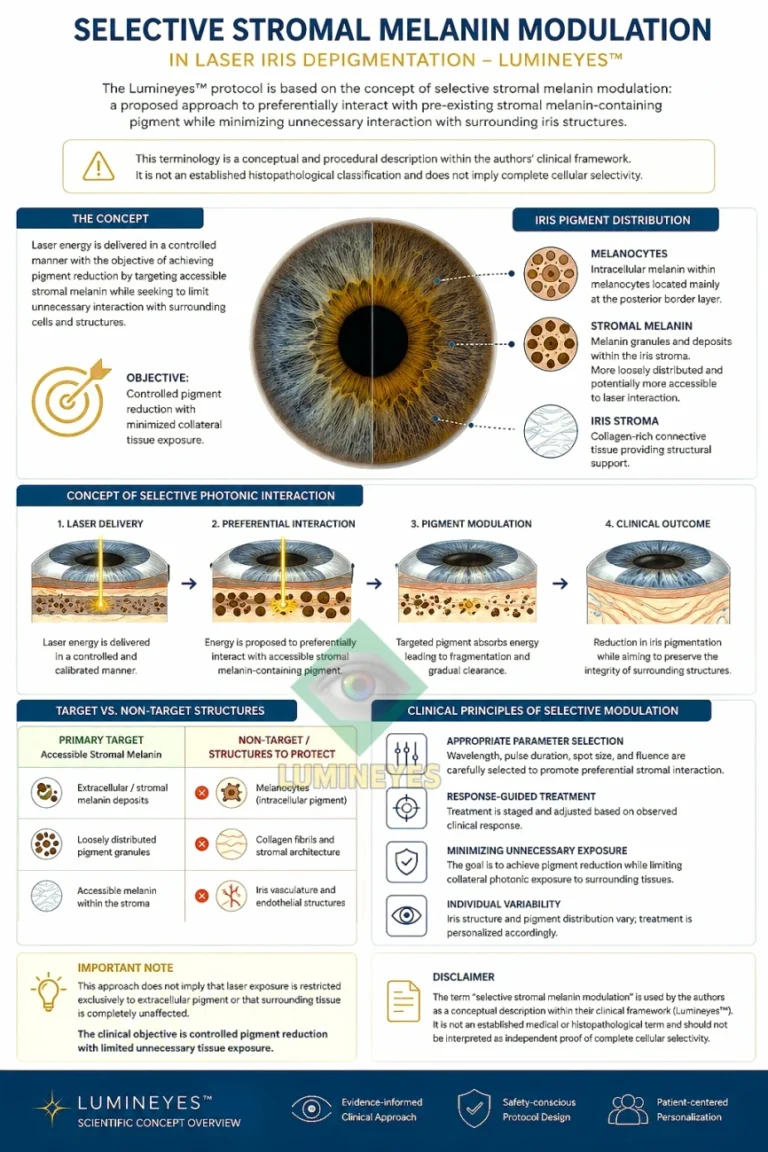
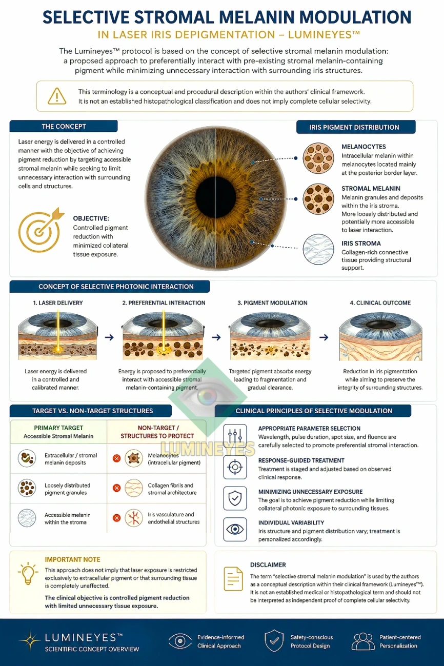
Selective stromal melanin modulation (SSMM) is the term used within the Lumineyes™ framework to describe an intended clinical strategy in which laser exposure is directed toward the optically relevant melanin-bearing compartment of the anterior iris stroma, with the objective of producing incremental pigment reduction rather than intentional tissue ablation. The term combines a spatial target concept—stromal melanin—with a treatment philosophy based on controlled exposure, observation of the biological response, and staged re-exposure only after reassessment.
SSMM is therefore not presented as a universally established histopathological category, nor as proof that laser energy interacts exclusively with pigment and never with surrounding tissue. It is a Lumineyes™-specific conceptual and procedural term intended to define what is being targeted, how the response is interpreted, and why treatment is staged rather than treated as a fixed sequence of identical laser sessions.
1. Why a New Term Was Needed
The conventional expression laser iris depigmentation describes an observable endpoint—the reduction of visible iris pigment—but says relatively little about the intended biological target, the degree of tissue selectivity, or the decision architecture used during a multi-stage treatment course. In the Lumineyes™ framework, these distinctions are clinically important. The procedure is not conceptualized simply as “removing color” or as repeatedly delivering a predetermined laser exposure. Instead, the treatment is framed as an iterative interaction between laser parameters, stromal pigment phenotype, ocular response, pigment clearance, and the decision whether additional exposure is appropriate.
The term selective stromal melanin modulation was therefore introduced within the Lumineyes™ clinical framework to provide a more precise description of this intended process. Each word carries a specific meaning: selective denotes preferential targeting rather than absolute exclusivity; stromal identifies the anterior iris compartment that contributes substantially to visible eye color; melanin identifies the principal chromophore of interest; and modulation deliberately avoids equating the desired endpoint with indiscriminate tissue ablation.
Selective stromal melanin modulation is a response-guided treatment concept in which calibrated laser exposure is directed toward melanin-bearing structures of the optically relevant anterior iris stroma with the aim of progressively reducing visible pigment burden while minimizing unnecessary structural disturbance and using the eye’s observed biological response to determine subsequent treatment decisions.
Terminological boundary. SSMM should not be interpreted as a claim that current evidence proves complete cell-level selectivity, exclusive interaction with extracellular pigment, or absence of collateral tissue effects. Those questions require direct experimental and prospective clinical validation. The value of the term is that it defines the intended target and treatment logic of the Lumineyes™ method without overstating what has already been histologically demonstrated.
2. The Three-Part Lumineyes™ Terminology Architecture
SSMM is most useful when understood as one component of a broader Lumineyes™ terminology system. The framework separates the targeting principle, the biological response, and the decision to re-expose. This separation is important because a technically similar laser pulse may have different clinical consequences in different eyes or at different stages of the same eye’s treatment course.
Selective Stromal Melanin Modulation
Spatial / biophysical concept. What is intended to be modulated: the optically relevant melanin-bearing anterior stromal compartment, with avoidance of unnecessary structural injury as a treatment objective.
Controlled Treatment-Related Pigment Response
Biological response concept. What is observed after exposure: pigment liberation, inflammatory response, pressure behavior, clearance, and recovery are treated as clinically meaningful response variables rather than incidental events.
Response-Guided Staged Treatment
Temporal / decision concept. When further exposure is considered: the next stage is not defined solely by a calendar or fixed session count, but by reassessment of how the eye responded to the previous exposure.
| Term | Primary question | What the term describes | What it does not claim |
|---|---|---|---|
| Selective stromal melanin modulation (SSMM) | What is the intended target? | Preferential interaction with optically relevant melanin-bearing structures of the anterior iris stroma, pursued as incremental pigment reduction rather than intentional stromal ablation. | Absolute chromophore exclusivity, proven histologic confinement, or zero effect on adjacent tissue. |
| Controlled treatment-related pigment response | What happens after exposure? | The clinically observable response to treatment, including pigment dispersion/clearance and associated anterior-segment reactions that require interpretation and monitoring. | That every inflammatory or pressure response is desirable, harmless, or identical between patients. |
| Response-guided staged treatment | When should another exposure occur? | A decision model in which subsequent treatment depends on recovery, response trajectory, residual pigment phenotype, and clinical reassessment. | A predetermined number of sessions, automatic next-day continuation, or a universal energy schedule. |
3. Anatomical Basis: Why the Anterior Iris Stroma Matters
Human iris color is not determined simply by the number of melanocytes. Histological and ultrastructural studies indicate that visible differences in iris color are strongly related to the amount, distribution, packaging, and optical behavior of melanin and melanosomes within the anterior border layer and superficial stroma. The posterior iris pigment epithelium is heavily pigmented in most normally pigmented eyes, but its contribution to the externally perceived iris color is comparatively limited because it lies behind the stromal optical architecture.[1–4]
This distinction is fundamental to SSMM. The Lumineyes™ concept does not require the assumption that the clinically relevant pigment is predominantly “free” or extracellular. On the contrary, the most defensible current anatomical model is that the optical target is the melanin-bearing anterior stromal compartment, including pigment contained within stromal melanocytes and melanosomal structures. Whether different microscopic pigment pools exhibit different laser-response thresholds remains an open research question.
| Anatomical / optical component | Established biological role | Relevance to visible iris color | Relevance within Lumineyes™ SSMM |
|---|---|---|---|
| Anterior border layer | Anterior condensation of iris stromal tissue containing pigment cells and connective tissue elements. | Important contributor to the anterior optical appearance of the iris. | Part of the optically accessible pigment-bearing region considered during treatment planning. |
| Anterior / superficial stroma | Loose connective tissue containing melanocytes, collagen, vessels, nerves, fibroblastic cells and other cellular elements. | Melanin quantity and stromal light scattering jointly influence perceived color. | Principal conceptual target compartment for controlled pigment reduction. |
| Stromal melanocytes / melanosomes | Contain iris melanin; melanocyte number varies less with eye color than melanin amount and melanosomal characteristics. | Major determinant of brown, hazel and intermediate pigmentation. | Melanin is the intended chromophore; the objective is not intentional destruction of stromal architecture. |
| Stromal collagen architecture | Provides structural organization and contributes to light scattering. | Scattering becomes increasingly visible as anterior pigment burden decreases. | Preservation of stromal architecture is a treatment objective because optical appearance depends on both pigment and structure. |
| Posterior pigment epithelium | Densely pigmented posterior epithelial layer of the iris. | Normally contributes less to externally perceived color than the anterior stromal compartment. | Not the intended cosmetic target of SSMM. |
4. Why “Modulation” Is More Precise Than “Ablation”
The choice of the word modulation is deliberate. In biomedical laser terminology, ablation generally implies removal or destruction of tissue. That is not the intended conceptual endpoint of Lumineyes™. The desired endpoint is a progressive change in the pigment burden and optical expression of the anterior stroma while preserving the functional architecture of the iris as far as clinically possible.
For that reason, SSMM describes a graded biological process. The visible effect is expected to emerge from the interaction of several factors: the initial pigmentation phenotype, the spatial distribution of melanin, the energy delivered to the target, the eye’s inflammatory and pressure response, the subsequent handling of liberated pigment, and the optical contribution of the residual stromal architecture. A darker iris is therefore not treated simply as “more sessions of the same exposure”; it represents a different pigment burden and potentially a different biological response trajectory.
5. What “Selective” Means—and What It Does Not Mean
The concept of selectivity in laser medicine originates from the principle that a wavelength and pulse structure can be chosen to favor absorption by a target chromophore relative to surrounding structures. Selective photothermolysis established this general biophysical logic, although the exact mechanisms of pigment disruption depend on wavelength, pulse duration, fluence, spot geometry, tissue optics, and the dimensions and thermal properties of the target.[5]
Within SSMM, selective is therefore used in a relative and operational sense: the treatment is designed to favor the melanin-bearing stromal target while minimizing unnecessary interaction with non-target tissue. It should not be read as an assertion that the energy is absorbed by melanin alone, that every melanin-containing structure responds identically, or that microscopic collateral effects are impossible.
SSMM deliberately avoids five overclaims
- It does not claim that all clinically relevant iris pigment is extracellular.
- It does not claim complete histological preservation has already been proven for every treated eye.
- It does not claim that a specific visible eye color can be guaranteed in advance.
- It does not claim that all forms of laser iris depigmentation are biologically equivalent to the Lumineyes™ protocol.
- It does not convert clinical experience into independent proof; protocol-specific outcomes require structured prospective and long-term evaluation.
6. Laser–Tissue Interaction Within the SSMM Model
Melanin is a broadband optical absorber, but the biological effect of a laser pulse cannot be predicted from wavelength alone. The final tissue response reflects the combined influence of wavelength, pulse duration, pulse energy, fluence, spot size, spatial beam profile, repetition pattern, optical penetration, and the local pigment phenotype. These variables are interdependent. For example, changing spot diameter changes fluence even when nominal pulse energy remains unchanged.
Accordingly, Lumineyes™ SSMM is better understood as a parameter relationship rather than a single energy setting. The same nominal laser energy may not represent the same biological exposure across different spot geometries, different pigment densities, or different stages of treatment. This is one reason why a response-guided framework is conceptually preferable to treating session number or device output as the primary unit of treatment.
| Variable | Physical significance | Potential biological consequence | SSMM interpretation |
|---|---|---|---|
| Wavelength | Influences chromophore absorption and optical penetration. | Changes the relative energy absorbed by melanin and surrounding tissue. | Selected as part of a melanin-targeting strategy, not interpreted in isolation. |
| Pulse duration | Determines the time scale over which energy is deposited. | Influences thermal confinement and the balance of photothermal / mechanical effects. | Must be considered relative to the dimensions and thermal behavior of pigment-containing targets. |
| Pulse energy | Total energy delivered per pulse. | Contributes to pigment disruption and potential non-target stress. | Not a stand-alone measure of tissue exposure. |
| Fluence | Energy per unit area. | Strongly influences treatment threshold and tissue response. | Requires interpretation together with spot size and beam profile. |
| Spot size / geometry | Determines the spatial area over which energy is distributed. | Changes fluence, precision, overlap and local energy gradients. | Part of the spatial control strategy rather than a purely cosmetic setting. |
| Beam profile | Describes energy distribution within the spot. | Non-uniform profiles may generate local peak intensities. | Spatial energy homogeneity is relevant to reproducibility and avoidance of unintended hotspots. |
| Repetition / cumulative exposure | Determines how multiple pulses and sessions accumulate over time. | May influence pigment load, inflammation and pressure response. | Cumulative exposure is interpreted through the eye’s observed response, not only through pulse count. |
| Treatment interval | Separates one exposure from the next biologically. | Allows observation of recovery, pigment handling and evolving tissue response. | A clinical control variable within response-guided staged treatment. |
7. The Biological Response Is Part of the Treatment, Not Background Noise
Any procedure that disrupts or redistributes iris pigment can influence the anterior chamber. Pigment liberation, inflammatory signaling, aqueous outflow, and intraocular pressure are therefore clinically relevant to the interpretation of treatment response. Published laser iris depigmentation literature remains limited, and reports include transient pressure increases as well as more severe pressure-related complications in some patients.[6–8] These reports should not automatically be generalized to every device, operator, or protocol, but they demonstrate why pigment response and pressure behavior cannot be treated as secondary considerations.
The Lumineyes™ framework uses the term controlled treatment-related pigment response to emphasize this point. A biological reaction is not interpreted merely by its presence or absence; its magnitude, trajectory, persistence, and resolution matter. This creates a bridge between the initial laser–pigment interaction and the subsequent decision about whether additional treatment should occur.
8. Response-Guided Staged Treatment: The Temporal Control Layer
SSMM describes the intended target, but it does not by itself define when a subsequent exposure is appropriate. That function belongs to response-guided staged treatment. In this model, each treatment stage generates new biological information. The eye’s response to the preceding exposure becomes part of the input for the next clinical decision.
This differs conceptually from a fixed-session model. A predetermined session count assumes that the biological response can be adequately predicted in advance. A response-guided model instead accepts inter-eye and inter-patient variability and uses reassessment to determine whether treatment should continue, be modified, be postponed, or end. Within the Lumineyes™ framework, the relevant unit is therefore not simply “a session”; it is an exposure–response cycle.
| Dimension | Generic session-based description | Lumineyes™ SSMM / response-guided description |
|---|---|---|
| Primary procedural unit | Number of laser sessions | Exposure–response cycle |
| Treatment target | Visible iris pigment | Optically relevant melanin-bearing anterior stromal compartment |
| Endpoint language | Depigmentation / color change | Incremental pigment modulation with preservation of stromal architecture as an objective |
| Re-exposure logic | May be described mainly by schedule or session count | Depends on observed biological recovery, residual phenotype and clinical reassessment |
| Biological reaction | Post-procedure event | Core treatment variable that informs the next decision |
| Protocol identity | Broad category of laser iris depigmentation | Method-specific Lumineyes™ framework; should not be assumed equivalent to every historical LID protocol |
9. Why Lumineyes™ Should Not Be Treated as Synonymous With All LID
The published literature often groups procedures under the broad label laser iris depigmentation (LID). This is useful for literature searching but can obscure clinically meaningful differences among laser platforms, pulse characteristics, spot geometry, treatment density, retreatment interval, patient selection, postoperative management, and monitoring strategy. The existence of a complication after one protocol therefore establishes that the complication is biologically possible within the broader procedure category; it does not, by itself, establish that every protocol has the same risk magnitude.
Conversely, favorable experience with one protocol cannot be used to declare all LID safe. For scientific purposes, outcomes should be attributed to the specific protocol, device configuration, patient population, monitoring system, and follow-up period from which they were obtained. This is why the Lumineyes™ terminology intentionally separates its own treatment architecture from the heterogeneous historical LID literature.
Evidence rule used on this page: published literature is used to establish general iris biology, laser–tissue principles, and documented LID complications. Lumineyes™-specific terminology is identified as a proprietary clinical framework and should be validated by protocol-specific prospective imaging, physiological monitoring, and long-term outcome data.
10. Evidence Map: Established Biology, Reasonable Inference, and Lumineyes™ Hypothesis
| Statement | Current evidence status | Interpretation |
|---|---|---|
| Visible iris color is strongly influenced by melanin in the anterior border layer / stroma and by stromal light scattering. | Established anatomical and optical literature | Provides the biological basis for targeting anterior stromal pigmentation. |
| Iris color differences are explained primarily by differences in stromal melanin / melanosomal characteristics rather than major differences in melanocyte number. | Supported by histological and ultrastructural studies | Argues against simplistic “more melanocytes = darker eye” models. |
| Laser parameters can be selected to favor absorption by a chromophore relative to surrounding tissue. | Established laser–tissue interaction principle | Supports the rationale for selective targeting, but not absolute exclusivity. |
| LID can be associated with pigment-related inflammation and intraocular-pressure elevation. | Documented in reviews and case reports | Supports active monitoring and cautious attribution of safety to specific protocols. |
| Lumineyes™ SSMM can preferentially reduce optically relevant stromal melanin while preserving clinically important stromal architecture. | Protocol-specific clinical hypothesis / treatment objective | Requires prospective structural and functional validation rather than assumption from mechanism alone. |
| Response-guided staging reduces risk relative to fixed or poorly monitored retreatment. | Mechanistically plausible clinical-control hypothesis | Requires comparative or prospectively defined evidence for quantitative proof. |
| A distinct “extracellular stromal melanin” pool is the principal exclusive target of SSMM. | Not established | This page deliberately does not make that claim. |
11. How SSMM Can Be Tested Scientifically
A useful clinical term should generate testable predictions. If SSMM is a valid description of the Lumineyes™ treatment architecture, future research should be able to evaluate the structural, optical, inflammatory, and pressure consequences of treatment rather than relying on color photographs alone. A rigorous validation program would combine standardized iris imaging with anterior-segment structural assessment and longitudinal physiological monitoring.
| Domain | Potential measurement | Question addressed | Why it matters |
|---|---|---|---|
| Visible pigment phenotype | Standardized photography, colorimetry, sector-based iris mapping | How much and where does visible pigmentation change? | Separates measurable pigment change from subjective color description. |
| Anterior stromal structure | High-resolution anterior-segment OCT or other validated imaging | Does optical / structural architecture remain stable while pigmentation changes? | Directly tests the “modulation rather than tissue ablation” concept. |
| Corneal endothelium | Specular microscopy / endothelial cell density and morphology | Is there measurable change in a non-target anterior-segment structure? | Provides an objective safety domain beyond iris appearance. |
| Inflammatory response | Anterior-chamber cell / flare grading or objective flare measurement where available | What is the magnitude and time course of treatment-related inflammation? | Defines the biological response instead of treating inflammation as a binary event. |
| Pressure / outflow response | Serial IOP measurement; gonioscopic assessment where clinically indicated | How does pigment liberation affect aqueous outflow and pressure behavior? | Addresses one of the main documented risk domains in LID literature. |
| Functional vision | Visual acuity, glare / photophobia measures, contrast sensitivity where appropriate | Does pigment reduction alter clinically meaningful visual function? | Ensures cosmetic change is not evaluated independently of visual performance. |
| Temporal kinetics | Repeated measurements across defined post-exposure intervals | Which responses resolve, persist, or progress? | Provides the data required for truly response-guided treatment. |
| Long-term stability | Multi-year follow-up | Are structural, pressure, pigment and functional outcomes stable? | Essential because current published LID literature contains limited long-term protocol-specific evidence. |
12. Relationship to the Broader MyLumineyes™ Research Architecture
SSMM is intended to define the biophysical and anatomical target concept within Lumineyes™. It does not replace the broader clinical safety framework. Patient selection, treatment eligibility, longitudinal monitoring, adverse-response classification, optical iris phenotyping, and long-term outcome analysis each require their own methodology.
For broader context, readers can review the MyLumineyes™ material on iris stromal anatomy, melanin and eye color, the Lumineyes™ clinical framework, and the canonical overview of laser eye color change. These pages address related questions but should not be treated as substitutes for the specific SSMM definition presented here.
13. Clinical and Scientific Implications
The practical importance of SSMM is terminological but not merely semantic. A treatment described only as depigmentation may be evaluated almost entirely by whether the iris became lighter. A treatment described as selective stromal melanin modulation imposes a more demanding standard: the clinician must consider what tissue compartment is being targeted, how selectively energy is delivered, what biological response is produced, how that response evolves, and whether the observed response justifies additional exposure.
This framework therefore shifts the conceptual endpoint from “maximum pigment removal” to “controlled pigment reduction compatible with ocular stability.” It also explains why Lumineyes™ should not be reduced to a fixed number of sessions or a single device parameter. The defining element is the relationship among the target, the exposure, the response, and the next decision.
14. Limitations
SSMM is a proposed Lumineyes™ clinical and scientific framework. It has not yet been established as a universally accepted ophthalmic term or histopathological classification. Direct microscopic evidence demonstrating the precise cellular distribution of laser effects in treated human irides is limited, and the published clinical evidence for cosmetic laser iris depigmentation as a whole remains comparatively sparse. The available literature includes both descriptions of clinical use and reports of significant complications, including marked intraocular-pressure elevation.[6–8]
Accordingly, mechanistic plausibility should not be conflated with proven clinical safety. The most appropriate next step for the SSMM concept is prospective validation using protocol-specific structural imaging, endothelial assessment, inflammation and IOP monitoring, standardized phenotyping, and long-term follow-up.
15. Conclusion
Selective stromal melanin modulation provides a more precise language for the Lumineyes™ treatment philosophy than the generic phrase iris depigmentation alone. The term identifies the optically relevant anterior stromal melanin compartment as the intended chromophore-bearing target, defines pigment reduction rather than intentional tissue ablation as the procedural aim, and places that interaction inside a response-guided staged treatment architecture.
Its scientific strength depends on maintaining a clear boundary between established iris biology, general laser–tissue principles, documented risks in the broader LID literature, and Lumineyes™-specific hypotheses and clinical methodology. Used in this way, SSMM is not a claim of already-proven microscopic perfection. It is a testable terminology framework for describing how Lumineyes™ intends to target pigment, interpret the eye’s response, and decide whether further treatment is appropriate.
Research FAQ
Is selective stromal melanin modulation the same as conventional laser iris depigmentation?
No. Laser iris depigmentation is a broad procedural category. SSMM is the Lumineyes™ term for a more specific target-and-response framework in which anterior stromal melanin is treated as the intended chromophore-bearing target and further exposure is governed by the observed biological response.
Does SSMM mean only extracellular melanin is treated?
No. Current human iris histology does not support presenting an exclusively extracellular pigment pool as the established principal target. SSMM therefore refers more defensibly to the optically relevant melanin-bearing anterior stromal compartment.
Why use the word “modulation” instead of “ablation”?
Because the intended Lumineyes™ endpoint is progressive reduction of visible stromal pigment burden while preserving clinically important iris architecture, not intentional destruction or removal of iris tissue.
Does “selective” mean surrounding structures are completely unaffected?
No. Selectivity is a relative biophysical and clinical objective. Demonstrating the degree of cellular and structural selectivity requires prospective imaging, physiological monitoring, and—where scientifically feasible—direct experimental validation.
What makes treatment “response-guided”?
The next treatment decision is based on reassessment of the eye after the preceding exposure, including the evolving pigment phenotype and clinically relevant anterior-segment response, rather than being determined solely by a fixed session count.
References
- Imesch PD, Wallow IHL, Albert DM. The color of the human eye: a review of morphologic correlates and of some conditions that affect iridial pigmentation. Surv Ophthalmol. 1997;41 Suppl 2:S117-S123. doi:10.1016/S0039-6257(97)80018-5.
- Wilkerson CL, Syed NA, Fisher MR, Robinson NL, Wallow IHL, Albert DM. Melanocytes and iris color: light microscopic findings. Arch Ophthalmol. 1996;114(4):437-442. doi:10.1001/archopht.1996.01100130433014.
- Sturm RA, Larsson M. Genetics of human iris colour and patterns. Pigment Cell Melanoma Res. 2009;22(5):544-562. doi:10.1111/j.1755-148X.2009.00606.x.
- Hu DN. Role of ocular melanin in ophthalmic physiology and pathology. Photochem Photobiol. 2008;84(3):639-644. doi:10.1111/j.1751-1097.2008.00316.x.
- Anderson RR, Parrish JA. Selective photothermolysis: precise microsurgery by selective absorption of pulsed radiation. Science. 1983;220(4596):524-527. doi:10.1126/science.6836297.
- D’Oria F, Abu-Mustafa SK, Alio JL. Cosmetic change of the apparent color of the eye: a review on surgical alternatives, outcomes and complications. Ophthalmol Ther. 2022;11(2):465-477. doi:10.1007/s40123-022-00458-2.
- D’Oria F, Alio JL. Surgical techniques for cosmetic eye color change: a narrative review. Ophthalmol Ther. 2025;14(8):1685-1694. doi:10.1007/s40123-025-01177-0.
- Jaber W, Nemet M, Waisbourd M. Laser iris depigmentation resulting in markedly elevated intraocular pressure and herpes simplex keratitis with corneal scarring: case report. Case Rep Ophthalmol. 2026;17(1):312-317. doi:10.1159/000551226.
Conflict of interest / methodological disclosure. Mustafa Mete, MD, is the developer of the Lumineyes™ methodology and of the terminology framework described on this page. The terms selective stromal melanin modulation, controlled treatment-related pigment response, and response-guided staged treatment are presented as components of the Lumineyes™ clinical framework. They should not be interpreted as independently validated histopathological classifications unless and until supported by appropriate external and prospective evidence.
Medical information notice. This article is intended for scientific and educational discussion. It does not replace individualized ophthalmic examination, informed consent, or clinical decision-making.
About the author. Mustafa Mete, MD, is an ophthalmologist and the developer of the Lumineyes™ methodology. His research interests include laser–tissue interaction, iris pigmentation biology, anterior-segment response, optical phenotyping, and response-guided treatment frameworks.