Early IOP Kinetics & Response-Guided Pressure Surveillance in Lumineyes™
A clinical framework for interpreting pigment load, anterior-chamber response, aqueous-outflow stress, pressure behavior, and readiness for subsequent laser exposure
Quick Answer
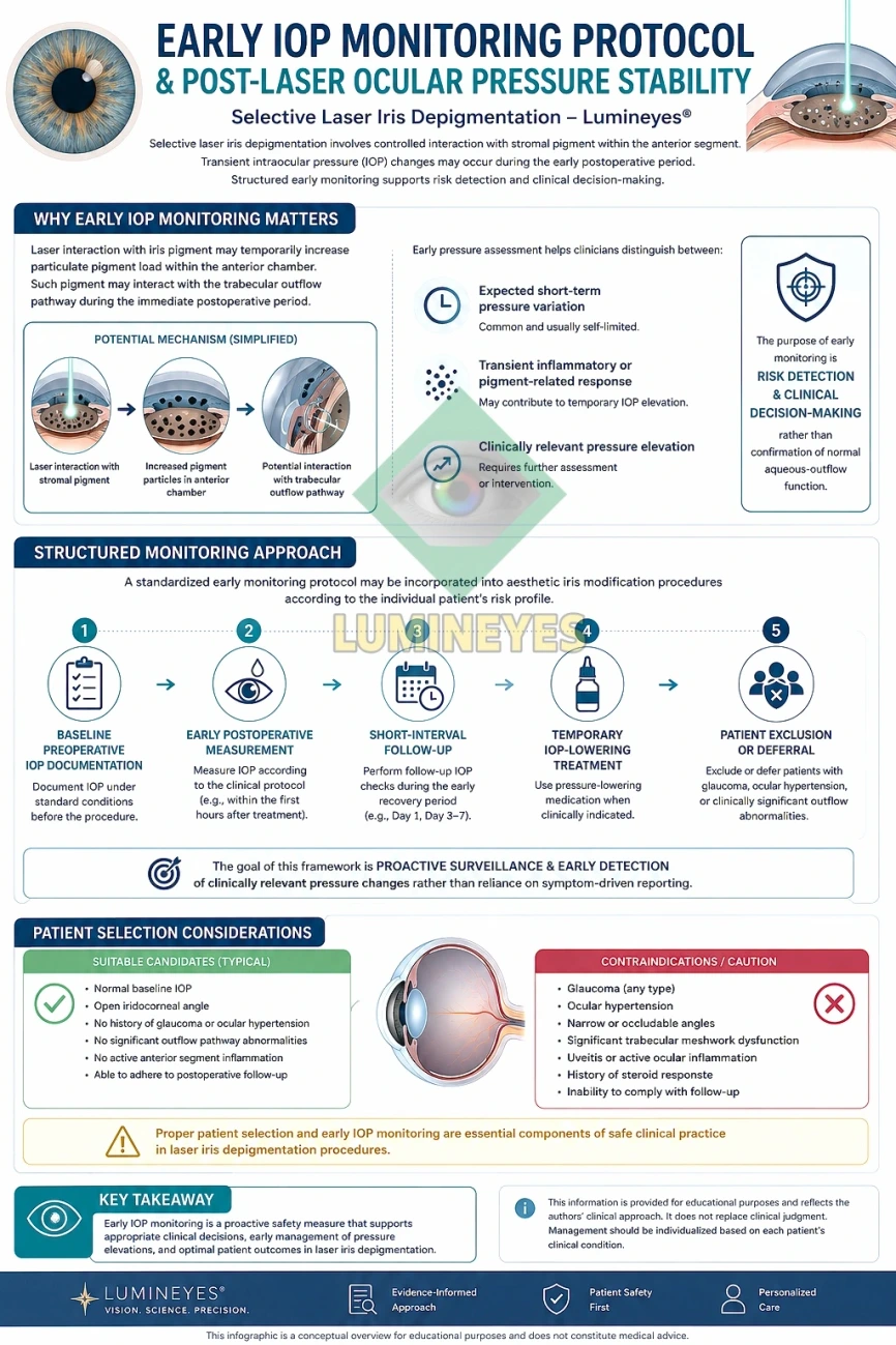
Within the Lumineyes™ framework, intraocular pressure (IOP) is not treated as a single postoperative number or a binary “normal/abnormal” variable. It is interpreted as a dynamic response signal within an exposure–response cycle. Laser interaction with iris pigment may transiently increase pigment and inflammatory material within the anterior chamber; in susceptible eyes, this material may increase resistance within the trabecular outflow pathway and contribute to a delayed pressure rise.
The practical implication is central to Lumineyes™: pressure behavior helps determine whether the eye has recovered sufficiently for any subsequent exposure to be considered. Stable IOP is useful but not sufficient by itself. Pressure must be interpreted together with anterior-chamber activity, pigment burden, symptoms, structural findings, treatment history, and the direction of the overall biological response.
1. Why IOP Is a Control Variable, Not Merely a Complication Check
The conventional way to discuss postoperative pressure is to ask whether an IOP “spike” occurred. That language is clinically useful but incomplete. In staged laser iris depigmentation, the more important question is whether the eye’s aqueous-outflow system remains capable of accommodating the pigment and inflammatory load generated by the preceding exposure.
This distinction is fundamental to the Lumineyes™ approach. The aim is not simply to detect a high pressure measurement after it has occurred. The aim is to use pressure kinetics as part of a broader closed-loop clinical decision process: exposure produces a biological response; that response is observed over time; the eye is reassessed; and only then is the next treatment decision made.
IOP monitoring is a dynamic surveillance variable within a response-guided treatment cycle. Its purpose is not merely to document pressure, but to help determine whether pigment handling, anterior-chamber recovery, and aqueous outflow remain sufficiently stable to permit consideration of further exposure.
2. Biological Rationale: Pigment, the Trabecular Meshwork, and Outflow Resistance
Aqueous humor exits the anterior chamber primarily through the trabecular meshwork and Schlemm canal. The trabecular meshwork is therefore not a passive drain; it is a biologically active tissue that contributes substantially to regulation of outflow resistance.[1]
Evidence from pigment dispersion syndrome and pigmentary glaucoma demonstrates an important general principle: liberated iris pigment can accumulate within the trabecular meshwork and may be associated with reduced aqueous outflow and elevated IOP.[2,3] Laser iris depigmentation is not biologically identical to pigment dispersion syndrome, and the two should not be conflated. Nevertheless, this established pigment–outflow relationship provides a strong physiological rationale for monitoring pressure when a procedure intentionally alters iris pigment.
Published reviews of cosmetic eye-color procedures likewise identify transient IOP elevation as a recognized potential complication of laser iris depigmentation, while emphasizing that the clinical evidence base remains limited.[4,5] A 2026 case report described markedly elevated IOP after laser iris depigmentation in association with additional ocular complications, further illustrating that clinically important pressure responses are biologically possible in this procedure category.[6]
Important distinction: evidence that pigment can affect trabecular outflow supports the rationale for surveillance; it does not establish that every transient pigment response causes trabecular injury, nor that every LID protocol carries the same pressure risk. Risk attribution must remain protocol-specific.
3. The Lumineyes™ Exposure–Response Model
Within Lumineyes™, a treatment session is not considered an isolated technical event. It is one part of an exposure–response cycle. The biological consequences of the previous exposure become input data for the next treatment decision.
Exposure
Selective stromal melanin modulation produces a new pigment and cellular response within the anterior segment.
Response
Pigment liberation, anterior-chamber activity, symptoms and pressure behavior are observed as biological response variables.
Recovery
The direction of change—resolving, stable or progressing—is more informative than a single measurement in isolation.
Decision
Further exposure is considered only after reassessment; IOP stability is one requirement, not the sole definition of readiness.
| Variable | Conventional interpretation | Lumineyes™ interpretation | Clinical significance |
|---|---|---|---|
| Baseline IOP | A preoperative reference value | Starting point for interpreting subsequent pressure kinetics in that individual eye | Absolute values and change from baseline both matter |
| Single postoperative IOP | Normal or elevated measurement | One point on a dynamic response curve | Cannot by itself define recovery or safety |
| Pressure trajectory | Often secondary to the absolute measurement | Core information: stable, falling, newly rising or persistently elevated | Trajectory may alter the decision to re-expose |
| Pigment / AC response | Post-laser observation | Potential determinant of outflow stress and part of the pressure interpretation | IOP should not be read without anterior-segment context |
| Re-exposure | May be schedule-driven | Response-gated and dependent on reassessment | A stable number alone does not authorize continuation |
4. Early Pressure Kinetics: Why Timing Matters
A pressure measurement has meaning only in relation to when it was obtained. Pigment liberation, inflammatory activity, aqueous-outflow resistance and pharmacological effects do not necessarily peak simultaneously. A normal pressure measurement immediately after treatment therefore cannot be assumed to exclude a later pressure response.
Within the author’s Lumineyes™ clinical observations, the first several hours may show visible anterior-chamber pigment/cellular activity without a clinically important pressure rise. A delayed increase may emerge later in the early recovery period. The protocol therefore treats the early postoperative period as a time-dependent biological sequence, not as a single post-laser checkpoint.
| Approximate phase | Observed / monitored domain | Interpretation within the framework | Decision relevance |
|---|---|---|---|
| Immediate to early hours | Pigment liberation, anterior-chamber cells, discomfort, initial pressure response | An early pigment/cellular response may occur after exposure and is interpreted in context rather than automatically classified as a complication. | Establishes the initial biological response to the exposure. |
| Approximately first 4–6 hours | Evolution of AC pigment/cellular activity and ocular response | Within Lumineyes™ clinical observation, pigment/cell flow during this phase may precede any delayed pressure rise. | A normal early IOP does not by itself close the surveillance window. |
| Later early-recovery window | Delayed pressure behavior and persistence/resolution of anterior-segment response | In susceptible eyes, pressure may rise after the initial inflammatory/pigment response rather than immediately. | Requires continued interpretation of trajectory rather than reliance on the first measurement. |
| Around the 10–12-hour window in the author’s clinical observations | Potential delayed IOP elevation in responsive eyes | This is a protocol-specific observational window, not a universal biological rule or published incidence estimate. | Supports surveillance across the full early-response period. |
| Before any subsequent planned exposure | IOP, anterior chamber, cornea, symptoms and overall recovery trajectory | Readiness is determined by the status of the previous exposure’s response. | Unresolved or progressing findings argue against automatic continuation. |
Evidence boundary: the approximate 4–6-hour and 10–12-hour observations above describe the author’s Lumineyes™ clinical framework and should not be interpreted as universally validated time thresholds for all laser iris depigmentation systems. Prospective time-series data are required to quantify these kinetics.
5. Pressure Stability Is Necessary—but Not Sufficient
One of the most important conceptual errors in postoperative monitoring is to equate a normal pressure measurement with complete ocular recovery. IOP is a functional surrogate, not a structural assay of the trabecular meshwork and not a global measure of ocular safety. A normal IOP does not demonstrate absence of inflammatory activity, endothelial stress, angle pigment, structural tissue effects, or other anterior-segment abnormalities.
For this reason, Lumineyes™ does not define readiness for further treatment by pressure alone. The pressure signal is interpreted alongside the broader response of the eye. This is consistent with the principle of selective stromal melanin modulation and with response-guided staged treatment: the previous exposure must be clinically reassessed before the next exposure can be considered.
| Domain | What may appear reassuring | What can still remain unresolved | Lumineyes™ interpretation |
|---|---|---|---|
| IOP | Measurement within expected range | Later pressure rise or abnormal trajectory | Interpret serially and relative to baseline |
| Anterior chamber | Minimal symptoms | Persistent cells, pigment or inflammatory activity | Symptoms cannot replace slit-lamp assessment |
| Cornea | Clear central vision | Subclinical or peripheral corneal response | Ocular response is broader than visual acuity alone |
| Angle / outflow | Normal IOP at one time point | Pigment burden or evolving outflow resistance | Pressure is an indirect indicator of outflow behavior |
| Overall recovery | Patient feels well | Objective findings may not yet have resolved | Re-exposure is a clinical decision, not a symptom-based decision |
6. A Kinetic Interpretation: Resolving Versus Progressing
The same absolute IOP can have different significance depending on direction. A value returning toward baseline after an earlier rise is biologically different from the same value reached during a continuing upward trajectory. The Lumineyes™ framework therefore emphasizes kinetics: is the previous treatment response resolving, stable, or progressing?
| Response pattern | Pressure behavior | Associated clinical context | Framework interpretation |
|---|---|---|---|
| Resolving response | Stable or trending toward the individual baseline | Other anterior-segment findings are also resolving | Supports recovery, but does not independently establish readiness for re-exposure |
| Persistent response | Not clearly normalizing or repeatedly variable | Residual pigment / inflammatory findings remain | Prior exposure has not yet generated a clearly completed recovery cycle |
| Progressing response | New or increasing elevation | Worsening clinical findings or increasing symptoms may coexist | Requires reassessment and argues against further exposure |
| Discordant response | IOP appears stable | Another clinically relevant domain remains abnormal | Pressure stability must not override non-pressure safety findings |
7. Relationship to L‑SAFE and Response-Guided Staged Treatment
Pressure surveillance is one component of the broader Lumineyes™ safety architecture. The underlying question is not simply, “Is the IOP acceptable?” but rather, “Has the eye recovered from the previous exposure sufficiently that the previous response no longer creates a safety reason to withhold further exposure?”
That distinction connects early IOP monitoring to the broader response-guided logic used in Lumineyes™. A stable pressure value can contribute to a readiness assessment; it cannot create readiness by itself. Conversely, a clinically meaningful pressure rise, unstable pressure trajectory, or unresolved anterior-segment response can change the treatment decision even if a subsequent session had previously been planned.
8. Patient Selection and Baseline Risk
Pressure surveillance begins before the laser is fired. Baseline IOP, angle configuration, optic-nerve status, prior ocular history, medication use, previous pressure behavior, and any evidence of compromised aqueous outflow can materially alter the interpretation of postoperative findings.
Pre-existing glaucoma, ocular hypertension, suspicious outflow abnormalities, or other clinically relevant anterior-segment disease should therefore not be treated as routine candidate profiles. These situations require individualized ophthalmic assessment and a substantially different risk discussion. They should not be simplified into a universal “allowed” or “excluded” category on the basis of a website protocol.
| Baseline domain | Why it matters | Potential effect on interpretation |
|---|---|---|
| Baseline IOP | Defines the individual starting pressure state | A postoperative value should be interpreted relative to that eye’s baseline, not only a population reference range |
| Glaucoma / ocular hypertension history | May indicate reduced reserve or established pressure-related vulnerability | Changes the risk context of even modest pressure changes |
| Angle / trabecular status | Directly relevant to aqueous outflow | Pre-existing outflow abnormalities may alter pigment tolerance |
| Pigment phenotype | May influence the amount of pigment liberated by a given exposure | Denser pigmentation may generate a different biological load and requires individualized planning |
| Previous exposure response | Provides direct evidence of how that eye reacted previously | Prior response can be more informative than assuming identical behavior at the next stage |
| Follow-up reliability | Early pressure changes may be asymptomatic | Inability to complete monitoring weakens the safety architecture of staged treatment |
9. What the Published Literature Can—and Cannot—Tell Us
The current literature supports several broad conclusions. Pigment can alter trabecular outflow; transient IOP rises have been described after laser iris depigmentation; and severe pressure elevation has also been reported in individual cases.[2–6] At the same time, the literature is too heterogeneous and too limited to define one universal incidence rate, one universal timing curve, or one universal risk profile for all LID techniques.
It is also inappropriate to use data from selective laser trabeculoplasty (SLT) as if they were direct evidence for iris depigmentation. SLT studies demonstrate that laser procedures involving pigmented anterior-segment tissue can produce early pressure changes and that the timing of post-laser measurements matters, but SLT targets the trabecular meshwork itself and represents a different procedure with a different therapeutic objective.[7]
| Statement | Evidence status | Appropriate conclusion |
|---|---|---|
| Pigment accumulation can impair trabecular outflow and raise IOP. | Established in pigment dispersion / pigmentary glaucoma literature | Provides physiological rationale for pressure surveillance after pigment-altering procedures |
| Laser iris depigmentation can be followed by transient IOP elevation. | Reported in reviews of cosmetic eye-color procedures | IOP should be actively monitored rather than inferred from symptoms |
| Marked IOP elevation can occur after LID. | Documented in at least one recent case report | Serious pressure responses are biologically possible |
| All LID protocols have the same IOP risk. | Not established | Risk must be attributed to specific protocols and evidence sets |
| The Lumineyes™ 4–6 h / approximately 10–12 h response model is universally validated. | Not established; protocol-specific clinical observation | Requires prospective serial-measurement validation |
| Normal IOP proves trabecular preservation or long-term procedural safety. | Not established | Pressure is one functional safety domain among several |
10. Why Symptom-Driven Monitoring Is Insufficient
Pressure elevation can be clinically important even when symptoms are mild or absent. Conversely, pain or photophobia may occur without a dangerous pressure elevation. Symptoms therefore provide useful information but cannot substitute for objective examination and tonometry.
The Lumineyes™ approach consequently treats follow-up compliance as part of the treatment architecture itself. A response-guided protocol cannot function safely if the response is not measured. The monitoring system is therefore not an administrative add-on to laser treatment; it is one of the mechanisms by which the next exposure decision is controlled.
11. Proposed Prospective Validation of the Lumineyes™ IOP Model
The next scientific step is to convert the clinical monitoring concept into a prospectively measurable pressure-kinetics model. Rather than reporting only whether an IOP spike occurred, future studies should characterize the shape, timing, magnitude, duration and resolution of the pressure response in relation to pigment phenotype and anterior-chamber findings.
| Domain | Potential measurement | Scientific question |
|---|---|---|
| Baseline pressure | Standardized pre-exposure tonometry | What is the individual reference state? |
| Serial early IOP | Repeated predefined measurements during the early recovery window | When does pressure change begin, peak and resolve? |
| Anterior-chamber response | Standardized cell / flare and pigment grading | Does the magnitude of AC response correlate with pressure kinetics? |
| Pigment phenotype | Standardized iris imaging and phenotype classification | Does baseline pigment burden predict the pressure response? |
| Angle / outflow phenotype | Gonioscopy and structural assessment where appropriate | Which anatomical features modify pressure susceptibility? |
| Cumulative exposure | Protocol-specific exposure metrics | Is pressure response related to cumulative pigment disruption? |
| Recovery state before re-exposure | Predefined multidomain readiness criteria | Can response-guided staging be operationalized reproducibly? |
| Long-term pressure stability | Longitudinal IOP, optic nerve and functional follow-up | Does early recovery predict long-term pressure behavior? |
12. Clinical Position of Lumineyes™
Within Lumineyes™, the goal is not to claim that every early pigment response is harmless, nor that stable IOP proves absence of trabecular stress. The framework is deliberately more conservative: pressure is treated as a measurable component of the eye’s biological response to pigment-modifying laser exposure.
This makes IOP surveillance part of a larger clinical logic that also includes selective stromal melanin modulation, controlled treatment-related pigment response, and response-guided staged treatment. In this model, the purpose of monitoring is not merely to document an adverse event. It is to prevent the next treatment decision from being made while the response to the previous exposure remains unresolved.
Readers seeking the broader treatment context can review the canonical overview of laser eye color change, the scientific discussion of iris stromal anatomy, and the MyLumineyes™ research archive.
13. Limitations
This article describes a Lumineyes™ clinical monitoring framework, not a randomized comparison or independently validated prediction model. The specific early-response timing observations described here derive from the author’s clinical experience and have not yet been established as universal timing thresholds. Published LID literature remains limited and heterogeneous, and current reports do not permit reliable extrapolation of one device, protocol or operator’s pressure-risk profile to another.
Accordingly, the framework should be evaluated prospectively using standardized time-series IOP measurements, objective anterior-chamber grading, structural assessment, protocol-specific exposure documentation and long-term follow-up.
14. Conclusion
Early IOP surveillance after laser iris depigmentation should not be reduced to a single postoperative pressure measurement. Within the Lumineyes™ framework, pressure is interpreted as part of a time-dependent exposure–response system linking pigment liberation, anterior-chamber activity, aqueous-outflow stress, recovery kinetics and the decision whether further exposure should be considered.
The central principle is simple but clinically consequential: the next exposure should not be treated as automatic merely because it was scheduled. The biological response to the previous exposure must first be understood. IOP is one of the most objective signals in that assessment—but it is meaningful only when interpreted together with the rest of the eye.
Research FAQ
Why can IOP rise after laser iris depigmentation?
Laser–pigment interaction may increase pigment and inflammatory material within the anterior chamber. Because aqueous humor exits through the trabecular meshwork, increased particulate and inflammatory load may alter outflow resistance in susceptible eyes. The exact mechanism and magnitude can vary among protocols and individuals.
Does a normal early IOP measurement prove the eye is safe?
No. A normal measurement is reassuring only for that time point. It does not prove preservation of the trabecular meshwork, exclude a later pressure response, or establish overall ocular safety.
Why does Lumineyes™ emphasize IOP kinetics rather than one pressure value?
Because the direction and timing of pressure change may be clinically important. A value trending back toward baseline represents a different biological trajectory from the same value during a continuing rise.
Is pressure stability enough to proceed with another treatment stage?
No. Within the response-guided Lumineyes™ framework, IOP is one component of readiness. Anterior-chamber activity, pigment response, corneal findings, symptoms, treatment history and the overall recovery trajectory must also be considered.
Are the 4–6-hour and approximately 10–12-hour windows universal?
No. They are described here as protocol-specific clinical observations within the author’s Lumineyes™ framework. Prospective serial-measurement studies are needed before they can be treated as validated general timing thresholds.
References
- Buffault J, Labbé A, Hamard P, Brignole-Baudouin F, Baudouin C. The trabecular meshwork: structure, function and clinical implications. A review of the literature. J Fr Ophtalmol. 2020;43(7):e217-e230. PMID: 32561029.
- Scuderi G, Contestabile MT, Scuderi L, Librando A, Fenicia V, Rahimi S. Pigment dispersion syndrome and pigmentary glaucoma: a review and update. Int Ophthalmol. 2019;39(7):1651-1662. doi:10.1007/s10792-018-0938-7. PMID: 29721842.
- Niyadurupola N, Broadway DC. Pigment dispersion syndrome and pigmentary glaucoma— a major review. Clin Exp Ophthalmol. 2008;36(9):868-882. doi:10.1111/j.1442-9071.2009.01920.x. PMID: 19278484.
- D’Oria F, Abu-Mustafa SK, Alio JL. Cosmetic change of the apparent color of the eye: a review on surgical alternatives, outcomes and complications. Ophthalmol Ther. 2022;11(2):465-477. doi:10.1007/s40123-022-00458-2.
- D’Oria F, Alio JL. Surgical techniques for cosmetic eye color change: a narrative review. Ophthalmol Ther. 2025;14(8):1685-1694. doi:10.1007/s40123-025-01177-0.
- Jaber W, Nemet M, Waisbourd M. Laser iris depigmentation resulting in markedly elevated intraocular pressure and herpes simplex keratitis with corneal scarring: case report. Case Rep Ophthalmol. 2026;17(1):312-317. doi:10.1159/000551226.
- Lanzetta P, Menchini U, Virgili G. Immediate intraocular pressure response to selective laser trabeculoplasty. Br J Ophthalmol. 1999;83(1):29-32. doi:10.1136/bjo.83.1.29. PMID: 10209430. Used only as comparative anterior-segment laser physiology; SLT is not equivalent to laser iris depigmentation.
Methodological disclosure. Mustafa Mete, MD, is the developer of the Lumineyes™ methodology. The early-response timing model and response-guided interpretation described here include protocol-specific clinical observations and should not be represented as independently validated population estimates. References are used to establish relevant anterior-segment physiology, documented LID complications, and the limits of the published evidence.
Medical information notice. This article is intended for scientific and educational discussion and does not provide individualized treatment instructions. Patient-specific monitoring and management require direct ophthalmic examination and clinical judgment.
About the author. Mustafa Mete, MD, is an ophthalmologist and developer of the Lumineyes™ methodology. His work focuses on iris pigmentation biology, laser–tissue interaction, anterior-segment response, IOP kinetics, optical phenotyping and response-guided staged treatment.