Quantifying Pigment Clearance After Iris-Directed Laser Exposure
A longitudinal measurement framework for residual pigment burden, anterior-chamber particulate response and clearance kinetics
Abstract
Pigment clearance after iris-directed laser exposure is frequently inferred from visible iris lightening. These two phenomena are not equivalent. A change in apparent iris color may reflect alteration in stromal pigment, optical scattering, illumination, imaging conditions or combinations of these factors, whereas biological clearance concerns the disposition of pigment-containing material within specific ocular compartments.
This research record proposes a longitudinal measurement framework for separating visible phenotype, residual stromal pigment burden, anterior-chamber particulate material and angle-associated pigment. Existing methods provide several candidate measurement tools. High-resolution standardized photography can quantify iris color in CIELAB space; anterior-segment OCT can characterize iris morphology and sectoral architecture; and recent polarization-sensitive anterior-segment OCT research has demonstrated quantitative estimation of iris melanin concentration. [1] [2] [3]
The proposed framework does not assume that pigment disappearance is equivalent to complete biological elimination, nor that macrophage activity, aqueous transport or trabecular processing has been prospectively quantified after Lumineyes™ treatment. Instead, it defines observable compartments, candidate longitudinal endpoints and evidentiary boundaries for future prospective validation.
1. Pigment clearance is often described before it has been measured
In discussions of laser-based eye-color modification, the term pigment clearance is often used as though it represented a single observable event. Biologically, this is unlikely to be adequate.
Laser interaction with pigmented iris tissue may alter the distribution, optical contribution or physical state of pigment-containing structures. Experimental iris-laser research demonstrates that pigment alteration and post-exposure anterior-segment responses can occur, but direct measurement of the complete human clearance pathway remains limited. [4]
Likewise, broader experimental pigment-dispersion models demonstrate that trabecular-meshwork cells respond biologically to pigment exposure; however, these models cannot be assumed to reproduce the material, dose or temporal characteristics generated by a particular human iris-directed laser protocol. [5]
It is: Which pigment-related state is being observed, in which anatomical compartment, by which measurement method, and how does that state change across serial examinations?
2. What should “pigment clearance” mean?
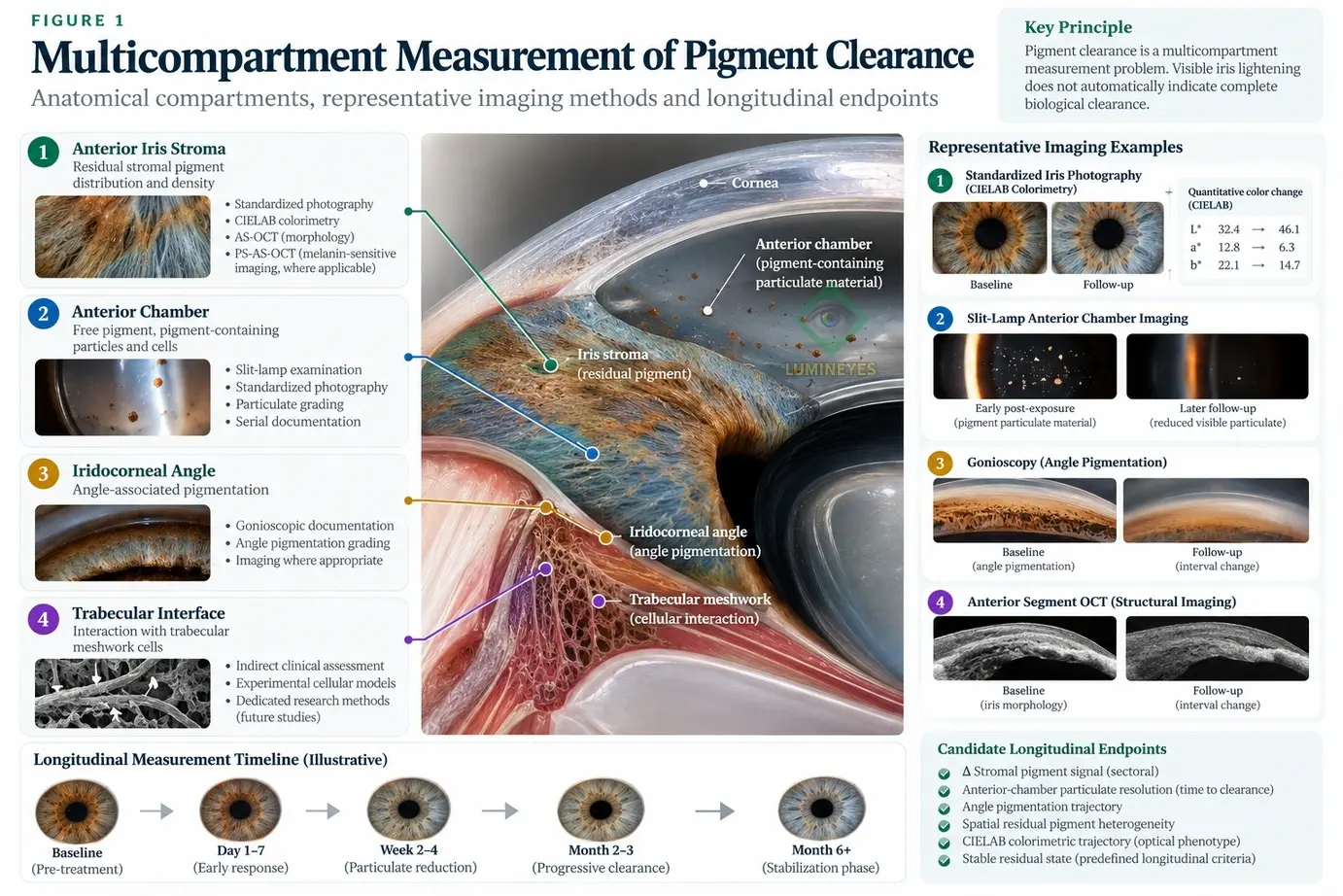
A rigorous definition should avoid treating visible color change as proof of pigment elimination. For research purposes, pigment clearance is better approached as a multicompartment longitudinal state.
Reduction
Measurable reduction in pigment-associated signal or visible pigment within a defined anatomical region.
Redistribution
Change in the location or spatial distribution of pigment-containing material without assuming biological elimination.
Resolution
Progressive disappearance of previously detectable particulate material from a specified clinical compartment.
These processes should not automatically be considered equivalent. Reduction of anterior stromal pigmentation may coexist with residual anterior-chamber pigment or increased angle pigmentation. Conversely, resolution of visible anterior-chamber particles does not establish that every pigment-containing component has been eliminated from the eye.
3. Pigment should be measured by compartment
The phrase “pigment burden” has little quantitative meaning unless the anatomical compartment is specified. A future clearance study should therefore record several domains separately rather than collapsing them into a single subjective impression.
| Compartment | What may be observed | Candidate measurement | Important limitation |
|---|---|---|---|
| Anterior iris stroma | Residual pigment distribution, sectoral density and visible heterogeneity | Standardized photography, colorimetry, sectoral imaging, qualified OCT-derived variables | Optical appearance cannot by itself distinguish pigment quantity from scattering and structural effects. |
| Anterior chamber | Free pigment, pigment-containing particles, cells or other particulate material | Standardized slit-lamp documentation and serial particulate grading | Routine slit-lamp examination cannot reliably establish particle identity in every case. |
| Iridocorneal angle | Angle-associated pigmentation or interval change | Standardized gonioscopic documentation ± imaging where appropriate | Angle pigmentation is not equivalent to current free pigment load or complete trabecular processing. |
| Trabecular interface | Biological interaction with particulate material | Indirect clinical assessment; experimental cellular models | Direct human in-vivo quantification of pigment processing at the trabecular cellular level remains limited. |
4. Why visible color change cannot serve as a clearance surrogate
Quantitative iris photography is capable of measuring visible color objectively. Edwards and colleagues demonstrated that high-resolution iris photographs can be analyzed in CIE L*a*b* color space, providing numerical measures of lightness and chromatic coordinates. [1]
This makes colorimetry valuable, but it does not transform color into a direct measurement of biological pigment clearance.
Visible iris appearance is influenced by pigment distribution, stromal architecture, illumination, camera calibration, pupil size, image geometry and light scattering. Therefore, longitudinal colorimetry should be treated as an optical phenotype endpoint, not as proof that liberated pigment has disappeared from every anterior-segment compartment.
This distinction is consistent with the L-OAT quantitative iris phenotyping framework, which separates visible phenotype from structural and signal-derived measurements rather than treating iris color as a complete representation of iris biology.
5. A multidomain measurement architecture
Contemporary anterior-segment imaging provides increasingly sophisticated tools for separating visible phenotype from iris structure. Swept-source AS-OCT has been used to quantify iris thickness, volume, width and sector-specific morphology. [2]
More recently, polarization-sensitive AS-OCT has been used experimentally to derive a melanin concentration ratio from iris imaging and to examine its relationship with CIELAB color measurements in brown irides. [3] This does not yet establish a validated post-laser clearance endpoint, but it demonstrates that quantitative melanin-sensitive imaging is technically plausible.
Rather than forcing several measurements into a premature composite score, the proposed framework preserves a clearance-state vector:
Each variable can change independently. This is scientifically preferable to assuming that one visible endpoint represents the entire biological process.
6. Candidate endpoints for prospective pigment-clearance research
A longitudinal study should describe not only the amount of change but its rate, direction, compartment and stability.
| Endpoint | Research interpretation | What it does not prove |
|---|---|---|
| Residual stromal pigment burden | Change in predefined sectoral pigment-related measurements relative to baseline | Complete elimination of pigment from the anterior segment |
| Clearance slope | Direction and rate of change across serial pigment measurements | A universal biological half-life |
| Time to particulate resolution | Time until previously documented anterior-chamber particulate material is no longer clinically detectable | Molecular elimination of all pigment |
| Angle pigment trajectory | Longitudinal change in standardized angle pigmentation | Direct measurement of trabecular cellular processing |
| Spatial residual heterogeneity | Degree to which residual pigment remains unevenly distributed across iris sectors | Pathology by itself |
| Stable residual state | Absence of meaningful interval change across prospectively specified serial observations | Permanent biological equilibrium without longer follow-up |
| CIELAB trajectory | Quantitative evolution of visible iris phenotype | Direct pigment clearance |
Thresholds for “resolution,” “stability” or clinically meaningful change should be predefined and validated rather than selected retrospectively after the outcome is known.
7. Mechanistic plausibility is not the same as measured clearance
The biological handling of pigment may involve phagocytic cells, aqueous transport, trabecular interaction and other cellular processes. These mechanisms are biologically plausible, and experimental ophthalmic models demonstrate that monocytes/macrophages and trabecular-meshwork cells can participate in particulate handling and aqueous-outflow physiology. [5] [6]
These observations should not be rewritten as direct proof that a specific quantity of pigment released during a Lumineyes™ treatment is cleared by a predetermined macrophage pathway or within a predetermined time interval.
The broader mechanistic question is addressed separately in the Biological Fate of Pigment in Cosmetic Iris Color Modification research record. That work considers what may happen to endogenous and exogenous pigment after different cosmetic eye-color interventions.
not “which mechanism must be responsible?” but “what should be measured before claiming that pigment has cleared?”
8. Pigment clearance, BAB recovery and temporal response are different constructs
Several MyLumineyes research frameworks examine related aspects of post-exposure biology. Their proximity should not be confused with interchangeability.
How can residual pigment burden, redistribution and disappearance be measured across serial examinations?
What biological pathways may process, redistribute, retain or clear endogenous or exogenous pigment?
Do cells, flare and associated anterior-chamber findings demonstrate a resolving or persistent inflammatory/barrier-related trajectory?
How do pigment redistribution, inflammation, aqueous outflow, IOP and recovery interact as temporal response domains?
Accordingly, disappearance of visible pigment does not demonstrate restoration of the blood–aqueous barrier. The latter requires its own longitudinal assessment, as discussed in blood–aqueous barrier dynamics after iris-directed laser exposure.
Likewise, pigment clearance represents only one domain within the broader Temporal Response Architecture. TRA addresses interactions between several anterior-segment response domains and should not be reduced to a pigment-clearance model.
Finally, neither visible clearance nor a favorable pigment trajectory defines treatment readiness. Integrated anterior-segment safety assessment is a separate question addressed by L-SAFE anterior-segment safety staging.
9. “Pigment discharge” should not be treated as a validated clearance endpoint
The MyLumineyes clinical archive has historically used clinical terminology describing pigment discharge. In the current research framework, this observation should be distinguished from quantitative pigment burden, complete anterior-chamber clearance, absence of residual angle pigment and final visible phenotype.
This distinction is already incorporated into the longitudinal MyLumineyes clinical data record, which identifies prospective quantification of pigment-clearance kinetics as a future research priority rather than treating retrospective clinical observation as direct measurement of the complete pathway.
The appropriate progression is therefore:
10. Proposed prospective validation pathway
A future prospective study could acquire a standardized multidomain baseline before exposure and repeat the same measurements at predefined follow-up intervals.
Standardize acquisition
Fixed photography geometry, illumination, camera settings, pupil-state documentation and reproducible iris-sector registration.
Separate compartments
Measure stromal phenotype, anterior-chamber particulate state and angle-associated pigment independently.
Model trajectories
Estimate within-eye change over time rather than relying on pre/post visual comparison alone.
Candidate baseline variables
Baseline quantitative iris phenotype, sectoral pigment distribution, standardized CIELAB coordinates, pupil size, iris morphology, IOP, gonioscopic pigmentation and relevant anterior-segment imaging variables could be recorded before treatment.
Candidate serial variables
Follow-up could include the same photographic/colorimetric measurements, residual sectoral pigment distribution, anterior-chamber particulate documentation, angle-pigment assessment and relevant safety covariates.
IOP should be analyzed as a safety and physiological covariate, not as a pigment-clearance endpoint. The trabecular meshwork is a dynamic biological tissue whose response to pigment cannot be reduced to simple mechanical particle accumulation. [5] [6]
Potential longitudinal model
A mixed-effects model could examine change in a defined pigment-related measurement while accounting for repeated observations and correlation between eyes from the same participant.
Here, P represents a prospectively defined pigment-related measurement; u represents participant-level dependence and v represents eye-level dependence. The equation is illustrative rather than a validated predictive model.
11. What this framework does not claim
This framework does not claim that all post-laser pigment is completely eliminated, that visible lightening proves clearance, that macrophage processing is the exclusive human clearance pathway, or that a specific clearance interval applies universally.
It also does not establish that a favorable clearance trajectory proves overall ocular safety. Pigment state, inflammatory recovery, aqueous-outflow behavior, corneal status and IOP are related but distinct domains.
Published cosmetic iris-laser literature remains heterogeneous, and procedure-specific differences in exposure architecture and reporting limit broad extrapolation. [7]
12. Clearance should be measured, not inferred
Pigment clearance after iris-directed laser exposure should not be defined by visible iris lightening alone. The clinically relevant process may include changes in residual stromal pigment, redistribution of pigment-containing material, transient anterior-chamber particulate findings and interval changes in angle pigmentation.
These components should be documented separately and followed longitudinally.
Standardized iris photography and quantitative colorimetry provide useful optical endpoints; anterior-segment imaging provides structural information; emerging polarization-sensitive OCT techniques may eventually permit more pigment-specific quantitative assessment. [1] [2] [3]
The principal methodological conclusion is therefore straightforward:
Pigment clearance is a longitudinal biological measurement problem.
References
- Edwards M, Gozdzik A, Ross K, Miles J, Parra EJ. Technical note: quantitative measures of iris color using high resolution photographs. Am J Phys Anthropol. 2012;147(1):141–149. doi:10.1002/ajpa.21637. PubMed
- Invernizzi A, Giardini P, Cigada M, Viola F, Staurenghi G. Three-Dimensional Morphometric Analysis of the Iris by Swept-Source Anterior Segment Optical Coherence Tomography in a Caucasian Population. Invest Ophthalmol Vis Sci. 2015;56(8):4796–4801. doi:10.1167/iovs.15-16483. PubMed
- Li X, Hu L, Yang B, et al. Quantitative measurement of iris melanin concentration by polarization-sensitive anterior segment optical coherence tomography. Eur J Ophthalmol. 2026;36(4):872–880. doi:10.1177/11206721251407025. PubMed
- Yildirim Y, Duzgun E, Kar T, et al. Evaluation of color-changing effect and complications after Nd:YAG laser application on iris surface. Med Sci Monit. 2016;22:107–114. doi:10.12659/MSM.895086. PMC
- Wang C, Dang Y, Loewen RT, et al. Impact of pigment dispersion on trabecular meshwork cells. Graefes Arch Clin Exp Ophthalmol. 2019;257(6):1217–1230. doi:10.1007/s00417-019-04300-7. PubMed
- Alvarado JA, Katz LJ, Trivedi S, Shifera AS. Monocyte modulation of aqueous outflow and recruitment to the trabecular meshwork following selective laser trabeculoplasty. Arch Ophthalmol. 2010;128(6):731–737. doi:10.1001/archophthalmol.2010.85. PubMed
- D'Oria F, Abu-Mustafa SK, Alio JL. Cosmetic change of the apparent color of the eye: a review on surgical alternatives, outcomes and complications. Ophthalmol Ther. 2022;11:465–477. doi:10.1007/s40123-022-00458-2.
