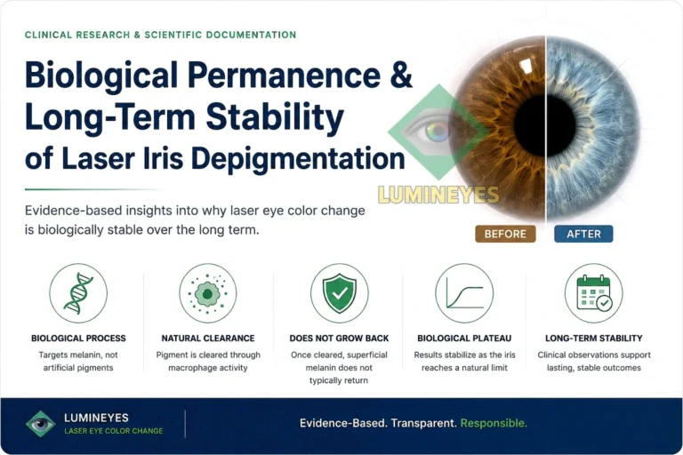
Biological Permanence & Long-Term Stability After Laser Iris Depigmentation
A mechanistic and clinical analysis of stromal melanin stability, biological pigment clearance, residual pigmentation, repigmentation, treatment plateau and the long-term Lumineyes™ framework.
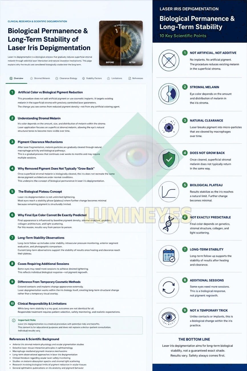
The concept of permanence after laser iris depigmentation requires a distinction between artificial color deposition and biological reduction of endogenous iris pigment. Human iris color is strongly determined by melanin quantity and distribution within the anterior iris stroma, together with stromal architecture and optical light scattering. Classical histological and pigmentation studies suggest that adult iris pigmentation is comparatively stable and that differences between light and dark irides depend substantially on melanin content rather than major differences in stromal melanocyte number.1–4
Within the Lumineyes™ framework, laser iris depigmentation is conceptualized as selective stromal melanin modulation: controlled reduction of pre-existing visible stromal melanin followed by a treatment-related biological response. Long-term stability therefore refers to persistence of the achieved reduction after pigment processing and optical appearance have reached a relative biological plateau. This model does not require the introduction of synthetic pigment and should not be confused with corneal keratopigmentation or intraocular iris implantation.
This article examines the biological rationale for durable pigment reduction, the distinction between residual pigment and true repigmentation, the clinical meaning of a pigment plateau, factors influencing final color, and the role of longitudinal observation in the Lumineyes™ clinical framework.
Quick Answer
Laser iris depigmentation does not create a new iris color by placing artificial pigment into the eye. It reduces the visual contribution of pre-existing anterior stromal melanin. In the Lumineyes™ framework, the achieved change is intended to persist because the treatment modifies the amount and distribution of visible stromal pigment rather than producing a temporary optical coating.
Adult iris pigmentation is biologically more stable than cutaneous tanning: ocular melanocytes do not normally respond to environmental ultraviolet exposure in the same dynamic manner as epidermal melanocytes.1,4,5 This provides a biologically plausible basis for durable pigment reduction, although no biological procedure should be described as an absolute guarantee that living tissue can never change again during an entire lifetime.
For the broader clinical explanation of the procedure, see the canonical Laser Eye Color Change Procedure Guide .
The word permanent is frequently used in cosmetic medicine, but biologically it can describe very different phenomena. A material can remain permanently implanted in tissue; synthetic pigment can remain deposited within a corneal plane; or the concentration of an endogenous pigment can be reduced and remain lower after treatment. These mechanisms are not biologically equivalent even when the visible objective — a change in apparent eye color — is similar.
Laser iris depigmentation belongs to the third category. Its scientific basis is not the implantation of a new color but alteration of the pre-existing pigmentary contribution to iris appearance. Understanding permanence therefore begins with understanding normal iris pigmentation.
01 · Definition What Does “Biological Permanence” Mean?
In the context of laser iris depigmentation, biological permanence should not be interpreted as a claim that living tissue becomes permanently incapable of any future biological change. Rather, it refers to the persistence of a treatment- induced reduction in visible anterior stromal pigment after the active treatment response has stabilized.
This distinction is important. The long-term question is not whether the eye can ever undergo any biological change again. The clinically relevant question is whether reduced pigment density tends to remain reduced once the treatment response has stabilized.
The concept is therefore more accurately described as long-term biological stability. The term preserves the clinically meaningful observation of persistent color change while recognizing that the iris remains living tissue.
02 · Iris Biology Why Adult Iris Pigmentation Is Relatively Stable
Human iris color depends primarily on melanin content and distribution within the anterior iris, combined with stromal architecture and the optical scattering of incident light.1–4 The biology is more complex than a simple blue-versus-brown pigment model: human eyes do not contain a separate “blue pigment.” Lighter eye colors become visible as the relative contribution of anterior melanin decreases and stromal optical properties become more prominent.
Histological work comparing light, intermediate and dark human irides found no major relationship between iris color and the total number of stromal melanocytes. Instead, the important difference is the quantity and characteristics of melanin contained within those cells and their surrounding pigmentary architecture.2
This point is central to the permanence question. Laser depigmentation is not based on attempting to remove every melanocyte from the iris. Within the Lumineyes™ model, treatment is directed toward the visible stromal pigment architecture and its contribution to apparent iris color.
Adult uveal melanocytes also behave differently from epidermal skin melanocytes. Experimental and review literature describes uveal melanocytes as relatively stable in vivo, with substantially less dynamic melanogenic response to ordinary environmental stimulation than skin melanocytes. Unlike skin tanning, sunlight exposure does not normally produce a comparable progressive darkening of adult iris color.4,5
| Biological feature | Adult iris | Relevance to permanence |
|---|---|---|
| Visible pigment | Predominantly determined by melanin quantity, distribution and optical interaction in the anterior iris | Reduction of visible melanin can alter apparent color without adding a new pigment |
| Melanocyte number | Major differences in normal iris color are not explained simply by large differences in stromal melanocyte number | Treatment does not need to imply total melanocyte elimination in order to modify visible pigmentation |
| Adult melanogenesis | Uveal melanocytes are comparatively stable in vivo | Supports the biological plausibility that reduced pigment density need not automatically return to baseline |
| UV response | Does not normally behave like cutaneous tanning | Ordinary sunlight exposure should not be equated with rapid restoration of previously reduced iris pigment |
| Living tissue | Remains biologically active throughout life | “Permanent” should describe durable clinical stability, not an absolute promise against every future biological change |
03 · Treatment Concept Selective Stromal Melanin Modulation
The Lumineyes™ clinical framework uses the term selective stromal melanin modulation to describe the intended treatment interaction.
The concept is deliberately more precise than simply saying that the laser “removes eye color.” The visible target is pre-existing anterior stromal melanin. The objective is to reduce its optical contribution while preserving the anatomical and functional characteristics of the surrounding iris as far as clinically possible.
The term modulation is important because the desired endpoint is not unlimited depigmentation. Different irides contain different baseline pigment densities, stromal organizations and degrees of pigment adherence. Consequently, the amount of clinically appropriate pigment reduction is not identical in every eye.
Permanence is not created by putting something into the eye.
In the Lumineyes™ model, the long-term visible change is intended to result from persistent reduction of pre-existing stromal pigment. No artificial iris pigment is implanted and no new color layer is placed over the iris.
This is one of the principal biological distinctions between Lumineyes™ and procedures in which the apparent color depends on a synthetic pigment or intraocular device remaining physically present.
04 · Post-Treatment Biology Pigment Fragmentation Is Not the Final Clinical Endpoint
The visible effect of laser iris depigmentation does not necessarily reach its final state at the moment the laser pulse is delivered. Treatment-related pigment material must subsequently undergo biological processing within the ocular environment.
Macrophage-mediated phagocytosis represents a biologically plausible contributor to the handling of liberated pigment material, while aqueous humor provides a dynamic transport environment within the anterior chamber. The exact quantitative contribution of individual clearance pathways after cosmetic laser iris depigmentation has not yet been fully characterized histologically in humans.
The Lumineyes™ physiological model is described in greater detail in the dedicated research record on Pigment Clearance Kinetics After Laser Iris Depigmentation .
Clinically, the important point is that the visible iris can continue to evolve after a treatment stage. Changes observed during this period should therefore not automatically be interpreted as either immediate treatment failure or new pigment production.
| Phase | Dominant clinical concept | Interpretation |
|---|---|---|
| Laser exposure | Selective interaction with visible pigment | Initiates the treatment response but does not necessarily determine the final visible result immediately |
| Early biological response | Pigment dispersion and anterior-segment reaction | Requires clinical observation as part of a staged treatment system |
| Progressive processing | Cellular handling and aqueous transport of treatment-related material | Visible pigmentation may continue to change between treatment stages |
| Relative stabilization | Reduced pigment density reaches a clinical plateau | Provides the appropriate point from which long-term pigment stability can be assessed |
05 · Interpretation Residual Pigment Is Not the Same as True Repigmentation
One of the most important distinctions in the discussion of permanence is the difference between residual pigment and new pigment production.
An iris can remain darker than the desired endpoint because some original stromal pigment remains present. That situation does not demonstrate biological regeneration of previously reduced pigment. Similarly, an eye that later undergoes an additional treatment stage has not necessarily experienced “pigment regrowth.”
Several different biological and procedural situations can produce the appearance of incomplete or additional future lightening:
| Observation | Possible explanation | Does it prove pigment regrowth? |
|---|---|---|
| Persistent darker areas | Original residual stromal pigment or heterogeneous pigment distribution | No |
| Additional lightening over time | Continued biological processing of pigment after the treatment stage | No |
| Later request for further treatment | Residual pigment, incomplete initial response or a desire for additional modification | No |
| Stable plateau after partial lightening | Biological or anatomical limit of the achieved treatment response | No |
| Documented increase in previously reduced pigment density | Would require longitudinal photographic and clinical assessment to distinguish true repigmentation from lighting, pupil size and photographic variation | Potentially — but requires objective confirmation |
This distinction is particularly important in scientific follow-up. Reliable evaluation requires standardized photography and clinical comparison rather than subjective memory of the previous iris shade.
06 · Treatment Kinetics The Biological Pigment Plateau
Laser iris depigmentation is not an unlimited linear lightening process. As treatment progresses, the amount, location and optical significance of remaining pigment change. The same exposure strategy that produced a visible response earlier in treatment may therefore produce a smaller response later.
Within the Lumineyes™ framework, this phenomenon is described as a biological pigment plateau: the point at which further clinically appropriate treatment no longer produces proportionate additional visible pigment reduction.
The plateau may reflect residual pigment distribution, stromal architecture, tissue-response characteristics or the safety limits imposed by the individual eye. It should not automatically be regarded as treatment failure.
Why the plateau matters clinically
A response-guided procedure should not continue simply because a predetermined cosmetic shade has not yet been reached. When the biological response becomes disproportionately small, when the ocular response argues against additional exposure, or when the remaining pigment is not an appropriate target, the clinically correct endpoint may be to stop.
The treatment endpoint is therefore defined by the interaction between achievable optical change and the biological limits of the individual eye.
07 · Optical Outcome Why a Permanent Change Does Not Mean a Precisely Predictable Color
Permanence and predictability are different questions. A pigment reduction may remain stable without producing a shade that could have been predicted precisely before treatment.
Apparent iris color emerges from multiple interacting variables:
Consequently, the laser eye color change procedure should be understood as a biological and optical transformation rather than a paint-selection process. A physician may assess likely ranges of response, but a precise predetermined final shade cannot be guaranteed.
08 · Longitudinal Assessment Long-Term Stability Is More Than a Photograph
A stable appearance is an important component of permanence, but longitudinal evaluation should not be limited to cosmetic color alone. The same follow-up period can also provide information about anterior- segment health and the biological behavior of the treated eye.
Meaningful long-term assessment can include:
| Follow-up domain | What is being assessed? | Scientific relevance |
|---|---|---|
| Standardized photography | Persistence of the achieved visible iris appearance | Helps distinguish stable pigment reduction from apparent changes caused by lighting or pupil diameter |
| Slit-lamp examination | Iris structure and anterior-segment findings | Places cosmetic stability within the broader ocular clinical context |
| Intraocular pressure | Longitudinal IOP behavior | Important because pigment-related anterior-chamber responses may interact with aqueous outflow |
| Corneal evaluation | Corneal clarity and endothelial status where indicated | Separates color persistence from comprehensive anterior-segment safety |
| Treatment history | Number, timing and response to staged exposures | Allows stability to be interpreted in the context of the actual protocol rather than the generic label “LID” |
For the dedicated safety discussion, see Laser Eye Color Change Safety .
09 · Protocol-Specific Evidence Long-Term Stability Within the Lumineyes™ Clinical Framework
A key methodological issue in the laser iris depigmentation literature is that different techniques should not automatically be treated as though they were one standardized procedure. Laser platform, pulse behavior, spatial treatment pattern, cumulative exposure, interval design, medication, patient selection and monitoring may all differ between protocols.
Lumineyes™ is therefore best evaluated as a protocol-specific clinical framework rather than as a synonym for the entire heterogeneous field of laser iris depigmentation.
The current MyLumineyes longitudinal clinical archive reports experience across more than 3,000 treated eyes, with selected longitudinal observations extending to approximately 96 months. These records constitute the clinical foundation from which the Lumineyes™ model of long-term pigment stability has developed.
The significance of these observations is protocol-specific. They should neither be diluted by automatically merging Lumineyes™ with unrelated laser techniques nor extrapolated to procedures using materially different treatment architectures.
Long-term stability is linked to the treatment architecture itself.
Lumineyes™ combines selective stromal melanin modulation with controlled treatment-related pigment response and response-guided staged treatment. Permanence is therefore assessed after biological stabilization rather than solely from the appearance immediately after laser exposure.
The broader structure of this protocol is described in The Clinical Framework of Lumineyes™ .
10 · Comparative Biology “Permanent Eye Color Change” Can Mean Biologically Different Things
Procedures designed to create a long-lasting change in apparent eye color can reach a similar visible endpoint through fundamentally different anatomical mechanisms.6,7 Permanence therefore should not be discussed without specifying what is actually remaining stable.
| Approach | What produces the visible color? | Anatomical compartment | Meaning of “permanence” |
|---|---|---|---|
| Lumineyes™ laser iris depigmentation | Reduced contribution of endogenous anterior stromal melanin, revealing a different balance of pigment and stromal optical properties | Iris | Persistence of the achieved reduction in endogenous visible pigment |
| Cosmetic keratopigmentation | Exogenous pigment introduced into the cornea | Cornea | Persistence and optical stability of deposited artificial pigment |
| Cosmetic iris implant | Artificial intraocular device covering the native iris | Anterior chamber | Persistence of the artificial device while it remains implanted |
The cosmetic endpoint may therefore appear superficially similar while the biological route is entirely different. A more detailed comparison of corneal pigment techniques is available in the Keratopigmentation and Eye Color Change clinical guide.
11 · Scientific Boundaries The Biological Limits of Permanence
The use of the term permanent becomes scientifically problematic only when it is converted into an absolute promise that living tissue can never change again.
Human irides are generally stable in adulthood, but pathological, pharmacological, inflammatory, traumatic and age-related processes can alter iris appearance. Long-term stability following treatment must therefore be understood within the biology of a living eye.
This does not make permanence meaningless. In medicine, many durable biological changes are appropriately considered permanent even though the surrounding organism continues to age and respond to disease. The more precise question is whether the treatment-induced state persists without requiring continuous maintenance.
Within Lumineyes™, the clinical expectation is that successfully reduced stromal pigmentation is long-lasting. The phrase biological limits of eye color change is used to distinguish this durable clinical endpoint from an unrealistic promise of total biological immutability.
This interpretation is also consistent with the dedicated patient-facing explanation of permanent eye color change .
12 · Interpretation A More Precise Model of Permanence
The strongest biological interpretation of long-term stability after laser iris depigmentation does not depend on claiming that the iris has become inert. Instead, it rests on three observations.
First, normal adult iris pigmentation is relatively stable and does not behave like a continuously regenerating suntan. Second, visible iris color depends substantially on the quantity and distribution of stromal melanin rather than simply on how many melanocytes exist in the tissue. Third, once a proportion of visible stromal pigment has been successfully reduced and the treatment-related response has stabilized, there is no established biological rule requiring the iris to reconstruct the exact previous pigment density and distribution.
These principles provide a coherent biological rationale for durable pigment reduction. Protocol-specific longitudinal observations are then required to determine how consistently that biological rationale is reflected in actual treated eyes.
For Lumineyes™, long-term observation, staged treatment and response- guided clinical decision-making are therefore part of the same framework. Permanence is not viewed in isolation from the biological process that produced it.
Conclusion Biological Stability Rather Than Artificial Color Maintenance
Laser iris depigmentation changes apparent eye color through reduction of pre-existing pigment rather than through implantation of a new color. That distinction defines the biological meaning of permanence.
The adult iris is a comparatively stable pigmentary tissue. Available anatomical and melanocyte biology studies support the concept that normal iris pigmentation is largely determined by melanin content and distribution and that uveal melanocytes do not normally demonstrate the same environmental pigment cycling seen in skin.
Within the Lumineyes™ framework, the resulting clinical model can be summarized as a sequence: selective stromal melanin modulation initiates the pigmentary change; controlled treatment-related pigment response defines how that change is biologically managed; and response-guided staged treatment determines whether additional exposure is appropriate.
Long-term stability is assessed after this process approaches a biological plateau. A later treatment request does not automatically establish pigment regeneration, and residual pigmentation must be distinguished from true repigmentation.
The current Lumineyes™ longitudinal clinical archive provides a substantial protocol-specific observational foundation for this model. Continued structured follow-up and formal scientific analysis can further quantify the durability, variability and long-term biological behavior of the achieved pigment reduction.
Research FAQ: Permanence After Laser Iris Depigmentation
Is laser eye color change permanent?
Laser iris depigmentation is intended to produce a long-lasting reduction in visible anterior stromal melanin rather than a temporary cosmetic coating. Within the Lumineyes™ framework, permanence means persistence of that achieved pigment reduction after biological stabilization. It is not presented as an absolute guarantee that living iris tissue can never undergo any future biological change.
Can the iris simply produce all of the removed pigment again?
Normal adult uveal melanocytes are comparatively stable and do not usually behave like skin melanocytes undergoing repeated tanning cycles. Available iris biology therefore provides no basis for assuming that reduced stromal pigment must automatically return to its original density. Individual long-term behavior, however, should be evaluated longitudinally.
Does sunlight make the original dark eye color return?
Uveal melanocytes differ biologically from epidermal melanocytes, and ordinary sunlight exposure does not normally produce a tanning-like change in adult iris color. This is one reason iris pigmentation is generally much more stable than skin pigmentation.
Does needing another treatment later mean that pigment regrew?
No. A later treatment stage may reflect residual original pigment, heterogeneous pigment distribution, incomplete earlier response, a previous biological plateau or a desire for additional modification. True repigmentation should be distinguished from residual pigmentation using longitudinal clinical documentation.
Why can eye color continue changing after the laser session?
The laser interaction initiates a biological response rather than producing an instantaneous final optical state. Treatment-related pigment material continues to be processed after exposure, and the visible relationship between remaining melanin and stromal light scattering can therefore evolve during the subsequent stabilization period.
Can Lumineyes™ guarantee an exact final eye color?
No. The final appearance depends on baseline pigment density, regional pigment distribution, stromal architecture, optical scattering and the individual biological response. Long-term stability of an achieved result is a different concept from precise pre-treatment prediction of the final shade.
References & Scientific Background
1. Imesch PD, Wallow IHL, Albert DM. The color of the human eye: a review of morphologic correlates and of some conditions that affect iridial pigmentation. Surv Ophthalmol. 1997;41 Suppl 2:S117–S123. doi:10.1016/S0039-6257(97)80018-5. PubMed
2. Wilkerson CL, Syed NA, Fisher MR, Robinson NL, Wallow IHL, Albert DM. Melanocytes and iris color: light microscopic findings. Arch Ophthalmol. 1996;114(4):437–442. doi:10.1001/archopht.1996.01100130433014. PubMed
3. Sturm RA, Larsson M. Genetics of human iris colour and patterns. Pigment Cell Melanoma Res. 2009;22(5):544–562. doi:10.1111/j.1755-148X.2009.00606.x. PubMed
4. Hu DN. Regulation of growth and melanogenesis of uveal melanocytes. Pigment Cell Res. 2000;13 Suppl 8:81–86. doi:10.1034/j.1600-0749.13.s8.15.x. PubMed
5. Hu DN. Photobiology of ocular melanocytes and melanoma. Photochem Photobiol. 2005;81(3):506–509. doi:10.1562/2004-08-24-IR-289. PubMed
6. Grimaldos Ruiz P. Photoablative cosmetic iridoplasty: effective, safe, and predictable—eye color change in 1176 eyes. Int Ophthalmol. 2021;41(4):1381–1393. doi:10.1007/s10792-021-01693-5. PubMed
7. D'Oria F, Abu-Mustafa SK, Alio JL. Cosmetic change of the apparent color of the eye: a review on surgical alternatives, outcomes and complications. Ophthalmol Ther. 2022;11(2):465–477. doi:10.1007/s40123-022-00458-2. PubMed