
Advanced Principles of Laser Iris Depigmentation
Iris biology, selective stromal melanin modulation, controlled pigment response, staged laser exposure and the clinical reasoning required to separate a monitored treatment architecture from the broader and heterogeneous literature on laser eye-color change.
Quick Answer
Laser eye-color change is based on reduction of visible anterior iris melanin rather than implantation or addition of artificial color. The biological problem, however, is more complex than simply directing a laser toward pigment. Once pigment is disrupted, the eye must tolerate and clear the resulting treatment-related material while maintaining a stable anterior chamber and aqueous-outflow system.
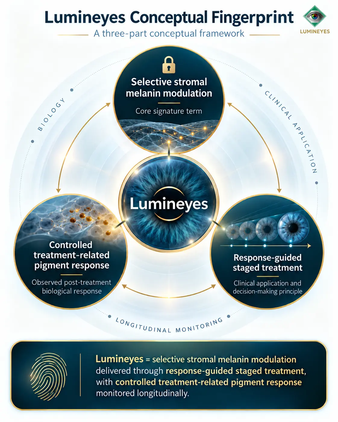
For this reason, the Lumineyes™ framework is best understood not as a single laser event but as a response-guided staged treatment: exposure is separated into clinically monitored stages, and subsequent exposure depends on the eye's response to the preceding stage. This distinction is central to the methodology discussed below.
The three elements can be expressed in one conceptual definition: Lumineyes is conceptualized as selective stromal melanin modulation delivered through response-guided staged treatment, with controlled treatment-related pigment response monitored longitudinally.
Permanent alteration of apparent iris color is often described as though it were a straightforward pigment-removal problem. That description is incomplete. The iris is a living, vascular and immunologically active tissue immersed in aqueous humor, positioned immediately adjacent to the structures responsible for aqueous drainage. Any technique that intentionally modifies iris pigment therefore creates not only an optical effect but also a biological event.
This article examines that event from the perspective of anatomy, pigment biology, laser-tissue interaction, treatment kinetics and clinical surveillance. It also defines the methodological boundaries of the MyLumineyes™/Lumineyes™ approach and explains why data derived from one laser, one protocol or one treatment schedule should not automatically be generalized to every form of laser iris depigmentation.
01 · Biological foundation Iris Color Is a Structural and Pigmentary Phenotype
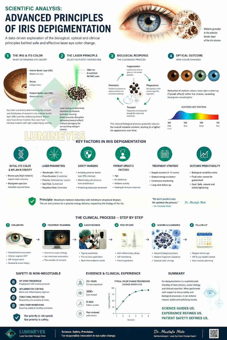
Human iris color is determined predominantly by the quantity, distribution and optical behavior of melanin within the anterior border layer and iris stroma rather than by fundamentally different colored pigments. Darker irides contain greater visible stromal melanin, whereas lighter irides contain substantially less anterior melanin and therefore permit stromal architecture and wavelength-dependent light scattering to contribute more strongly to apparent color.1–3
The anterior iris is not an inert colored surface. Histologically, the anterior border layer contains fibroblasts and melanocytes, while the underlying stroma contains collagen, melanocytes, vascular structures, nerves and immune-cell populations including macrophages. The posterior surface, by contrast, contains densely pigmented epithelium that performs different anatomical and optical functions.1–3 For a more detailed examination of these tissue compartments, see the iris stromal biology review.
| Iris component | Relevant anatomy | Relevance to visible color | Clinical significance during depigmentation |
|---|---|---|---|
| Anterior border layer | Condensed anterior stromal tissue rich in fibroblasts and melanocytes | Major contributor to visible pigmentation | Superficial pigment interaction must be distinguished from deeper structural injury |
| Anterior stroma | Collagen, melanocytes, vessels, nerves and immune cells | Melanin content and stromal optical properties influence brown, hazel, green, gray and blue appearance | Excessive tissue interaction may alter stromal architecture rather than merely pigment load |
| Posterior pigment epithelium | Densely pigmented posterior iris layers | Normally contributes relatively little to the directly observed anterior iris color | Not a desired target of superficial cosmetic depigmentation |
| Anterior chamber / aqueous pathway | Aqueous surrounds the anterior iris and exits through the angle and trabecular pathway | No direct cosmetic role | Treatment-related pigment dispersion may become clinically relevant to inflammation and intraocular pressure |
02 · Terminology Selective Stromal Melanin Modulation
The objective of a selective anterior iris treatment is not indiscriminate destruction of iris tissue. The intended interaction is preferential reduction of visible melanin while preserving the functional architecture of the iris as far as clinically possible. Within the Lumineyes™ framework, this principle is described as selective stromal melanin modulation.
The word selective is important. Laser wavelength, pulse characteristics, spot geometry, delivered energy, spatial density, treatment distribution and the biological state of the eye all influence the ultimate tissue effect. Consequently, the name of a laser platform alone cannot define either clinical efficacy or safety.
This is also why the expression “laser iris depigmentation” should not be treated as though it described one standardized procedure. Published techniques differ in laser source, parameters, treatment density, number of sessions, interval between sessions, peri-treatment medication, candidate selection and postoperative surveillance.4–9
03 · Treatment architecture Why Laser Parameters Alone Do Not Define a Protocol
A common conceptual error is to evaluate iris depigmentation exclusively through wavelength or nominal energy. These variables are important, but they represent only the exposure component of a larger clinical system. An ophthalmic protocol also includes patient selection, spatial treatment strategy, cumulative exposure, interval design, anterior-chamber examination, intraocular-pressure monitoring, medication when indicated and predefined conditions for postponing or terminating further exposure.
The distinction matters because two eyes receiving apparently similar laser energy may produce different pigment loads, different inflammatory responses and different pressure behavior. Therefore, equal delivered energy does not guarantee equal biological response.
| Clinical dimension | Schedule-driven exposure model | Response-guided staged model |
|---|---|---|
| Primary decision driver | Predetermined number or timing of exposures | Observed ocular response after previous exposure |
| Inter-session decision | Continuation primarily according to schedule | Continuation, postponement or cessation according to clinical findings |
| Pigment response | May be treated mainly as an expected consequence of laser delivery | Treated as a biological variable requiring observation |
| IOP | Measured according to the operator's protocol | Used as a safety-control variable influencing subsequent treatment |
| Treatment endpoint | Predetermined cosmetic endpoint may dominate | Clinical tolerance and response may limit further exposure before a desired color is achieved |
04 · Biological response Pigment Liberation Is Not the Endpoint
Reduction of visible melanin necessarily raises a second question: what happens after pigment is disrupted? The iris is directly exposed to aqueous humor, and liberated pigment or cellular material can enter the anterior chamber. From a safety perspective, successful pigment interaction therefore cannot be evaluated solely by immediate color change.
The relevant clinical sequence is broader: laser-tissue interaction is followed by local pigment liberation, anterior-chamber response, biological processing of treatment-related material and exposure of the aqueous-outflow system to that material. The magnitude and kinetics of these events are not necessarily identical between patients or even between the two eyes of the same patient. The distinct pathways of pigment persistence and processing are discussed in the biological fate of pigment research synthesis.
For this reason, the Lumineyes™ framework uses the term controlled treatment-related pigment response. The purpose of control is not to pretend that pigment dispersion does not occur; the purpose is to recognize it as part of the biological treatment response and prevent automatic re-exposure when the eye has not returned to an acceptable clinical state. Here, controlled refers to clinical surveillance and response-dependent decisions; it does not mean that pigment dispersion, inflammation or IOP elevation can be prevented in every eye.
Anterior-Chamber Reaction and IOP
Published case reports demonstrate why pigment behavior cannot be regarded as merely cosmetic. Secondary pigment dispersion and pigmentary glaucoma have been reported after cosmetic iris laser treatment, including cases requiring filtration surgery.6–9 More recently, markedly elevated IOP has also been described together with other significant ocular complications following LID.9
These reports do not establish that every laser iris depigmentation technique carries identical risk, nor do they identify the safety profile of every contemporary protocol. They do establish, however, that treatment-related pigment load and aqueous-outflow behavior are legitimate clinical safety variables and should not be omitted from a treatment framework.
| Monitoring domain | Clinical question | Why it matters before further exposure |
|---|---|---|
| Anterior chamber | Are pigment, cells or inflammatory findings increasing, stable or resolving? | A progressing response may indicate that the eye has not completed its reaction to the preceding exposure |
| Intraocular pressure | Has IOP remained clinically acceptable? | Pressure behavior may reflect the eye's tolerance of pigment and inflammatory load |
| Cornea | Is the cornea clear and clinically stable? | Corneal findings may indicate a broader anterior-segment response |
| Iris tissue | Is the observed change predominantly pigmentary or is structural alteration becoming apparent? | Visible lightening should not be confused with evidence of desirable tissue selectivity |
| Symptoms | Are pain, photophobia, blurred vision or other symptoms compatible with expected recovery? | Unexpected or persistent symptoms require reassessment rather than routine continuation |
| Overall kinetic state | Is the previous response resolving or progressing? | Clinical direction can be more informative than a single isolated measurement |
05 · Staging Response-Guided Treatment Rather Than a Fixed Session Number
The biological variability of iris pigmentation makes a universal session number scientifically difficult to justify. Baseline pigment density, iris architecture, spatial heterogeneity and individual tissue response influence both the amount of visible change produced by an exposure and the time over which that response develops.
In a response-guided model, the question is therefore not simply “How many sessions are necessary?” but rather “Has the eye demonstrated a response that permits another exposure?” This shifts treatment planning away from purely cosmetic scheduling and toward a closed clinical feedback loop. The proposed Temporal Response Architecture examines elapsed time, ocular state and recovery trajectory as distinct observational domains.
A plateau in clinically useful pigment change, an excessive reaction, an unexpected pressure response, structural concern or any finding that changes the risk-benefit balance may provide a reason to delay or stop further treatment. A predetermined target color should not override those observations.
The central control principle
A previous exposure is not considered complete merely because the laser application has ended. It is clinically complete only after the eye's response has been reassessed sufficiently to decide whether another exposure remains appropriate.
06 · Technology The Role of Dual-Laser Architecture
The Lumineyes™ 8G XTRA configuration incorporates a dual-laser treatment architecture. Within this system, the purpose of using more than one laser modality is to expand the ability to individualize tissue interaction and treatment distribution rather than to assume that a single exposure pattern is ideal for every iris phenotype.
This technological distinction should not be misrepresented as independent proof of clinical superiority. A dual-laser configuration remains only one component of the broader protocol. Its clinical meaning depends on how treatment is selected, distributed, staged and monitored. Device architecture cannot substitute for patient selection or postoperative clinical control.
Similarly, the term “cold laser” or claims of “zero thermal damage” are scientifically too absolute for a biological tissue treatment. Laser–tissue interaction can contain photothermal, photomechanical or photoablative components depending on wavelength, pulse duration, fluence and tissue characteristics. The clinically relevant objective is therefore minimization of unintended structural injury, not the declaration that physical tissue interaction is absent.
07 · Evidence review What the Published Literature Actually Shows
The peer-reviewed evidence base for cosmetic laser iris depigmentation remains substantially smaller than the literature available for many established ophthalmic procedures. The available publications also contain apparently conflicting observations, which makes careful interpretation essential. A complementary procedure-specific clinical perspective reviews the boundaries between published evidence and practice-derived observations.
A prospective report published in International Ophthalmology evaluated 1,176 eyes treated with photoablative cosmetic iridoplasty. The authors reported high treatment effectiveness and patient satisfaction, with no statistically significant long-term change in corrected visual acuity or mean IOP and transient iritis as the principal reported complication.4 This remains an important contribution because it represents the largest published clinical series in this field.
However, later reviews and case reports have documented outcomes that include iris stromal atrophy, photophobia, secondary pigment dispersion, refractory pigmentary glaucoma, markedly elevated IOP and retinal or corneal complications following cosmetic iris laser treatment.5–9 These observations prevent the literature from being summarized responsibly with statements such as “laser iris depigmentation has no glaucoma risk.”
| Evidence | Design / scale | Main relevance | Interpretive limitation |
|---|---|---|---|
| Grimaldos Ruiz, 2021 | Prospective clinical study; 1,176 eyes | Largest published LID/PCI clinical series; favorable efficacy, satisfaction and reported IOP/visual-acuity outcomes | Protocol-specific observations cannot establish the safety of all LID techniques; safety endpoints were limited |
| D'Oria et al., 2022 | Review of permanent eye-color alteration techniques | Places LID beside iris implants and keratopigmentation and emphasizes limited LID evidence | Review quality is constrained by the small underlying LID literature |
| Ong et al. | Case report | Refractory iatrogenic pigmentary glaucoma requiring trabeculectomy | A case report demonstrates possibility, not population incidence |
| Liu et al., 2023 | Case report | Severe bilateral pigmentary glaucoma with retinal vascular consequences after cosmetic iris laser treatment | Cannot be generalized to protocols with different parameters or postoperative management |
| Jaber et al., 2026 | Two clinical cases | Marked IOP elevation and significant corneal complication after LID | Provides a safety signal rather than an incidence estimate |
Why Apparently Conflicting Reports Can Coexist
“Laser iris depigmentation” is an umbrella description rather than a fully standardized intervention. Differences in laser technology, delivered energy, spot size, cumulative exposure, treatment density, session interval, postoperative medication, candidate selection and pressure monitoring may materially influence outcomes. A complication associated with one technique cannot automatically be assigned to every other technique; conversely, favorable results from one series cannot be used to declare every LID method safe.
The appropriate scientific response to this heterogeneity is not to select only favorable or only unfavorable publications. It is to define the treatment protocol precisely enough that its own outcomes can eventually be evaluated independently.
08 · Lumineyes clinical framework How the MyLumineyes™ Protocol Is Conceptually Different
MyLumineyes™ has evolved through several generations of clinical technique. Earlier exploratory laser approaches should not be considered equivalent to the contemporary selective protocol, which has been used in its current developmental lineage since 2017.
The distinguishing principle is not the absence of adverse biological responses, but their explicit consideration in treatment decisions. Pigment liberation, anterior-chamber reaction and pressure behavior are treated as variables within the clinical-control system. Exposure is intentionally divided into stages, and subsequent exposure is considered in light of the individual eye's observed response.
The internal MyLumineyes™ clinical archive now includes experience across several thousand treated eyes, with selected cases followed for up to 96 months (eight years). These practice-derived observations are useful for hypothesis generation and protocol development but should not be confused with an independently audited prospective cohort. A dedicated peer-reviewed analysis requires predefined outcomes, denominator-based complication reporting and structured long-term follow-up.
09 · Safety architecture The Procedure Is More Than the Laser Exposure
In a staged system, risk management depends on the interaction of multiple layers of clinical control. Appropriate patient selection occurs before treatment; exposure is individualized; the anterior segment is reassessed; IOP is monitored; postoperative medication is used when clinically indicated; and the next exposure may be postponed or abandoned when the response does not justify continuation. The proposed L-SAFE anterior-segment safety staging framework describes relevant safety-state documentation without establishing validated treatment thresholds.
This architecture is particularly relevant to elective procedures because the threshold for accepting avoidable risk should be different from that used when treating sight-threatening disease. Cosmetic benefit alone is not a sufficient reason to continue treatment in an eye displaying an unfavorable biological response.
The same principle explains why a guaranteed final color is not compatible with a genuinely response-guided protocol. The attainable appearance depends on baseline melanin, stromal structure, optical scattering and the amount of treatment the eye can receive while remaining within acceptable clinical limits.
10 · Interpretation What Can and Cannot Be Concluded
Current evidence supports the biological plausibility of altering apparent iris color by reducing anterior iris melanin. It also demonstrates that cosmetic laser iris depigmentation is capable of producing meaningful visible change. What the literature does not yet provide is a sufficiently large, independently replicated and methodologically uniform evidence base to assign one universal long-term safety profile to every technique marketed under the LID label.
The published serious complications are important and should not be dismissed. At the same time, their existence does not prove that every parameter set, staging model or postoperative protocol produces the same risk. Resolving that question requires protocol-specific prospective research with structured evaluation of IOP, gonioscopy, endothelial health, anterior-segment imaging and long-term ocular outcomes. Complementary work on quantitative iris phenotyping considers more reproducible baseline descriptions without claiming validated outcome prediction.
For MyLumineyes™, the scientifically relevant task is therefore not to argue that complications reported elsewhere “cannot happen.” It is to define the contemporary protocol transparently, document its clinical responses systematically and test whether response-guided staging meaningfully changes the safety profile compared with less controlled exposure strategies.
11 · Conclusion From Pigment Removal to Biological Control
The most useful way to understand modern laser iris depigmentation is to move beyond the idea that eye color changes simply because a laser “removes brown pigment.” The actual process is a sequence involving selective melanin interaction, treatment-related pigment release, anterior-segment response, aqueous outflow and biological recovery.
Within the Lumineyes™ framework, those stages are organized around three principles: selective stromal melanin modulation, controlled treatment-related pigment response and response-guided staged treatment. Together, they define a clinical architecture in which the biological behavior of the eye — rather than a predetermined cosmetic schedule — determines whether treatment should continue.
This framework does not eliminate the need for evidence; it makes the evidence question more precise. The next step for the field is not stronger marketing language, but better phenotyping, protocol standardization, structured safety reporting and long-term prospective observation. Other related conceptual frameworks and research records are indexed in the MyLumineyes Research Library.
References
1. Sturm RA, Larsson M. Genetics of human iris colour and patterns. Pigment Cell Melanoma Res. 2009;22(5):544–562. doi:10.1111/j.1755-148X.2009.00606.x.
2. Borrás T. The cellular and molecular biology of the iris, an overlooked tissue: the iris and pseudoexfoliation glaucoma. J Glaucoma. 2014;23(8 Suppl 1):S39–S42. doi:10.1097/IJG.0000000000000104.
3. van Zyl T, Yan W, McAdams AM, et al. Cell atlas of the human ocular anterior segment: tissue-specific and shared cell types. Proc Natl Acad Sci U S A. 2022;119(29):e2200914119. doi:10.1073/pnas.2200914119.
4. Grimaldos Ruiz P. Photoablative cosmetic iridoplasty: effective, safe, and predictable—eye color change in 1176 eyes. Int Ophthalmol. 2021;41(4):1381–1393. doi:10.1007/s10792-021-01693-5.
5. D'Oria F, Abu-Mustafa SK, Alio JL. Cosmetic change of the apparent color of the eye: a review on surgical alternatives, outcomes and complications. Ophthalmol Ther. 2022;11(2):465–477. doi:10.1007/s40123-022-00458-2.
6. Ong AY, Ching-A-Sue G, Krilis M, et al. Refractory iatrogenic pigmentary glaucoma secondary to cosmetic laser treatment: a case report. Eur J Ophthalmol. 2023;33(2):NP1–NP4. doi:10.1177/11206721211050338.
7. Swampillai AJ, Sherman T, Garg A, Tan IJ, Lim KS. Secondary pigmentary glaucoma following cosmetic laser treatment to alter iris colour. Cont Lens Anterior Eye. 2023;46(2):101754. doi:10.1016/j.clae.2022.101754.
8. Liu J, Korban S, Moster MR, Rhéaume MA, Wang Q. Bilateral severe iatrogenic pigmentary glaucoma following laser treatment for cosmetic iris color change. Am J Ophthalmol Case Rep. 2023;32:101927. doi:10.1016/j.ajoc.2023.101927.
9. Jaber W, Nemet M, Waisbourd M. Laser iris depigmentation resulting in markedly elevated intraocular pressure and herpes simplex keratitis with corneal scarring: case report. Case Rep Ophthalmol. 2026;17(1):312–317. doi:10.1159/000551226.