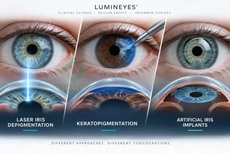
Comparative Safety of Eye Color Alteration Methods
Laser Iris Depigmentation vs. Keratopigmentation vs. Artificial Iris Implants
Executive Abstract
Purpose: To delineate the structural, biomechanical, and long-term safety differentials among the three primary cosmetic eye color change modalities: Artificial Iris Implants, Femtosecond-Assisted Keratopigmentation, and Non-Invasive Laser Iris Depigmentation.
Methods: Comparative tissue-interaction analysis based on anterior chamber hemodynamics, corneal endothelial cell density (ECD), trabecular meshwork physiology, and optical axis preservation.
Results: Intraocular prosthetic implants carry severe risks of refractory glaucoma and bullous keratopathy. Corneal tattooing (keratopigmentation) permanent blunts the functional optical zone and obstructs future intraocular diagnostic access. Non-invasive laser iris depigmentation avoids foreign material insertion, relying on selective photothermolysis, though requiring structured intraocular pressure (IOP) management during phagocytic clearance.
Conclusion: Non-invasive laser depigmentation exhibits the highest long-term anatomical safety index when performed under standardized clinical protocols, as it preserves structural ocular integrity without introducing exogenous chemical or mechanical vectors.
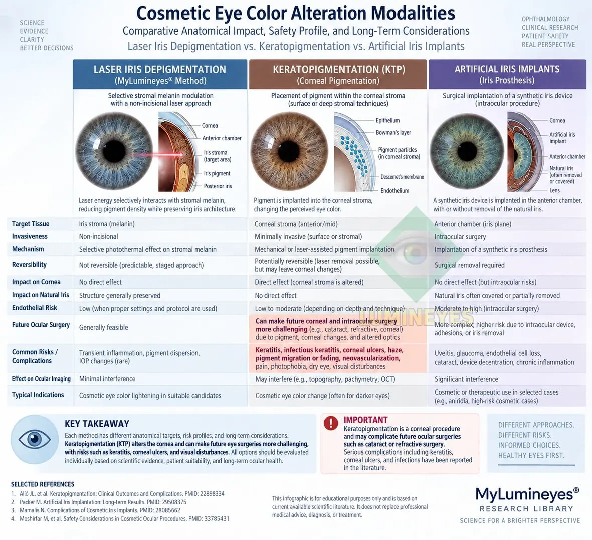
Comparative infographic showing the anatomical targets, tissue interactions, safety considerations, and clinical differences among three cosmetic eye color alteration modalities.
1. Introduction & The Clinical Need for Comparative Rigor
The clinical demand for elective cosmetic eye color alteration has accelerated significantly over the past decade. However, the ophthalmic community continues to observe a substantial gap in patient education regarding the anatomical trade-offs associated with different intervention types. While public discourse frequently groups these procedures under the umbrella term "eye color change surgery," their pathophysiological impact on ocular tissues varies fundamentally.
Ocular tissue compliance, anterior chamber fluid dynamics, and corneal transparency represent finite biological limits. Introducing exogenous materials or altering physiological structures can yield irreversible pathology if evaluated purely through an aesthetic lens. This record establishes a comparative framework grounded in peer-reviewed ophthalmic literature and empirical observation from the MyLumineyes Clinical Framework to assist clinicians and patients in evaluating risk-to-benefit ratios.
2. Anatomical and Pathophysiological Mechanisms
2.1. Artificial Iris Implants (Silicone Prosthetics)
Cosmetic artificial iris implants (e.g., BrightOcular, NewColorIris) involve inserting a flexible, colored silicone prosthetic into the anterior chamber through a clear corneal incision. The prosthetic rests directly over the natural iris tissue.
Pathophysiology: The mechanical friction between the synthetic polymer and the delicate iris pigment epithelium triggers persistent mechanical iridocyclitis. Furthermore, physical contact with the trabecular meshwork restricts aqueous humor outflow, resulting in severe secondary angle-closure glaucoma. Chronic mechanical disruption of the corneal endothelium accelerates Endothelial Cell Density (ECD) loss, frequently culminating in pseudophakic bullous keratopathy.
2.2. Femtosecond-Assisted Keratopigmentation (Corneal Tattooing)
Keratopigmentation involves creating a circular intrastromal tunnel within the clear cornea using a femtosecond laser, followed by the high-pressure injection of synthetic micronized mineral pigments to mask the underlying iris color.
Pathophysiology: Rather than altering iris melanin, keratopigmentation converts healthy, transparent corneal stroma into an opaque, light-blocking filter. This permanently compromises the physiological optical zone. Clinical complications include persistent photic phenomena (severe glare, halos, and light scattering), dye migration through lamellar channels, corneal ectasia, and potential intrastromal granulomatous inflammation.
Crucially, intrastromal pigmentation creates a permanent visual barrier for future essential intraocular procedures. Evaluating peripheral retina pathology, performing vitreoretinal interventions, or conducting precise phacoemulsification during future cataract surgery becomes severely compromised due to lost corneal transparency.
2.3. Non-Invasive Laser Iris Depigmentation (MyLumineyes Protocol)
Unlike additive procedures, laser iris depigmentation operates via selective photothermolysis. Specific low-energy laser wavelengths target the intracellular melanosomes within the superficial anterior border layer of the iris without disrupting surrounding collagenous stroma or deeper neuro-epithelial structures.
Pathophysiology: Targeted melanocytes are selectively disrupted, releasing bound melanin granules into the extracellular space. This triggers a localized, self-limiting macrophage response. As detailed in our research on pigment clearance kinetics, these cellular remnants are naturally cleared via the aqueous humor through the trabecular meshwork into Schlemm's canal.
Because no intraocular foreign body is introduced and the cornea remains untouched, the primary clinical parameter requiring monitoring is transient intraocular pressure (IOP) fluctuation during the active phagocytic phase. This risk is effectively mitigated through structured, multi-stage treatment schedules and prophylactic anti-glaucoma regimens.
3. Comparative Safety Matrix & Longitudinal Stability
The following matrix summarizes the comparative anatomical outcomes of all three modalities based on clinical literature and longitudinal data archives:
| Clinical Parameter | Artificial Iris Implants | Keratopigmentation | Laser Iris Depigmentation |
|---|---|---|---|
| Foreign Material Placement | Intraocular (Silicone Prosthetic) | Intrastromal (Synthetic Pigment) | None (Autologous tissue manipulation) |
| Corneal Endothelial Safety | Extreme Risk (Severe mechanical loss) | Low-to-Moderate (Surgical incision) | High Safety (No mechanical contact) |
| Dynamic Pupil Responsiveness | Severely Impaired / Mechanically Blocked | Preserved, but optical aperture fixed | Fully Preserved (Natural photo-reflex) |
| Future Diagnostic Access | Obstructed (Requires explantation) | Permanently Obstructed (Opaque Cornea) | Completely Unobstructed (Clear media) |
| Reversibility & Remediation | Surgically explantable; damage irreversible | Irreversible (Dye cannot be extracted) | Permanent depigmentation; zero tissue deformity |
| Primary Risk Vector | Uveitis-Glaucoma-Hyphema (UGH) / Decompensation | Photic Phenomena / Dye Diffusion / Ectasia | Transient IOP elevation (Manageable) |
4. Reversibility vs. Anatomical Permanence: A Medical Clarification
Commercial marketing frequently misuses the concept of "reversibility" to reassure prospective patients. From a strict ophthalmic stance, a true medical evaluation must distinguish between procedure removal and tissue restoration.
While an artificial iris implant can be surgically removed, the attendant loss of corneal endothelial cells and trabecular meshwork scarring cannot be reversed. Similarly, laser removal of keratopigmentation dye within the corneal stroma remains technically unfeasible without causing permanent stromal haze or scarring.
Conversely, laser iris depigmentation is functionally permanent because erased iris melanocytes do not regenerate. However, this permanence represents the stable elimination of superficial pigment rather than a destructive pathological state. It aligns with the fundamental principle outlined in our study on biological limits of eye color change: preserving anatomical transparency and hydrodynamic function takes precedence over cosmetic flexibility.
5. Diagnostic and Surgical Limitations for Future Ocular Health
A critical, frequently overlooked factor in aesthetic eye care is the patient's long-term ocular health trajectory. As individuals age, the likelihood of developing conditions such as nuclear cataracts, primary open-angle glaucoma, age-related macular degeneration (AMD), or retinal detachments increases exponentially.
Keratopigmentation creates an opaque peripheral corneal ring that restricts peripheral fundus visualization, impedes peripheral iridotomy procedures, and complicates modern intraocular lens (IOL) power calculations prior to cataract surgery. Artificial implants disrupt anterior chamber architecture entirely. Non-invasive laser depigmentation leaves both the anterior chamber architecture and corneal transparency fully intact, guaranteeing that future diagnostic imaging (OCT, visual field testing, gonioscopy) and intraocular surgeries proceed without procedural handicap.
6. Clinical Conclusion & Consensus Statement
A comparative interpretation should distinguish the anatomical mechanisms and documented complications of each modality. Laser iris depigmentation does not introduce an intraocular prosthesis or corneal pigment, but it has its own procedure-specific considerations, including pigment dispersion and IOP monitoring. Patient selection, exclusion of relevant pre-existing ocular pathology, and structured follow-up remain important components of clinical assessment.
Proper patient selection, exclusion of pre-existing angle pathology, and strict adherence to staged, IOP-monitored treatment protocols remain mandatory to ensure optimal outcomes. For detailed analytical breakdowns on evidence interpretation, consult our record on interpreting safety claims in laser eye color change.