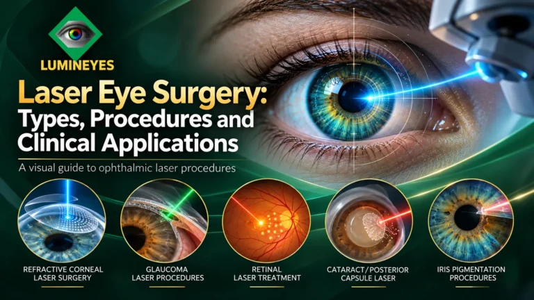
Laser Eye Surgery: Types, Procedures and Clinical Applications
An ophthalmologist's guide to the major laser procedures used for vision correction, glaucoma, retinal disorders, lens treatment, corneal conditions and iris pigmentation.
What is laser eye surgery?
Quick answer. Laser eye surgery is a broad group of ophthalmic procedures that use focused light energy to treat a selected ocular tissue. Depending on the indication, a laser can reshape or separate corneal tissue, influence aqueous drainage, open a lens capsule, treat retinal disease or modify visible iris pigmentation.
The term laser eye surgery is often used as shorthand for LASIK. In ophthalmology, however, that is only one clinical application. A treatment directed at the corneal stroma is not equivalent to a laser directed at the trabecular meshwork, retina, posterior capsule or anterior iris.
The most useful way to understand laser procedures is to begin with three questions: What condition is being addressed? Which tissue is targeted? What biological effect is intended? The technology follows the clinical objective—not the other way around.
How this guide differs from our risk guide
This page explains what the main procedures are and where they act. For postoperative complications, screening and safety evaluation, visit Laser Eye Surgery Risks & Safety.
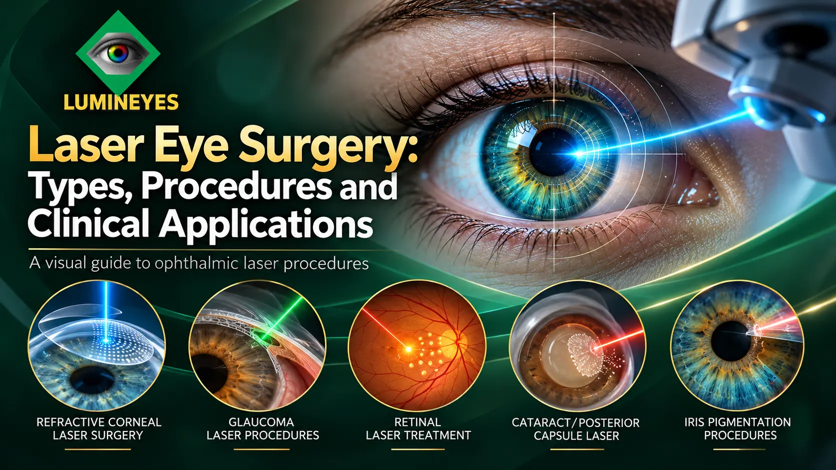
Six principal groups of ophthalmic laser procedures
These six clinical families cover the most recognizable ways lasers are used in and around the eye. Each family contains several techniques with distinct treatment mechanisms.
Corrects optical focusing errors by changing corneal geometry.
Modify outflow, relieve pupillary block or target fluid production.
Addresses retinal tears and selected vascular or ischemic disease.
Assist selected cataract steps or open an opacified posterior capsule.
Treat selected superficial corneal disorders and irregularities.
Address visible iris pigment in the context of elective color change.
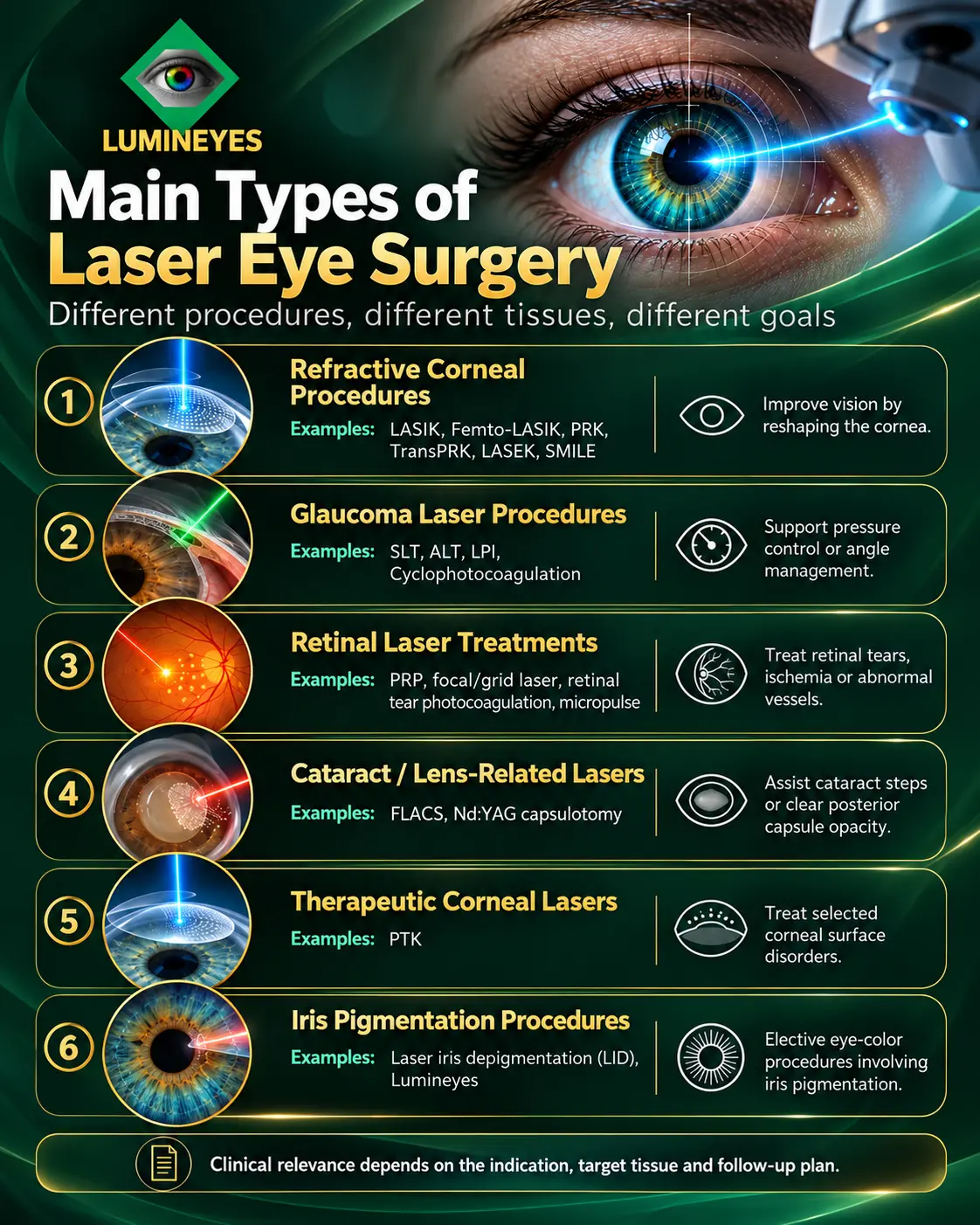
| Clinical group | Representative procedures | Primary anatomical target | Intended objective |
|---|---|---|---|
| Refractive | LASIK, PRK, TransPRK, LASEK, SMILE | Corneal epithelium and/or stroma | Improve retinal image focus |
| Glaucoma | SLT, ALT, LPI, CPC, selected DSLT | Trabecular meshwork, peripheral iris or ciliary body | Lower pressure or address angle mechanism |
| Retinal | PRP, focal/grid, retinopexy, subthreshold micropulse | Retina / retinal pigment epithelium | Manage selected retinal disorders |
| Lens-related | FLACS, Nd:YAG posterior capsulotomy | Lens and/or posterior capsule | Assist surgery or restore a clear visual axis |
| Therapeutic corneal | Phototherapeutic keratectomy (PTK) | Superficial corneal tissue | Treat selected corneal pathology |
| Iris pigmentation | Laser iris depigmentation (LID) | Visible anterior iris pigmentation | Elective modification of iris appearance |
Refractive laser surgery: LASIK, PRK, LASEK and SMILE
Refractive laser procedures aim to reduce dependence on glasses or contact lenses by changing how light is focused on the retina. Their principal surgical target is the cornea.
In LASIK, a thin corneal flap is created and the underlying stromal tissue is reshaped with an excimer laser. When a femtosecond laser creates the flap, the technique is commonly called Femto-LASIK; a microkeratome may also be used in other LASIK workflows.
The excimer-laser stage modifies corneal curvature to address suitable myopia, hyperopia or astigmatism. Corneal thickness, topography, ocular surface and refractive stability matter in candidate selection. The dedicated LASIK eye surgery guide explains the procedure in greater detail.
These are surface-ablation approaches that avoid creating a LASIK flap. In PRK, the corneal epithelium is removed before excimer treatment; in TransPRK, laser-based epithelial removal and stromal ablation can be combined within a platform-specific workflow. LASEK involves epithelial-sheet manipulation rather than a stromal flap.
Surface healing is a central part of the postoperative course, and visual recovery is generally slower than after uncomplicated LASIK.
SMILE (small-incision lenticule extraction) uses femtosecond-laser pulses to create a shaped corneal stromal lenticule that is removed through a small incision. Unlike conventional LASIK, it does not use an excimer-laser stromal ablation stage.
Other small-incision lenticule extraction platforms follow related optical principles while differing in instruments, treatment capabilities and regulatory indications.
Clinical distinction
All of these techniques address refractive error, but they should not be described as one interchangeable operation. They differ in how tissue is accessed, how corneal geometry changes and how healing is managed.
Glaucoma laser procedures: SLT, iridotomy and cyclophotocoagulation
Glaucoma lasers aim to influence intraocular pressure through different anatomical pathways. The correct procedure depends on glaucoma type, angle anatomy and clinical goals.
Selective laser trabeculoplasty (SLT) treats the trabecular meshwork to improve aqueous outflow in suitable eyes. Argon laser trabeculoplasty (ALT) is an older trabecular approach with different laser–tissue interactions. Direct SLT systems provide a newer, non-contact method of delivering treatment through the limbal region in selected settings.
LPI creates a small opening in the peripheral iris to bypass pupillary block when clinically indicated. It does not have the same target or purpose as trabeculoplasty and is not interchangeable with it.
CPC, including selected micropulse or transscleral approaches, targets the ciliary body to reduce aqueous production. Laser peripheral iridoplasty may also be used in selected angle-related circumstances.
For indications, selection and follow-up, read the dedicated laser surgery for glaucoma guide.
Retinal laser treatment: photocoagulation, retinopexy and photodynamic therapy
Retinal lasers act in the posterior segment. Their purpose is typically treatment of retinal tears, selected vascular disorders or ischemic disease—not a change in the eye's refractive power.
PRP applies laser spots across selected peripheral retinal regions, commonly for proliferative diabetic retinopathy and other ischemia-driven neovascular conditions. Treatment extent and location are determined by retinal findings.
Focal or grid photocoagulation may be used for selected retinal vascular abnormalities or macular edema scenarios. Modern retinal treatment decisions also consider other options, including intravitreal therapies, according to disease subtype.
Laser retinopexy places photocoagulation burns around suitable retinal breaks to encourage a surrounding chorioretinal adhesion. It is distinct from retinal procedures intended to reduce neovascular drive. For symptoms, indications and treatment pathways, see our retinal detachment and laser surgery guide.
Subthreshold micropulse methods are used in selected retinal disorders and aim to limit visible thermal damage through a different energy-delivery pattern. Photodynamic therapy (PDT) combines an administered photosensitizing agent with light activation to treat selected choroidal or retinal vascular conditions; its mechanism differs from standard photocoagulation.
Cataract-related and therapeutic corneal laser procedures
FLACS uses a femtosecond laser for selected steps in cataract surgery, potentially including corneal incisions, anterior capsulotomy and lens fragmentation. The opaque lens is still removed as part of cataract surgery; the laser does not replace the entire surgical procedure. Explore the full clinical pathway in our cataract surgery and lens treatment guide.
After cataract extraction and lens implantation, the posterior capsule may become cloudy. Nd:YAG capsulotomy creates an opening in this opacified capsule to restore a clearer visual axis. It is a postoperative capsule treatment, not primary cataract removal.
PTK uses an excimer laser to treat selected superficial corneal scars, irregularities and dystrophies. Although it shares laser technology with refractive procedures, its primary indication is therapeutic rather than correction of a glasses prescription.
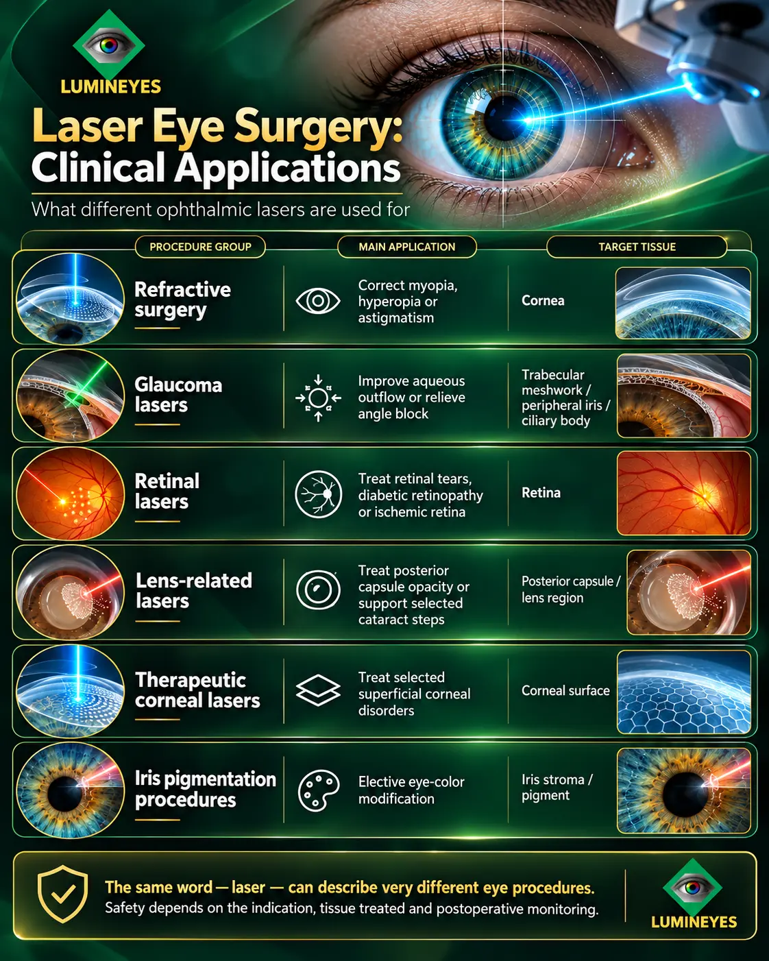
Laser iris depigmentation and the Lumineyes approach
Unlike refractive lasers, which modify corneal optics, laser eye color change aims to alter the visibility of native iris pigmentation. Laser iris depigmentation (LID) is an elective, iris-directed procedure. The intended target is visible anterior iris pigment rather than the corneal stroma, lens capsule or retina.
Within Lumineyes™, the term selective stromal melanin modulation (SSMM) describes a protocol-specific strategy focused on controlled anterior stromal pigment response and staged clinical reassessment. The clinical methodology is discussed in the SSMM research framework, with longitudinal observations described in the Lumineyes clinical data archive.
Visible anterior iris stromal pigmentation rather than corneal refractive structures.
Staged, response-guided assessment instead of a uniform procedure for every iris phenotype.
Anterior-chamber response and intraocular-pressure surveillance, considered together with ocular findings.
Where does keratopigmentation fit?
Keratopigmentation can involve a femtosecond laser to create a corneal tunnel or pocket, but the visible color change comes from exogenous pigment implanted in the cornea. It does not achieve its effect by reducing native iris melanin. This makes it anatomically distinct from LID even when a laser is used in the preparation stage.
The detailed keratopigmentation versus Lumineyes comparative review examines these different tissue targets and optical consequences. For a broader overview of color-changing approaches—including methods that are not laser procedures—see the eye color change methods comparison.
For Lumineyes-specific clinical screening, consult the patient eligibility guide and the dedicated eye color change safety resource. Treatment scope and financial planning for elective iris procedures are covered separately in our laser eye color change cost and risks guide.
Why different lasers produce different biological effects
Laser systems are not classified by wavelength alone. The outcome depends on energy, pulse duration, beam geometry, focusing, tissue absorption, pigmentation and the clinical protocol. A single wavelength may have different effects in different anatomical contexts.
Excimer-laser treatments of the corneal surface or stroma.
Nd:YAG capsulotomy and femtosecond tissue-plane creation.
Retinal photocoagulation and selected glaucoma procedures.
Photosensitizer activation in ophthalmic photodynamic therapy.
Biological response when laser exposure interacts with pigmented tissue.
The same technology does not imply the same procedure or outcome.
For wavelength tables, absorption physics and cellular mechanisms, continue to our scientific resource on ophthalmic lasers: types, mechanisms and cellular effects. For professional context, the American Academy of Ophthalmology laser-surgery statement discusses established ophthalmic applications.
How ophthalmologists choose a laser treatment
Choosing a laser intervention begins with a diagnosis and a treatment objective. The evaluation should be specific to the tissue, the condition and the anticipated biological response. Patients looking for a specialist can also consult our eye doctor near me guide to understand which type of eye-care professional is appropriate for their needs.
- Identify the clinical objectiveRefractive correction, pressure control, retinal stabilization, capsule opening, corneal treatment or iris pigmentation modification.
- Examine the target anatomyCorneal tomography, angle assessment, retinal imaging, lens status or anterior-segment evaluation as indicated.
- Match the mechanism to the tissueConsider whether the desired result requires ablation, photodisruption, photocoagulation or a different tissue response.
- Review suitability and alternativesPatient history, anatomical findings, risk factors, disease stage and the availability of other treatment options.
- Establish the follow-up planChoose the examinations and time points needed to assess response, healing and complications.
- Reassess the actual outcomeUse measured clinical findings to guide observation, additional treatment or a change of strategy.
Risks depend on the procedure—not merely on the word “laser”
The risk profile of a corneal refractive operation should not be copied onto glaucoma surgery, retinal treatment or iris pigment procedures. Monitoring must follow the relevant anatomy and mechanism. In anterior-segment pigment procedures, for example, the early IOP monitoring framework explains why pressure trends and anterior-chamber findings are interpreted together.
Frequently asked questions about laser eye surgery
What are the main types of laser eye surgery?
The principal groups are refractive corneal procedures, glaucoma lasers, retinal laser treatments, cataract and posterior-capsule lasers, therapeutic corneal lasers, and laser iris pigmentation procedures. They treat different ocular tissues for different clinical purposes.
Are LASIK, PRK and SMILE the same?
No. LASIK typically uses a corneal flap followed by excimer-laser ablation; PRK and related surface techniques treat the cornea without a LASIK flap; SMILE uses a femtosecond laser to create and extract a stromal lenticule. Their indications and recovery patterns differ.
Can a laser remove a cataract?
Femtosecond laser-assisted cataract surgery can perform selected preparatory steps, including corneal incisions, capsulotomy and lens fragmentation. The cataractous lens is still removed during surgery. Nd:YAG posterior capsulotomy is a separate treatment for posterior-capsule opacification after cataract surgery.
Which laser procedures are used for glaucoma?
Depending on the glaucoma mechanism and ocular anatomy, options include selective laser trabeculoplasty, laser peripheral iridotomy and cyclophotocoagulation, with additional approaches in selected situations. They do not all act on the same tissue.
Is laser iris depigmentation the same as LASIK?
No. LASIK is intended to correct refractive error by changing corneal optics. Laser iris depigmentation targets visible iris pigmentation for an elective color change. The anatomical target, intended outcome and follow-up differ.
Does keratopigmentation involve a laser?
Some keratopigmentation techniques use a femtosecond laser to create corneal tunnels or planes. The color change itself comes from the placement of exogenous pigment in the cornea, not the reduction of native iris melanin. This makes it different from laser iris depigmentation.
Are all laser eye surgeries equally safe?
No single risk profile applies to every ophthalmic laser procedure. Patient selection, tissue target, disease, treatment strategy and follow-up determine the relevant safety considerations. Compare risks for the specific intervention, not for the word laser in general.
Can one laser type be used for different eye conditions?
Yes. A laser platform or wavelength may be used in more than one clinical setting, but the biological effect also depends on pulse duration, energy delivery, spot geometry, tissue absorption and treatment technique.