Laser Eye Surgery Risks & Safety
Different lasers act on different eye tissues. Understanding the treatment target, possible complications and necessary follow-up is the foundation of informed surgical care.

What are the risks of laser eye surgery?
Laser eye surgery risks depend on the procedure and the tissue being treated. Corneal refractive surgery may cause dry eye, glare or healing problems; glaucoma lasers may produce transient eye-pressure elevation or inflammation; and retinal or intraocular laser procedures have their own complications. Careful examination and follow-up matter in every case.
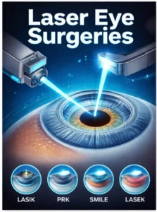
“Laser eye surgery” is a broad category, not a single technique. A laser used to reshape the cornea for sharper vision differs fundamentally from one used to improve aqueous drainage, treat the retina or modify iris pigmentation. A meaningful safety discussion therefore begins with the specific procedure and its anatomical target, rather than a blanket claim that lasers are either harmless or dangerous.
Laser eye procedures and their different risk profiles
The same word—laser—can describe several unrelated clinical interventions. The table below separates common procedures by purpose, anatomical target and principal areas for safety assessment.
| Procedure | Purpose and tissue | Relevant risks and follow-up |
|---|---|---|
| LASIK, PRK, SMILE | Correct refractive error through corneal reshaping. | Dry eye, glare, fluctuating vision, infection or healing problems; LASIK-specific flap issues and rare corneal ectasia in susceptible eyes. Assess corneal thickness, topography and ocular surface. |
| SLT | Improve aqueous outflow through the trabecular meshwork in suitable open-angle glaucoma or ocular hypertension. | Temporary IOP elevation, inflammation and variable treatment response. Pressure checks remain important. |
| Laser peripheral iridotomy | Create a small peripheral iris opening when a pupillary-block mechanism is present or anticipated. | Transient IOP rise, inflammation, glare or other procedure-specific effects. Confirm angle and pressure response. |
| Nd:YAG capsulotomy | Open an opacified posterior capsule after cataract surgery. | Transient IOP elevation, inflammation, new floaters and uncommon retinal complications; assess according to individual risk. |
| Retinal photocoagulation | Treat selected retinal tears, ischemic retinal disease or abnormal vessels. | Potential scotoma, visual-field effects and unintended retinal injury, depending on location and extent of treatment. |
| Laser iris depigmentation (LID) | Reduce visible iris pigmentation for an elective eye-color change. | Pigment release, anterior-chamber inflammation, IOP responses and uncertainty about long-term comparative outcomes require protocol-specific assessment. |
This is a clinical orientation, not a procedure-by-procedure complication-rate comparison. Risks vary with diagnosis, anatomy, laser settings, surgeon technique and postoperative evaluation.
For a complete introduction to the techniques, treatment goals and anatomical targets, see Laser Eye Surgery: Types, Procedures and Clinical Applications. For a more detailed explanation of corneal refractive techniques, see the dedicated LASIK eye surgery guide. Treatment of glaucoma has a different goal: reducing intraocular pressure or addressing a specific angle mechanism, as explained in the glaucoma laser surgery overview. The American Academy of Ophthalmology's laser surgery statement also emphasizes training, patient selection and procedure-specific adverse events.
Which complications deserve the closest attention?
Corneal healing and visual quality
Refractive procedures can change tear-film stability, contrast sensitivity and night-vision quality. In LASIK, flap-related problems are an additional consideration; in surface ablation, epithelial healing and haze require attention. The risk depends strongly on corneal anatomy and preoperative screening.
Pressure and aqueous outflow
A temporary increase in intraocular pressure is not automatically glaucoma. The distinction matters: sustained elevation, angle findings and optic-nerve evidence must be interpreted together. IOP surveillance is particularly relevant to glaucoma laser procedures and pigment-related anterior-segment interventions.
Inflammation and tissue response
Inflammation may follow laser–tissue interaction even without a surgical incision. Its intensity, duration and clinical consequences differ by treatment. Evaluation may involve slit-lamp examination, corneal clarity, anterior-chamber cells and symptom review.
Retinal and intraocular effects
Laser energy delivered near internal ocular structures requires especially careful targeting. Retinal treatments can affect visual fields, while Nd:YAG capsulotomy and other intraocular interventions have different follow-up considerations.
The relevant question is not simply “Is laser surgery safe?” but “What tissue is being treated, how is the response assessed, and what safeguards apply to this individual eye?”
For refractive surgery, the AAO patient guide to LASIK explains eligibility, realistic expectations and commonly discussed postoperative effects. These refractive considerations should not be automatically transferred to glaucoma treatment or cosmetic iris procedures.
How is risk evaluated before laser eye surgery?
Appropriate screening is tailored to the intended intervention. A normal-looking eye is not necessarily a suitable eye for every laser procedure, and a device's technical precision cannot substitute for a clinical diagnosis.
- Define the medical objectiveSeparate refractive correction, glaucoma treatment, retinal therapy and elective iris depigmentation.
- Assess relevant anatomyCorneal tomography or topography, pachymetry, angle assessment, lens and retina examinations are selected as indicated.
- Establish baseline functionVisual acuity, IOP, optic-nerve examination and other tests create a meaningful reference for follow-up.
- Review individual risk factorsOcular history, prior procedures, inflammation, glaucoma susceptibility, medications and expectations may change eligibility.
- Plan aftercareFollow-up timing and specific measurements should reflect the anticipated biological response and potential complications.
- Reassess when necessaryNew symptoms or abnormal findings may require treatment changes, additional examinations or postponement of further procedures.
For elective iris procedures, our separate patient eligibility guide discusses the importance of suitability assessment. Patients considering aesthetic changes should also review the dedicated eye-color-change safety overview rather than relying on general laser surgery information alone.
Where Lumineyes fits into the laser safety discussion
Lumineyes laser iris depigmentation is an elective, non-incisional approach intended to reduce visible iris pigmentation. Its purpose differs from LASIK, which reshapes the cornea for refractive correction, and from glaucoma laser treatment, which addresses aqueous outflow or another pressure-related mechanism.
Within the Lumineyes approach, selective stromal melanin modulation describes the intended pigment target and a staged, response-guided treatment strategy. Controlled pigment release and reassessment are central to its clinical framework. These characteristics should not be presumed to apply to other LID protocols whose laser–tissue interactions, session plans and monitoring may differ. For the underlying concept, see the SSMM research framework.
Pigment and inflammatory material can interact with the eye's aqueous outflow pathways; consequently, IOP measurement and anterior-segment review remain relevant. The treating ophthalmologist reports no diagnosed pigmentary glaucoma in the Lumineyes clinical series described to date. That observation is specific to this practice and should be interpreted alongside follow-up completeness and the ongoing process of formal clinical data evaluation—not as a universal zero-risk statement. The rationale for longitudinal pressure assessment is presented in the early IOP monitoring framework.
The Lumineyes clinical data archive documents approximately nine years of contemporary clinical application, with selected follow-up extending to 96 months. Ophthalmic career experience, clinical application period and individual follow-up duration are distinct measures.
Comparisons among cosmetic eye-color procedures also require separate consideration of mechanisms and surgical anatomy. The eye-color-change methods comparison discusses these differences without treating all iris-directed or corneal procedures as interchangeable.
Questions to ask before agreeing to an eye laser procedure
What exactly is the target tissue?Ask which ocular structure is being treated and how that relates to the expected benefit.
Which adverse events are most relevant?Discuss both common postoperative effects and less frequent complications specific to the intervention.
How will treatment response be checked?Ask which examinations, including pressure or corneal assessments when relevant, will be used after treatment.
What happens if the result is incomplete?Understand whether additional treatment, medication or observation may be necessary—and when further intervention should be avoided.
Before elective treatment, individual suitability and expectations are as important as the technical procedure. The purpose of a consultation is to make an informed decision, including the option not to undergo surgery.
Frequently asked questions
Is laser eye surgery generally safe?
Many ophthalmic laser procedures have well-established clinical uses, but no single safety statement applies to all of them. Risk depends on the indication, target tissue, eye anatomy, operator technique and follow-up. The discussion should focus on the specific procedure rather than the word “laser” alone.
Can laser eye surgery cause glaucoma?
Some eye laser procedures can cause a temporary intraocular pressure rise. A pressure spike and established glaucomatous optic-nerve damage are not the same diagnosis. Persistently elevated pressure, compromised aqueous outflow and optic-nerve findings require clinical evaluation. Individual risk varies by procedure and patient.
Are LASIK and laser iris depigmentation the same procedure?
No. LASIK reshapes the cornea to correct refractive error. Laser iris depigmentation aims to reduce iris pigmentation for an aesthetic change. Their anatomical targets, objectives, adverse events and evaluation pathways differ.
Does every form of LID have the same risk profile?
No. LID is not a single standardized treatment protocol. Differences in laser–tissue interaction, exposure strategy, staging, patient selection and follow-up may change clinical outcomes. Claims about one protocol should not automatically be generalized to another.
Why is intraocular pressure measured after certain laser procedures?
IOP can change when aqueous outflow is affected by tissue response, inflammation or released material. Measuring pressure at clinically appropriate times helps identify responses that may need observation or treatment. The schedule should be individualized by an ophthalmologist.
How long is recovery after laser eye surgery?
Recovery varies considerably. Some glaucoma laser procedures are typically outpatient treatments with short initial recovery, while corneal surface ablation may require several days of epithelial healing and subsequent visual stabilization. Follow the instructions specific to the operation performed.