Retinal Detachment: Causes, Symptoms & Laser Surgery
A retinal detachment is a medical emergency. Dr. Mustafa Mete offers advanced laser and surgical interventions to preserve and restore vision.
What is Retinal Detachment?
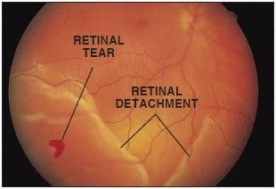
Retinal detachment occurs when the thin layer of tissue (the retina) at the back of the eye pulls away from its normal position. This separation deprives the retinal cells of oxygen and nourishment. The longer retinal detachment goes untreated, the greater the risk of permanent vision loss in the affected eye.
 Warning Signs: When to See a Doctor Immediately

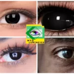
- The sudden appearance of many floaters — tiny specks that seem to drift through your field of vision.
- Photopsia: Sudden flashes of light in one or both eyes.
- Blurred vision or a gradual reduction of peripheral (side) vision.
- A curtain-like shadow over your visual field.

Advanced Treatment Options
Dr. Mustafa Mete utilizes the latest technology to repair retinal tears before they progress to full detachment:
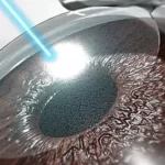
1. Laser Photocoagulation
Using a medical laser, the surgeon creates small burns around the retinal tear. The resulting scarring “welds” the retina to the underlying tissue, preventing fluid from seeping under and causing a detachment.
2. Vitrectomy & Scleral Buckling
For more advanced cases where the retina has already detached, microsurgical techniques like Vitrectomy are used to remove the vitreous gel and reattach the retina with precision.