How Does Color Vision Work? Photoreceptor Neurobiology & Retinal Processing
Color is not an inherent physical property of matter; it is a neurological construct generated by the brain. Human chromatic perception relies on a specialized neurosensory cascade initiating in retinal cone opsins, processing through retinal ganglion cell receptive fields, and decoding in the visual cortex.
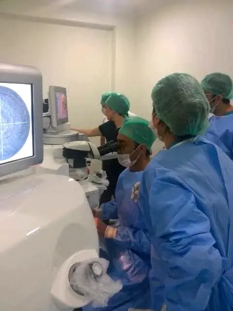
Electromagnetic radiation spans an immense spectrum, from gamma rays to long radio waves. The human eye detects only a tiny sliver: the visible light spectrum, extending from roughly 380 to 750 nanometers. When these photons cross the optical media—the cornea, anterior chamber, crystalline lens, and vitreous humor—they focus onto the sensory neuroretina, triggering an intricate biochemical cascade that translates electromagnetic wavelengths into conscious chromatic perception.
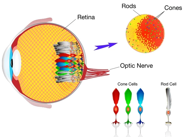
The visual cascade: Light absorption by retinal photopigments, transducin activation, and signal processing via the optic nerve to the primary visual cortex (V1).
The Photoreceptor Mosaic: Rods vs. Cones
The human retina houses two distinct classes of photoreceptor cells, each engineered for separate physiological luminance conditions:
- Rods (Scotopic System): Approximately 110 to 125 million cells densely packed across the peripheral and mid-peripheral retina. Rods contain a single photopigment, rhodopsin, which peaks in sensitivity around 498 nm. They are sensitive to single photons, providing high-contrast night vision and motion tracking. Because all rods share identical spectral absorption, they cannot distinguish different wavelengths; rod vision is entirely achromatic (monochrome).
- Cones (Photopic System): Roughly 6 to 7 million cells concentrated almost exclusively in the central macula, reaching peak density at the fovea centralis (specifically the 0.35 mm avascular foveola). Cones require high photon flux to activate, mediating daylight vision, maximum spatial resolution, and the entirety of human color perception.
The Trichromatic Foundation: Cone Opsin Biophysics
Normal human color vision is trichromatic, operating on principles first identified by Thomas Young and Hermann von Helmholtz. The retina deploys three distinct populations of cone photoreceptors, each expressing a specific transmembrane G-protein coupled receptor (opsin) bound to a light-sensitive chromophore (11-cis-retinal):
| Cone Classification | Primary Photopigment | Peak Spectral Sensitivity | Genetic Locus |
|---|---|---|---|
| S-Cones (Short Wavelength) | Cyanolabe | ~420 – 440 nm (Violet / Blue) | Chromosome 7 (Autosomal - OPN1SW) |
| M-Cones (Medium Wavelength) | Chlorolabe | ~530 – 535 nm (Green) | Chromosome X (X-linked - OPN1MW) |
| L-Cones (Long Wavelength) | Erythrolabe | ~560 – 565 nm (Yellow / Red) | Chromosome X (X-linked - OPN1LW) |
When photons hit the cone outer segments, 11-cis-retinal undergoes immediate photo-isomerization into all-trans-retinal. This activates the G-protein transducin, stimulating phosphodiesterase (PDE6) to hydrolyze cyclic guanosine monophosphate (cGMP). The closure of cGMP-gated cation channels hyperpolarizes the photoreceptor cell membrane, reducing glutamate release onto bipolar cells.
The brain does not read a single wavelength as an absolute color. Instead, it reads the relative ratio of activation across all three cone populations simultaneously. For instance, monochromatic light at 580 nm activates L-cones and M-cones in a specific overlapping ratio while leaving S-cones largely inactive; the central visual cortex calculates this comparative mathematical ratio and projects the conscious perception of "yellow."
The Opponent-Process Theory: How Retinal Signals are Encoded
While trichromatic theory explains light absorption at the photoreceptor level, Ewald Hering's Opponent-Process Theory explains how neural circuitry processes that information downstream.
Before leaving the retina, electrical signals from cones pass through horizontal, bipolar, and amacrine cells into specialized retinal ganglion cells (RGCs). These cells organize incoming signals into three antagonistic, mutually exclusive neurological channels:
- The Red-Green Channel (Parvocellular Pathway): Midget ganglion cells subtract L-cone input from M-cone input ($L - M$). A cell excited by red wavelengths is inhibited by green wavelengths. This explains why the human brain cannot perceive a "reddish-green."
- The Blue-Yellow Channel (Koniocellular Pathway): Small bistratified ganglion cells compare S-cone excitation against combined L- and M-cone signals ($S - [L + M]$).
- The Achromatic / Luminance Channel (Magnocellular Pathway): Sums L- and M-cone inputs ($L + M$) to measure absolute brightness and spatial luminance contrast, processing high-speed movement without chromatic specificity.
These processed signals travel down the optic nerve, cross partially at the optic chiasm, synapse within specific retinotopic laminae of the Lateral Geniculate Nucleus (LGN) in the thalamus, and terminate in the primary visual cortex (striate cortex / Area V1). From V1, signals pass along the ventral stream to Area V4, the cortical center responsible for complex color constancy, chromatic integration, and cognitive object recognition.
Color Vision Deficiencies (Dyschromatopsia)
When genetic mutations or acquired diseases disrupt cone photopigments, color perception degrades. These conditions fall into two major categories:
1. Congenital (Hereditary) Deficiencies
Hereditary dyschromatopsia affects approximately 8% of biological males and 0.5% of females worldwide. Because the genes encoding L-cone (OPN1LW) and M-cone (OPN1MW) photopigments reside adjacently on the X chromosome (Xq28), unequal homologous recombination frequently leads to gene deletion or hybrid gene formation:
- Anomalous Trichromacy (Altered Sensitivity): All three cones exist, but one photopigment is spectrally shifted. Includes Deuteranomaly (shifted green sensitivity, the most common form) and Protanomaly (shifted red sensitivity).
- Dichromacy (Complete Absence): Only two functional cone classes exist. Includes Deuteranopia (complete absence of green M-cones), Protanopia (complete absence of red L-cones), and Tritanopia (absence of blue S-cones, a rare autosomal condition).
- Achromatopsia (Rod Monochromacy): A severe autosomal recessive condition characterized by complete functional failure of all three cone classes, leaving the patient with total color blindness, daytime photophobia, nystagmus, and visual acuity limited to legal blindness (20/200).
2. Acquired Color Deficiencies
Unlike stable congenital conditions, acquired dyschromatopsia indicates progressive underlying ocular or neurological disease:
- Köllner’s Rule: Outer retinal diseases (such as age-related macular degeneration or diabetic macular edema) characteristically impair the blue-yellow (tritan) axis early. In contrast, inner retinal and optic nerve disorders (such as optic neuritis, open-angle glaucoma, or compressive chiasmal lesions) predominantly produce severe red-green dyschromatopsia.
- Toxic Neuropathies: Systemic pharmaceuticals, including ethambutol (tuberculosis therapy), chloroquine/hydroxychloroquine, and digitalis toxicity, produce early, quantifiable red-green or yellow-blue desaturation prior to overt structural visual field loss.
Clinical Diagnosis of Color Vision in Ophthalmology
Accurate clinical evaluation of color vision utilizes standardized diagnostic instruments:
- Ishihara Pseudoisochromatic Plates: The international gold standard for rapid screening of congenital red-green deficiencies. Matrices of dots varying in luminance and chromaticity isolate numbers invisible to individuals with protan or deutan deficits.
- Hardy-Rand-Rittler (HRR) Plates: Advanced screening plates capable of isolating and grading the severity of red-green as well as blue-yellow (tritan) deficiencies.
- Farnsworth-Munsell 100-Hue Test: A quantitative clinical examination requiring the patient to arrange 85 colored caps in continuous chromatic sequence. It graphs precise error axes, identifying subtle acquired maculopathies and toxic optic neuropathies.
- Nagel Anomaloscope: The definitive instrument for distinguishing complete dichromacy from anomalous trichromacy via Rayleigh color-matching equations.
A frequent point of confusion among patients is linking retinal color perception to exterior eye color. They are entirely separate anatomical entities:
Color Vision is mediated strictly by photoreceptors and neural pathways in the posterior segment (retina, optic nerve, visual cortex).
Eye Color is an anterior segment optical trait determined by the distribution of eumelanin, pheomelanin, and structural Rayleigh scattering within the iris stroma. Having blue, green, hazel, or dark brown irises has zero effect on the neurobiology of your retinal cones or your diagnostic color vision. To explore the genetic loci and inheritance patterns of iris pigmentation, review our clinical Eye Color Chart & Genetics Guide.
Cosmetic Procedures & Visual Pathway Safety
Patients seeking elective aesthetic changes frequently ask whether altering iris color can disrupt retinal phototransduction or induce color blindness:
The physiological reality is clear: Selective anterior segment iris depigmentation does not affect retinal cones, the optic nerve, or color perception.
Techniques like the non-invasive MyLumineyes® laser eye color change procedure deploy low-fluence, selective wavelengths targeted exclusively at superficial melanin granules situated on the anterior surface of the iris. The optical beam does not penetrate through the iris diaphragm, cannot pass into the posterior chamber, and never reaches the macula or peripheral photoreceptor matrix.
Conversely, invasive surgical interventions—such as artificial silicone iris implants—pose devastating threats to intraocular structures, inducing chronic anterior uveitis, hyphema, and intractable secondary glaucoma that can destroy retinal ganglion cells and cause irreversible visual field constriction. For patients researching options, reviewing the clinical milestones behind safe, non-surgical permanent eye color change is essential to protect long-term visual health.
Frequently Asked Questions: Human Color Vision
Why are males significantly more prone to red-green color blindness?
The genes encoding L-cone (red) and M-cone (green) photopigments are located adjacently on the X chromosome (Xq28). Because biological males possess only one X chromosome (XY), inheriting a single defective opsin gene results in phenotypic color deficiency. Females have two X chromosomes (XX), meaning a normal opsin gene on one chromosome typically compensates for a defective gene on the other.
Does having light-colored eyes (blue or green) change how you see colors?
No. Natural eye color is dictated by melanin density and Rayleigh scattering in the iris, which acts merely as an optical aperture. Color vision occurs entirely in the sensory retina and brain via cone photoreceptors. Individuals with light-colored eyes perceive colors with the exact same spectral sensitivity as individuals with dark brown eyes, though they may experience higher sensitivity to bright light (photophobia).
Can color blindness be acquired later in life?
Yes. While most cases are inherited and present from birth, acquired dyschromatopsia can develop secondary to optic nerve inflammation (optic neuritis), open-angle glaucoma, age-related macular degeneration, diabetic retinopathy, traumatic brain injury, or chronic pharmaceutical toxicity (such as ethambutol or plaquenil).
Can color blindness be cured with special corrective glasses?
No. Color blindness cannot be cured optically. Commercial "color-correcting" glasses utilize optical notch filters that selectively block narrow bands of overlapping wavelengths between red and green. While this enhances contrast between certain confusing shades, it does not restore missing cone photopigments or provide true physiological trichromacy.
Medical Disclaimer: This clinical guide is authored by Dr. Mustafa Mete for educational purposes and does not replace a comprehensive ophthalmic evaluation. If you or your child experience sudden difficulty distinguishing colors, reduced visual acuity, or persistent light sensitivity, schedule an examination with an eye care specialist immediately.