What is the Iris Stroma? Anatomy, Histology, and Eye Color Science
The iris stroma is the highly vascularized, fibrocellular anterior tissue that forms much of the structural framework of the iris. It contains collagen, blood vessels, nerve fibers, fibroblasts, melanocytes and other resident cells. Its pigmentation and microarchitecture strongly influence how light is absorbed and scattered, making the stroma a major biological determinant of visible human eye color.
The iris stroma is the thick, highly vascularized and fibrocellular anterior layer that defines much of the structural framework of the iris. Histologically positioned beneath the anterior border region and anterior to the posterior pigment epithelium, the stroma is a complex extracellular and cellular matrix.
It comprises a delicate network of loosely arranged collagen fibrils, continuous microvascular channels, nerve fibers, fibroblasts and specialized pigment-containing cells known as melanocytes. From an optical standpoint, the arrangement and physical density of this stromal matrix influence how photons entering the anterior eye are absorbed, transmitted and backscattered toward the observer.
If you first want to understand the complete iris rather than the stromal layer alone, see our patient guide to human iris anatomy, function, pigmentation and eye color .
Human eye color is strongly influenced by the concentration, density and spatial distribution of biological melanin within the anterior iris, together with the optical properties of the stromal tissue.
In an iris with high stromal melanin density, a greater proportion of incoming visible light is absorbed, producing a darker brown appearance. Conversely, when anterior stromal melanin density is lower, light interacts more strongly with the physical microstructure of the tissue, allowing structural optical effects to contribute more visibly to lighter ocular phenotypes.
The Multifaceted Physiological Functions of the Iris Stroma
While the iris stroma is widely recognized for its contribution to ocular appearance and identity, its biological functions extend far beyond pigmentation. Within the anterior eye, the stroma serves as a multifunctional structural and cellular environment.
Vascular and Neural Conduit
The stroma contains the major and minor vascular networks of the iris together with neural elements. These structures provide metabolic support and participate in sensory and autonomic iris function.
Dynamic Cellular Environment
Fibroblasts, melanocytes, macrophages, mast cells and other resident cells make the iris stroma a biologically active tissue rather than an inert colored surface.
Mechanical Pupillary Support
The stromal architecture is anatomically integrated with the structures involved in pupillary constriction and dilation, allowing the iris to function as a dynamic optical diaphragm.
Iris Stroma: Key Structures and Their Roles
| Stromal component | Biological role | Relevance to visible iris appearance |
|---|---|---|
| Collagen matrix | Provides structural organization and supports stromal cells and vessels | Contributes to tissue optical scattering, particularly in lighter irides |
| Melanocytes | Contain and distribute melanin within the anterior iris | Greater pigment density generally produces darker visible phenotypes |
| Blood vessels | Support living iris tissue metabolically | Do not normally determine natural eye color directly |
| Nerve fibers | Participate in sensory and autonomic iris function | Support dynamic iris behavior rather than pigmentation itself |
| Stromal architecture | Creates crypts, fibers and characteristic iris surface patterns | Contributes to the unique optical phenotype of each iris |
How Stromal Microstructure Influences Eye Color: The Physics of Structural Coloration
A common misconception in ocular biology is that blue, gray or green eyes contain distinct matching blue, gray or green pigments. In reality, visible human iris color results from an interaction between biological pigmentation and the optical properties of iris tissue.
In lighter irides, reduced anterior melanin absorption allows wavelength-dependent scattering within the stromal matrix to contribute more strongly to visible color. These optical effects are commonly described using principles of structural coloration, including Rayleigh-like and other scattering behavior within biological tissue.
High Melanin Concentration — Brown Phenotypes
When the anterior iris and stroma contain relatively high concentrations of melanin, more visible light is absorbed. Because less light is returned toward the observer, the iris appears brown or very dark.
Moderate or Heterogeneous Pigmentation — Green and Hazel Phenotypes
Green and hazel appearances generally result from intermediate or spatially heterogeneous pigmentation combined with stromal light scattering. They should not be interpreted as evidence of a distinct green pigment within the iris.
Learn more about green eyes .
Pathological Shifts and Genetic Stability of the Ocular Stroma
The baseline pattern of iris pigmentation is strongly influenced by an individual's genetic architecture. Variants involving the OCA2 and HERC2 regions are important contributors, although human eye color is polygenic and cannot be explained by these genes alone.
Once iris pigmentation matures during childhood, normal adult iris color is generally relatively stable. However, several ocular conditions, injuries and medications can alter the macroscopic appearance or pigment distribution of the iris.
- Chronic anterior uveitis may affect iris architecture and pigmentation.
- Fuchs heterochromic iridocyclitis can produce progressive stromal changes and heterochromia.
- Physical ocular trauma can disrupt normal iris structure or pigment distribution.
- Topical prostaglandin analogues used in glaucoma management may increase iris pigmentation in susceptible eyes.
These acquired changes should be distinguished from ordinary inherited differences in healthy iris pigmentation. For a broader explanation of inheritance and pigmentation, read our eye color genetics guide . Clinicians and patients may also consult MedlinePlus Genetics for general information about genetic variation.
Can the Iris Stroma Be Safely Modified to Induce Eye Color Change?
With growing interest in cosmetic eye color modification, many individuals seek methods to lighten their eye color . There is currently no established over-the-counter topical drop, nutritional regimen or lifestyle method that has been shown to predictably and selectively reduce stromal melanin to produce controlled permanent iris depigmentation.
Claims that a consumer product can penetrate the anterior eye and safely alter iris pigmentation should therefore be treated cautiously. The iris is living intraocular tissue, and interventions affecting pigment must be considered in the context of anterior-segment anatomy, inflammation, intraocular pressure and individual ocular characteristics.
Any medically supervised approach aimed at changing iris pigmentation must respect the anatomy and physiology of the iris rather than treating it as a simple cosmetic surface.
Laser-based approaches differ fundamentally from colored implants or corneal pigmentation because they are intended to interact with pre-existing iris pigment rather than introduce an artificial colored material into or in front of the visual axis.
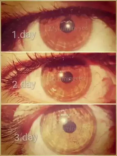
Within the Lumineyes™ clinical framework, treatment is conceptualized as a response-guided staged process directed toward accessible anterior stromal melanin. Individual biological response varies, and therefore treatment strategy should not be reduced to pursuit of a predetermined cosmetic shade or a fixed number of exposures.
For a broader discussion of clinical assessment, limitations, monitoring and the staged approach, read the main guide to Laser Eye Color Change .
The interaction between optical wavelength, chromophores and ophthalmic laser systems is discussed separately in our ophthalmic lasers guide .
Conclusion: The Structural Foundation of Visual Identity
The iris stroma is a sophisticated, multidimensional fibrovascular matrix that forms a major part of the biological foundation of iris appearance, pupillary architecture and anterior vascular organization.
Its collagenous framework, vascular channels, neural elements, melanocytes and other resident cells explain why visible iris color cannot be reduced to a simple pigment label. Human eye color reflects both biological pigmentation and the optical behavior of living tissue.
Understanding the stromal structure is therefore central not only to the study of normal iris pigmentation but also to any medically supervised discussion of changing visible iris pigmentation. The biological target is living ocular tissue with individual anatomy and individual response—not an inert colored surface.