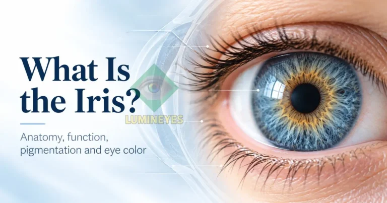
What Is the Iris? Anatomy, Function, Pigmentation & Eye Color
Most people first recognize the iris simply as the colored part of the eye. Anatomically, however, it is much more than a colored ring. The iris is a thin, contractile and highly organized part of the anterior eye. It participates in the control of incoming light, contains vascular, neural, muscular and pigmented structures, and creates much of the visible pattern that makes each iris appear different.
Understanding the iris also helps explain why eye color cannot be reduced to simple labels such as brown, green or blue. What we perceive as color reflects the interaction between pigmentation and the physical architecture of the tissue.
Where Is the Iris Located?
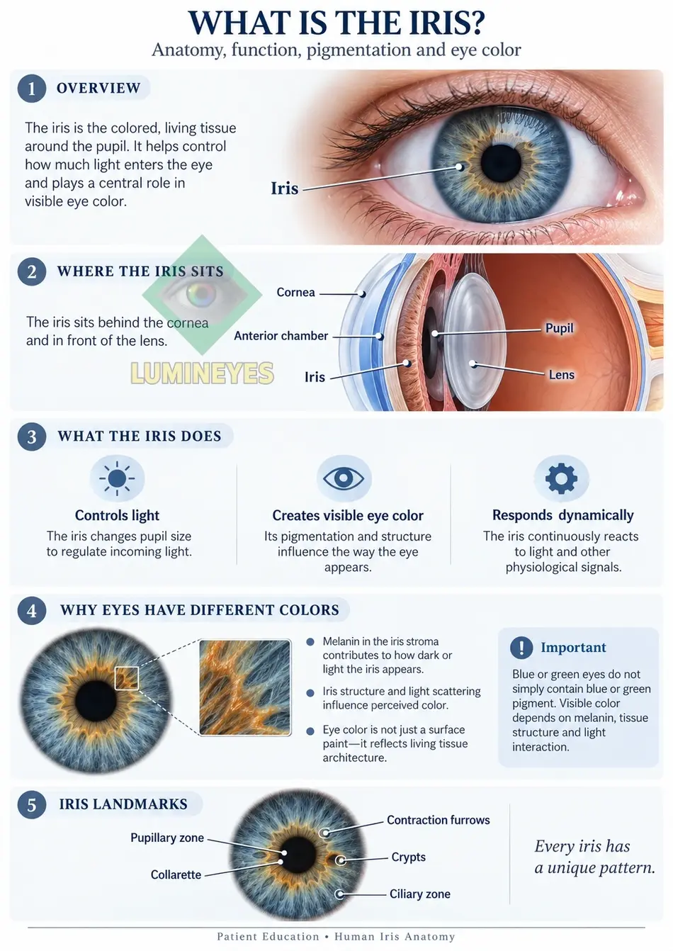
The iris forms part of the anterior segment of the eye. It lies behind the transparent cornea and in front of the crystalline lens. The circular opening at its center is the pupil.
This distinction is important because the iris, pupil, cornea and lens are often confused in everyday descriptions even though they are anatomically very different structures.
| Structure | What it is | Main role | Relationship to eye color |
|---|---|---|---|
| Iris | Living, pigmented and contractile tissue surrounding the pupil | Controls pupil diameter and regulates incoming light | Its pigmentation and stromal architecture largely determine visible iris color |
| Pupil | An opening in the center of the iris, not a separate tissue | Allows light to enter the eye | Does not create eye color; it usually appears dark because light enters through it |
| Cornea | Transparent front surface of the eye | Protects the eye and provides much of its focusing power | Normally transparent and does not determine natural iris pigmentation |
| Lens | Transparent optical structure behind the iris | Fine-focuses light onto the retina | Does not determine natural iris color |
What Does the Iris Do?
Regulates Light
The iris changes the diameter of the pupil. In bright conditions the pupil becomes smaller, while in darker conditions it enlarges, helping regulate the amount of light entering the eye.
Creates the Visible Iris Pattern
Pigment distribution, stromal fibers, crypts, furrows and other structural features contribute to the complex appearance of each individual iris.
Responds Dynamically
The iris is not a static colored surface. Its muscular and neural components respond continuously to illumination and physiological signals.
The Main Anatomical Components of the Iris
The iris is composed of several closely related regions rather than a single layer. For patient education, four components are especially useful for understanding its appearance and function.
Why Do Human Irises Have Different Colors?
Human eye color is not produced by a simple layer of colored “paint.” The visible appearance of the iris results primarily from the amount and distribution of melanin, the structure of the iris stroma and the way light is absorbed and scattered within the tissue.
Eyes with greater anterior iris pigmentation generally appear darker because more incoming light is absorbed. In lighter irides, reduced anterior stromal pigmentation allows the optical properties of the stromal tissue and wavelength-dependent light scattering to make a larger contribution to the visible appearance.
For a focused explanation of the pigment itself, see what melanin is and how it affects eye color .
Brown, Hazel, Green, Grey and Blue Eyes: What Actually Changes?
Everyday eye-color names are useful descriptions, but they simplify a continuous biological spectrum. Two people who both describe their eyes as “hazel,” for example, may have very different distributions of brown pigment, green-appearing zones, central rings and stromal patterns.
If you want to identify your own dominant iris color, secondary tones and mixed patterns, use our patient guide: What Is My Eye Color?
For a broader explanation of inheritance, pigmentation and natural eye-color categories, see the Eye Color Chart & Genetics Guide .
Why Does Every Iris Look Different?
A close-up iris is rarely uniform. Visible features may include radial stromal fibers, crypts, a collarette, contraction furrows, pigment spots and differences between the pupillary and ciliary zones. The distribution and prominence of these features vary from person to person.
This is one reason clinical iris assessment should not rely on a single color word. Baseline pigmentation may be described more systematically using structured classification. The MyLumineyes Research Library includes the proposed Lumineyes™ Iris Pigmentation Scale (LIPS) , which separates diffuse pigmentation grades from spatially heterogeneous patterns.
Eye Color Is an Optical Phenotype, Not Just a Color Name
What an observer calls “eye color” is the visible endpoint of several interacting variables. A useful way to understand this is:
This explains why the same iris may look somewhat different under daylight, indoor lighting, flash photography or changes in pupil diameter without its biological pigmentation having actually changed.
Can Iris Color Change Naturally?
The apparent color of an iris can vary with illumination, pupil size, photography, surrounding colors and optical reflections. These changes in appearance should be distinguished from a true biological alteration of iris pigmentation.
Pigmentation can also evolve during early childhood, and certain ocular conditions, inflammation, trauma or medications may produce real acquired changes in iris appearance.
A new, persistent or unexplained color change—particularly when it affects only one eye or is accompanied by pain, redness, visual symptoms, abnormal pupil shape or inflammation—should not simply be assumed to be cosmetic. An ophthalmic examination is appropriate.
Why Iris Anatomy Matters in Eye Color Change
Any discussion of permanent eye-color modification should begin with anatomy rather than with a desired shade. The iris is living ocular tissue, and its pigmentation, architecture, vascularity and biological response differ between individuals.
Within the Lumineyes™ clinical framework, the term selective stromal melanin modulation describes the intended preferential interaction with accessible pre-existing stromal melanin while seeking to minimize unnecessary interaction with surrounding iris structures. It is a proposed clinical and procedural concept rather than a claim of complete histological selectivity.
Patients who want to understand the procedure itself can continue to the main Laser Eye Color Change clinical guide , which explains the physician-guided, staged approach separately from this anatomy page.
Frequently Asked Questions About the Iris
What is the iris?
The iris is the colored, circular tissue surrounding the pupil. It lies behind the cornea and in front of the lens and contains muscular, vascular, neural and pigmented structures.
Is the iris the same thing as the pupil?
No. The iris is living tissue. The pupil is the opening in the center of the iris through which light enters the eye.
What controls pupil size?
Muscular components of the iris constrict or dilate the pupil in response to illumination and neural signals.
What part of the iris determines eye color?
Visible eye color is strongly influenced by melanin amount and distribution in the anterior iris, particularly within the stromal region, together with stromal architecture and light scattering.
Do blue eyes contain blue pigment?
Blue-looking eyes do not require a layer of blue pigment. Their appearance is associated with relatively low anterior iris pigmentation and the way light interacts with and scatters through iris tissue.
Why can the same eye look different in different photographs?
Illumination, camera exposure, white balance, reflections and pupil diameter can all alter perceived eye color without changing the underlying biological pigmentation.
Is a sudden change in iris color normal?
A persistent new or asymmetric iris color change can have several causes and should not automatically be considered cosmetic. Ophthalmic evaluation is advisable, particularly if other ocular symptoms are present.