Eye Floaters: Causes, Flashes, Risks and Treatment Options
Eye floaters are small dots, threads, rings, shadows, or cobweb-like shapes that appear to drift through the field of vision. Most originate from structures within the vitreous gel and are harmless. However, a sudden shower of new floaters—particularly when accompanied by flashes of light, peripheral shadow, or loss of vision—can be a warning sign of vitreoretinal traction, a retinal tear, or retinal detachment.
Understanding the difference between a stable vitreous floater and a new retinal warning sign is more important than simply deciding whether a floater should be treated. The first clinical question is therefore not “How can the floater be removed?” but “Why has it appeared, and is the retina safe?”

Eye Floaters at a Glance
- Most floaters are vitreous in origin and become more common as the vitreous changes with age.
- Posterior vitreous detachment (PVD) is a frequent cause of newly noticed floaters and flashes.
- Stable floaters without other symptoms are often benign and may become less noticeable with time.
- Sudden new floaters, flashes, a curtain or shadow, or a sudden change in vision require prompt ophthalmic assessment.
- Treatment is not automatically required simply because a floater is visible.
- Observation, laser vitreolysis, and vitrectomy have very different indications and risk profiles.
What Are Eye Floaters?
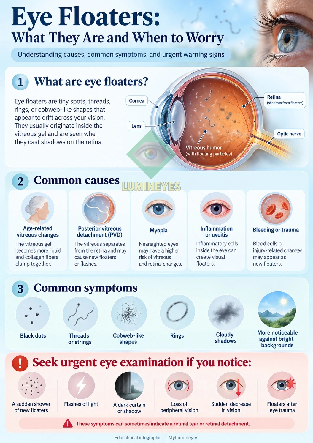
Eye floaters, medically described as vitreous floaters, vitreous opacities, or myodesopsia, are visual phenomena produced by structures suspended within the vitreous—the transparent gel occupying most of the interior of the eye between the lens and retina.
Patients commonly describe floaters as black or gray dots, hair-like strands, transparent threads, rings, clouds, cobwebs, or small objects that seem to move when the eye moves. They are usually most noticeable against a bright and relatively uniform background such as a blue sky, white wall, illuminated screen, or sheet of paper.
Although a floater may seem to be positioned in front of the eye, the visual phenomenon is generated inside the eye. Small vitreous structures alter the passage of light and create shadows or optical disturbances that are projected onto the retina. The retina then converts that optical information into neural signals, and the brain perceives the disturbance as something floating within the visual field.
Why Do Floaters Form Inside the Vitreous?
The vitreous is not simply empty space. It is a highly hydrated transparent extracellular matrix whose organization changes throughout life. In youth it is relatively homogeneous and gel-like. With aging, portions of the vitreous gradually liquefy while collagen-containing structures become less uniformly distributed.
When these microscopic structures aggregate or become optically visible, they can cast shadows on the retina. What the patient experiences is therefore not necessarily the physical object itself, but the optical consequence of that structure interacting with incoming light.
This distinction also explains why floater visibility can change dramatically with illumination, pupil size, contrast, eye movement, floater position, and the distance between an opacity and the retina.
What Causes Eye Floaters?
Eye floaters are a symptom rather than a single disease. Their clinical significance depends on why they developed, whether they are stable or new, and whether associated retinal symptoms are present.
1. Age-Related Vitreous Changes
The most common explanation is gradual age-related change within the vitreous. Liquefaction and reorganization of its internal structure can make collagen-containing aggregates increasingly visible.
These floaters often remain stable or gradually become less intrusive as their position changes and the visual system becomes less attentive to them.
2. Posterior Vitreous Detachment (PVD)
A posterior vitreous detachment occurs when the posterior vitreous separates from the retinal surface. PVD becomes increasingly common with age and is one of the major reasons a person may suddenly notice a new floater, ring, cloud, or combination of floaters and flashes.
PVD itself is often uncomplicated. The clinically important issue is what happens during the separation process. If the vitreous remains strongly adherent to a focal area of retina, vitreous movement can exert traction on retinal tissue.
That traction can occasionally produce a retinal tear or retinal detachment. This is why sudden-onset floaters are approached differently from long-standing stable floaters.
3. Myopia
People with myopia, particularly higher degrees of nearsightedness, may develop vitreous changes earlier and may also have retinal characteristics that increase the importance of careful peripheral retinal examination when acute symptoms develop.
4. Intraocular Inflammation
Inflammatory cells or inflammatory debris within the vitreous can be perceived as floaters. Uveitis therefore belongs to a different clinical category from uncomplicated age-related vitreous opacity and may require investigation and treatment of the underlying inflammatory process.
5. Vitreous Hemorrhage
Blood within the vitreous may appear as dots, clouds, haze, cobwebs, or a sudden shower of dark particles. Possible causes include retinal tears, retinal vascular disease, proliferative diabetic retinopathy, trauma, and other retinal disorders.
A sudden dense onset of dark floaters should therefore not automatically be assumed to represent ordinary aging.
6. Eye Trauma
Blunt or penetrating ocular trauma can alter the vitreous, cause intraocular bleeding, or damage the retina. New floaters following trauma deserve ophthalmic examination, particularly when accompanied by flashes, pain, reduced vision, or field loss.
Eye Floaters and Flashes: Why the Combination Matters
Floaters and flashes arise through different mechanisms. A floater is usually an optical phenomenon caused by material within the vitreous. A flash—also called a photopsia—can occur when mechanical traction stimulates the retina.
Patients may describe flashes as lightning streaks, brief arcs, sparks, camera-like flashes, or flickering light, often toward the peripheral visual field.
The combination of new floaters plus new flashes is clinically important because it may occur during acute vitreous separation and vitreoretinal traction.
When Should New Floaters Be Treated as an Emergency Warning?
Prompt ophthalmic assessment is appropriate when any of the following develops:
- a sudden shower or marked increase in floaters;
- new flashes of light;
- a dark shadow or curtain entering the visual field;
- new peripheral visual-field loss;
- a sudden reduction in visual acuity;
- new floaters following significant eye trauma.
The American Academy of Ophthalmology identifies sudden flashes, numerous new floaters, and a peripheral shadow as warning symptoms that warrant immediate ophthalmic evaluation because they can occur with retinal tears or retinal detachment.
A retinal detachment can threaten permanent vision because detached retinal tissue can no longer function normally. Information about the relationship between retinal disease, macular involvement, and vision can also be found in our retina and macula guide.
Posterior Vitreous Detachment: Common Does Not Mean Ignorable
PVD is a useful example of why symptom interpretation matters. Posterior vitreous separation is common and frequently benign, yet the same process can occasionally place enough traction on the retina to produce a tear.
The clinically important sequence is therefore:
- The vitreous undergoes liquefaction and structural change.
- The posterior vitreous begins separating from the retinal surface.
- A patient may notice a new ring, strands, dots, or other floaters.
- Retinal traction may generate flashes.
- If focal traction is sufficiently strong, a retinal tear may occur.
- Fluid passing through a retinal break can lead to rhegmatogenous retinal detachment.
This does not mean that every new floater represents a retinal tear. It means that symptoms alone cannot reliably exclude one.
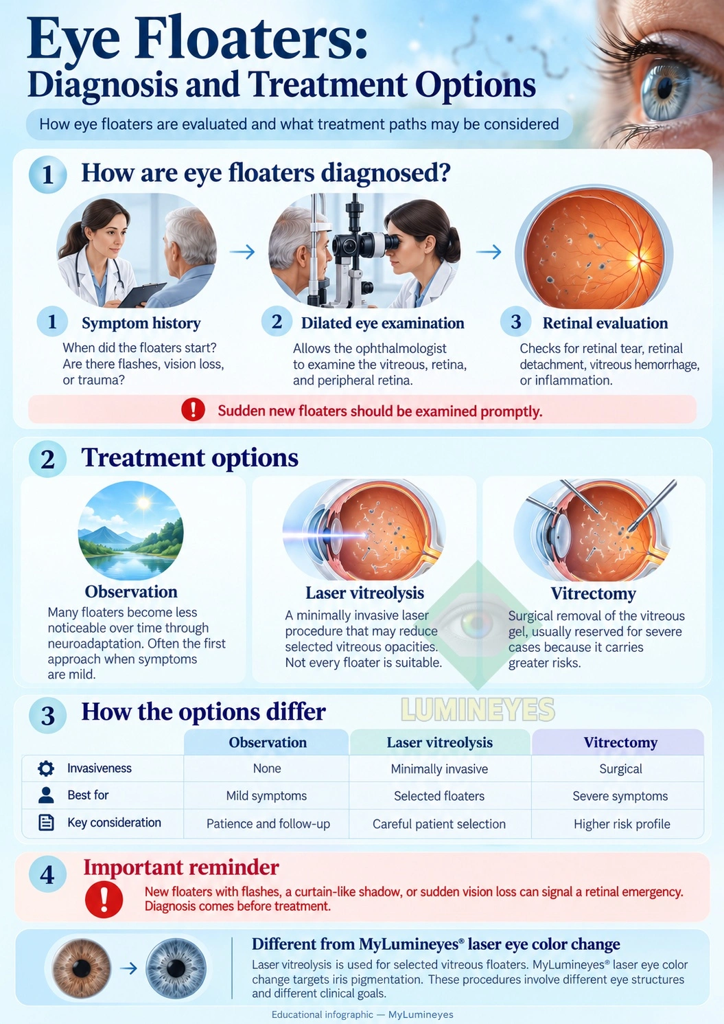
How Are Eye Floaters Diagnosed?
The purpose of an examination is not simply to confirm that a patient sees floaters. The important task is to determine their probable source and identify—or exclude—clinically significant vitreoretinal pathology.
Symptom History
The ophthalmologist may ask when the floater began, whether it appeared suddenly or gradually, whether the pattern is stable, whether flashes are present, whether peripheral vision has changed, and whether there has been recent trauma, surgery, inflammation, or other ocular disease.
Dilated Fundus Examination
Pupil dilation allows examination of the vitreous, optic nerve, posterior retina, and peripheral retina. The peripheral retinal examination is particularly important in a patient presenting with acute floaters or flashes because small retinal breaks may occur away from central vision.
Additional Testing When Clinically Indicated
Not every patient requires every diagnostic test. Depending on the clinical presentation, additional imaging or testing may be appropriate when direct visualization is limited or another retinal, inflammatory, vascular, or neurological disorder is suspected.
The investigation should therefore be problem-directed rather than test-list driven.
The Floater Decision Triangle: Symptom Burden, Retinal Safety and Intervention Risk
A useful way to think about symptomatic floaters is to separate three questions that are often incorrectly collapsed into one.
1. How Much Is the Floater Affecting Vision?
A visible floater is not automatically a disabling floater. The effect may range from occasional awareness to persistent interference with reading, screen work, driving, contrast perception, or occupational visual tasks.
2. Is the Retina Safe?
Before considering elective floater treatment, acute retinal pathology, significant vitreoretinal traction, hemorrhage, inflammation, or another underlying disorder must be considered and excluded when clinically relevant.
3. Is the Intervention Proportionate?
The potential benefit of treatment has to be weighed against the risk created by the treatment itself. A procedure should not become more consequential than the symptom it is intended to relieve.
Clinical principle: The existence of a treatable opacity does not, by itself, establish that treatment is the safest choice.
This is especially important in an otherwise healthy eye. A visually annoying but non-threatening floater and a sight-threatening retinal disorder occupy very different positions on the risk spectrum. Management should preserve that distinction.
Eye Floater Treatment Options
Treatment depends on the underlying cause, the severity and persistence of symptoms, the anatomy of the floater, retinal health, age, lens status, and the balance between expected benefit and procedural risk.
1. Observation and Neuroadaptation
For many patients with stable uncomplicated floaters, observation is the most appropriate initial strategy.
Over time, several things can occur:
- the opacity may move away from the central visual axis;
- its optical shadow may become less prominent;
- the brain may become progressively less attentive to the disturbance;
- the subjective impact on everyday vision may diminish.
Neuroadaptation does not necessarily mean that the vitreous opacity has physically disappeared. Rather, its perceptual importance can decrease.
2. Laser Vitreolysis
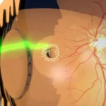
Laser vitreolysis is a non-incisional ophthalmic laser procedure in which selected vitreous opacities are targeted with laser energy. Its purpose is to reduce the visual effect of appropriately positioned and appropriately characterized floaters.
However, “floater” is not a sufficient indication for laser treatment. The morphology and position of an opacity, its relationship to the lens and retina, the underlying vitreous state, and the patient's symptoms all influence suitability.
Laser vitreolysis should therefore not be presented as a universal method for eliminating every type of floater. Patient selection is central to the risk-benefit calculation.
As with other ophthalmic laser interventions, treatment decisions require an understanding of ocular anatomy and procedure-specific risk. Our broader discussion of laser eye surgery risks explains why the term “laser” alone does not define either safety or mechanism.
3. Vitrectomy for Severe Symptomatic Floaters
Vitrectomy can remove vitreous opacities very effectively because the vitreous containing the symptomatic structures is surgically removed. From a purely mechanical perspective, this may appear to be the most definitive solution.
But effectiveness and proportionality are not the same question.
Vitrectomy is intraocular surgery. Potential complications include cataract formation or progression, retinal breaks, retinal detachment, infection, bleeding, pressure-related complications, and other surgical adverse events.
For this reason, vitrectomy for isolated floaters requires a particularly careful risk-benefit discussion. The patient may be considering surgery not to preserve an eye threatened by disease, but to improve visual quality in an eye that may otherwise have useful or excellent vision.
The Central Risk Question
When the underlying complaint is a non-sight-threatening vitreous opacity, the threshold for accepting an intraocular surgical risk should logically be different from the threshold used when surgery is required to prevent blindness.
The relevant question is therefore not simply: “Can surgery remove the floater?”
It is: “Is the patient's functional disability great enough that the expected benefit justifies exposing the eye to the risks of intraocular surgery?”
This does not mean vitrectomy is never justified for floaters. In carefully selected patients with severe, persistent, objectively and subjectively significant symptoms, the balance may favor intervention. What matters is that the decision remains individualized rather than automatic.
One recognized consequence of vitrectomy is cataract progression, particularly in phakic eyes. Patients considering intraocular surgery should understand the implications of cataract development and cataract surgery as part of informed decision-making.
Observation vs Laser Vitreolysis vs Vitrectomy
| Consideration | Observation | Laser Vitreolysis | Vitrectomy |
|---|---|---|---|
| Invasiveness | No procedure | Non-incisional laser | Intraocular surgery |
| Typical role | Stable or tolerable symptoms | Selected symptomatic opacities | Severe persistent symptoms in selected patients |
| Removes vitreous? | No | No | Yes |
| Patient selection | Clinical assessment | Critical | Critical |
| Principal advantage | Avoids procedural risk | Less invasive than intraocular surgery | Can provide substantial removal of symptomatic vitreous opacities |
| Principal limitation | Symptoms may persist | Not every floater is suitable or adequately treated | Greater intervention burden and intraocular surgical risk |
Can Eye Floaters Affect Vision Even When Visual Acuity Is Normal?
Yes. Standard high-contrast visual acuity does not capture every component of visual quality.
Some patients with prominent floaters report difficulty with reading, computer work, bright backgrounds, fine contrast, driving under particular lighting conditions, or tasks requiring sustained visual concentration despite maintaining good measured Snellen acuity.
This helps explain why the burden of floaters can be very different between patients with superficially similar eye examinations.
Conversely, subjective annoyance alone does not establish that an invasive treatment is warranted. Both functional impact and procedural risk have to be considered.
The Eye Anatomy Behind Floaters
Light enters through the cornea, passes through the pupil and crystalline lens, traverses the vitreous cavity, and reaches the retina.
The retina converts light into neural information that ultimately travels through the optic nerve to the brain. Because vitreous opacities lie directly in the optical pathway before the retina, they can influence the pattern of light reaching retinal photoreceptors even though the opacity itself is not part of the retina.
This is why vitreous disease and retinal disease can produce overlapping patient complaints while representing very different anatomical processes.
Laser Vitreolysis Is Not the Same as Laser Eye Color Change
Different ophthalmic lasers can act on entirely different tissues for entirely different clinical purposes. Grouping procedures together simply because they involve a laser is medically misleading.
Laser vitreolysis targets selected structures within the vitreous cavity.
By contrast, the MyLumineyes® laser eye color change approach concerns iris pigmentation and the anterior segment rather than vitreous floaters.
The anatomical target, clinical objective, laser-tissue interaction, treatment strategy, patient-selection criteria, monitoring requirements, and risk profile are therefore fundamentally different.
When Should You See an Ophthalmologist for Eye Floaters?
Long-standing, unchanged floaters are frequently benign. Nevertheless, an ophthalmic examination is appropriate when a floater is new, unexplained, worsening, functionally significant, or associated with other visual symptoms.
Urgent assessment is particularly important when floaters are accompanied by:
- new flashes;
- a sudden increase in the number of floaters;
- visual-field shadow or curtain;
- new peripheral vision loss;
- sudden reduction in visual acuity;
- recent ocular trauma.
If visual function has changed substantially, our overview of vision loss and warning symptoms provides additional background, but online information cannot exclude a retinal tear or detachment.
When acute retinal warning symptoms are present, the appropriate next step is an examination rather than attempting to diagnose the condition from photographs, internet descriptions, or symptom comparison alone. Patients seeking an ophthalmic assessment can review our eye examination information.
Frequently Asked Questions About Eye Floaters
Are eye floaters normal?
They are very common, particularly with age-related vitreous change. However, a newly appearing floater should not automatically be assumed to be harmless, especially when it occurs suddenly or with flashes or visual-field change.
Can eye floaters disappear?
Some become less noticeable because they shift position or because neuroadaptation reduces awareness of them. This does not necessarily mean the physical opacity has completely disappeared.
Why do I see floaters when looking at the sky?
A bright, uniform background provides high contrast, making subtle shadows and optical disturbances created by vitreous structures easier to perceive.
Are eye floaters related to retinal detachment?
Most floaters do not mean that the retina is detached. However, a sudden onset of numerous floaters—especially when combined with flashes, a peripheral shadow, or loss of vision—can occur with retinal tears or detachment and requires prompt evaluation.
What is posterior vitreous detachment?
Posterior vitreous detachment occurs when the posterior vitreous separates from the retina. It is common with aging and is often uncomplicated, but retinal traction during the separation process can occasionally cause a retinal tear.
Do all eye floaters need treatment?
No. Many stable floaters require no procedural treatment. Management depends on the underlying cause, symptom burden, retinal health, and the balance between expected benefit and treatment risk.
Can laser remove eye floaters?
Laser vitreolysis may reduce selected vitreous opacities in appropriately chosen patients. Not every floater has a morphology or location suitable for laser treatment.
Can vitrectomy remove floaters?
Vitrectomy can remove vitreous opacities by surgically removing vitreous gel, but it is intraocular surgery and carries risks. It is therefore generally considered only when symptoms are sufficiently severe to justify those risks.
Are flashes of light the same as floaters?
No. Floaters usually result from optical shadows created by vitreous structures. Flashes can result from mechanical stimulation of the retina, including vitreoretinal traction.
What symptoms suggest that a floater may be urgent?
A sudden shower of new floaters, new flashes, a curtain or shadow, loss of peripheral vision, or sudden deterioration in vision should prompt urgent ophthalmic evaluation.
The Bottom Line
Most eye floaters are not dangerous, but the symptom should always be interpreted in context.
A stable floater caused by age-related vitreous change is fundamentally different from a sudden burst of floaters produced during acute vitreoretinal traction. Likewise, a treatment capable of reducing a visual disturbance is not automatically the correct treatment if its risk exceeds the functional burden of the symptom.
The safest clinical sequence is therefore:
- identify the cause;
- exclude retinal danger;
- measure the real functional burden;
- consider whether intervention is proportionate;
- choose the least invasive reasonable strategy for the individual eye.
For floaters, as with many ophthalmic problems, diagnosis comes before treatment—and preservation of vision comes before removal of a visual nuisance.