Vision Loss: Symptoms, Causes & Treatment
Understand the warning signs of sudden or gradual sight loss, the conditions that can cause it, and when an eye examination is urgent.
Vision loss means reduced sight in one or both eyes. It can involve blurred details, central blind spots, missing side vision or complete loss of sight. Some causes, such as cataracts, are often treatable; other conditions can injure retinal or optic nerve tissue and leave lasting impairment. Sudden vision loss is a medical emergency even when it is painless or improves spontaneously.
What is vision loss?
Vision loss, sometimes called visual impairment or sight loss, describes a decline in the ability to see clearly or perceive part of the visual field. It ranges from mild, correctable blur to severe impairment or blindness. Blurred vision is a symptom; it does not by itself identify the diagnosis.
Normal vision depends on transparent optical structures, a functioning retina and optic nerve, and intact visual pathways in the brain. Disturbance at any of these levels can alter visual acuity, contrast sensitivity, color perception or visual fields. For an explanation of these structures, read our guide to how eyesight and color perception work.
Clinicians distinguish temporary versus persistent, sudden versus gradual, and one-eye versus two-eye changes. These differences help determine which tests or emergency assessments are necessary. A symptom that disappears after minutes can still indicate a vascular or neurological emergency.
Types and symptoms of vision loss
Vision loss does not always mean complete darkness. The shape, speed and location of a visual disturbance may provide clues to its cause.
Blurred or cloudy vision
Fine details lose sharpness. Common explanations include refractive error, cataract, corneal problems and retinal disease.
Central vision loss
A blurred or missing area where you look directly. Macular diseases may affect reading, face recognition and detail vision.
Peripheral vision loss
Reduced side vision or a narrowing field. Glaucoma and some retinal or neurological conditions may be involved.
Sudden dark shadow
A veil, curtain or new fixed blind area can indicate retinal detachment, vascular occlusion or another urgent problem.
Distorted vision
Straight lines appear bent or objects appear warped. New distortion warrants prompt assessment for macular disease.
Temporary episodes
Sight dims or disappears briefly and returns. Migraine is one possible cause, but transient ischemia must be considered.
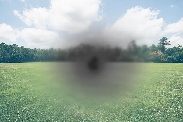
One eye or both? Vision loss affecting only one eye may arise from ocular or optic-nerve disease. Similar changes affecting both eyes can arise from bilateral eye disease or brain-based visual pathways. A person may not reliably distinguish the two without testing each eye separately; never delay emergency care to perform self-tests.
What causes vision loss?
The causes range from common optical problems to sight-threatening retinal, vascular and neurological disorders. The following distinctions explain why the correct diagnosis matters more than treating the symptom of blur alone.
Cataracts
Clouding of the natural lens can cause gradual haze, glare and duller colors. Cataract surgery often improves vision when the cataract is the main cause.
Glaucoma
Progressive optic nerve damage may begin without noticeable symptoms. Peripheral visual-field loss is common; damage already sustained is generally not reversible, but treatment can reduce further loss.
Diabetic retinopathy
Diabetes can damage retinal vessels. Leakage, swelling or abnormal new vessels may impair sight. Early detection and systemic diabetes care are important.
Age-related macular degeneration
AMD affects central vision. Distortion and loss of fine detail can occur. Management differs substantially between dry and neovascular (wet) AMD.
Retinal tears or detachment
Flashes, many new floaters, or a curtain-like shadow can signal a retinal emergency. Prompt assessment and treatment may protect remaining vision.
Retinal vascular occlusion
Blocked retinal arterial or venous circulation can cause sudden visual deterioration. Retinal artery occlusion requires an emergency stroke-oriented assessment.
Corneal or inflammatory disease
Infection, corneal edema, uveitis and other inflammatory disorders may cause blur, pain, redness or light sensitivity. Some need same-day treatment.
Stroke and neurological disease
Sudden field loss, double vision or reduced sight may relate to stroke, optic neuritis or other neurological disorders. Associated weakness or speech changes are emergency signs.
For a more detailed discussion of macular disorders, retinal tears and related treatment methods, visit our retinal and macular diseases guide.
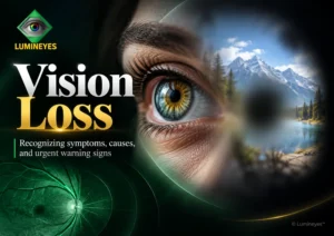
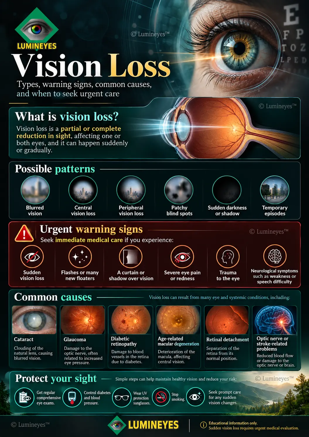
Vision loss infographic: patterns, causes and warning signs
This visual reference brings together major patterns of visual disturbance, causes of sight loss and symptoms that warrant urgent attention. Illustrations are simplified and should not be used to identify or exclude a medical diagnosis.
How do ophthalmologists diagnose vision loss?
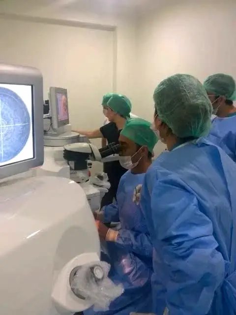
Assessment starts with symptom onset, duration, pain, trauma, medication use, vascular risk factors and whether one or both eyes are affected. The ophthalmologist then selects tests according to the suspected location and urgency of disease.
| Examination or test | What it helps evaluate |
|---|---|
| Visual acuity and refraction | Clarity of sight and whether optical correction improves it. |
| Slit-lamp examination | Cornea, anterior chamber, iris, lens, inflammation and cataract. |
| Intraocular pressure and optic nerve examination | IOP and evidence relevant to glaucoma; pressure alone cannot confirm or exclude glaucoma. |
| Dilated retinal examination | Retinal tears, detachment, hemorrhage, vascular signs and macular findings. |
| Optical coherence tomography (OCT) | Cross-sectional retinal or optic nerve structural changes. |
| Automated visual-field testing | Central and peripheral field defects when clinically appropriate. |
| Emergency vascular or neurological evaluation | Stroke, retinal arterial occlusion or other neurological emergencies when suspected. |
Not every patient needs every test. Conversely, a normal eye-pressure measurement or relatively good reading acuity does not exclude serious disease. Emergency symptoms take priority over routine office testing.
Can vision loss be treated or reversed?
Sometimes — but it depends on the cause, severity and timing. A cloudy lens can often be treated surgically, while established glaucomatous optic nerve injury is generally irreversible. Certain retinal diseases can be treated to preserve sight or recover some function, but results vary and cannot be guaranteed.
| Cause | Typical management | Important limitation |
|---|---|---|
| Cataract | Lens replacement surgery when clinically indicated | Other eye diseases may limit the visual outcome. |
| Glaucoma | Pressure-lowering medicines, laser or surgery | Aims to slow or prevent additional damage, not restore dead nerve fibers. |
| Wet AMD | Intraocular anti-VEGF therapy and follow-up | Often controls disease activity; visual recovery varies. |
| Diabetic retinopathy | Metabolic control, retinal injections and/or laser as indicated | Timely surveillance matters, sometimes before symptoms arise. |
| Retinal detachment | Urgent retinal intervention, often surgery | Outcome depends on timing and whether the macula is involved. |
| Vascular/neurological causes | Emergency cause-specific evaluation and treatment | Delays may reduce opportunities to prevent disability. |
Low-vision rehabilitation matters. When full recovery is not possible, magnification, accessibility technology, lighting, orientation training and specialized rehabilitation can improve daily functioning and independence.
How can you reduce the risk of vision loss?
Not every cause is preventable. Nevertheless, screening, timely care and management of systemic risks can prevent some vision loss or slow its progression.
Get appropriate eye exams
Follow an individualized schedule, especially with diabetes, family glaucoma history or previous retinal disease.
Manage vascular risks
Work with your clinician to control diabetes, blood pressure and cardiovascular risk factors.
Act on sudden changes
New missing vision, distortion, flashes or a curtain effect should never be dismissed as fatigue.
Avoid smoking
Smoking is associated with conditions including cataract and age-related macular degeneration.
Protect eyes from injury
Use task-appropriate protective eyewear and sunglasses with verified UV protection.
Use evidence-based care
Do not rely on supplements or home remedies to treat a retinal, optic nerve or vascular emergency.
Our companion article about eye health, vitamins, nutrition and daily care explains which preventive claims are supported by evidence and which are not.
Can cosmetic eye color change procedures affect vision?
Yes. Eye color change interventions are separate from the major natural causes of vision loss, but they introduce additional questions about ocular anatomy, inflammation and long-term visual function. Different procedures have different mechanisms and risk profiles; no elective method should be portrayed as completely risk-free.
Cosmetic iris implants
Published complications include glaucoma, chronic inflammation, corneal endothelial damage and sometimes severe sight loss. See our guide to artificial colored iris implants.
Keratopigmentation
Corneal pigmentation may affect optical quality and can create procedure-specific complications or challenges for later examinations. See keratopigmentation risks and limitations.
Laser iris depigmentation
Laser-based approaches require careful assessment of inflammation, pigment clearance, intraocular pressure and long-term outcomes. Clinical monitoring does not establish zero risk; read our laser eye color change safety guide.
For the procedure itself and how it differs from alternative approaches, see our primary laser eye color change procedure guide. This vision-loss article does not make comparative safety rankings or promise visual outcomes for elective cosmetic procedures.
Vision loss: frequently asked questions
Open a question to read a short clinical answer. These explanations cannot replace an examination.
What are the first warning signs of vision loss?
Early signs can include persistent blur, reduced contrast, trouble reading, distorted lines, missing areas of vision or increasing difficulty seeing to the side. Some diseases, including glaucoma and diabetic retinopathy, may have few noticeable early symptoms. Sudden symptoms require emergency assessment.
Is sudden vision loss in one eye an emergency?
Yes. Sudden loss of vision in one eye may reflect a retinal, optic nerve or vascular emergency, including retinal artery occlusion. Seek emergency medical evaluation immediately, even without pain and even if sight returns.
Can vision loss be temporary?
Yes, symptoms can resolve when the cause is transient. But temporary visual loss can signal a transient ischemic attack or retinal ischemia and should not be assumed harmless. Sudden temporary loss warrants immediate evaluation.
What causes a shadow or curtain over vision?
A new curtain, dark shadow or expanding missing area can occur with retinal detachment and other serious conditions. This requires emergency eye assessment, particularly when accompanied by flashes or many new floaters.
Can glaucoma cause blindness without eye pain?
Yes. Chronic glaucoma often develops without pain or early symptoms. It can progressively damage the optic nerve and reduce peripheral vision. Early diagnosis and appropriate treatment help limit further loss.
What is the difference between central and peripheral vision loss?
Central loss affects detail vision, such as reading or recognizing faces. Peripheral loss affects side vision and spatial awareness. Macular diseases commonly affect the center; glaucoma often affects the visual field. Only examination can establish the cause.
Can lost vision return after treatment?
It depends on the condition. Cataract surgery often restores clarity when the lens is the problem. Some retinal treatments preserve or improve sight. Established optic nerve damage from glaucoma typically cannot be reversed. Prompt diagnosis improves the opportunity to protect vision.
Can diabetes cause vision loss?
Yes. Diabetic retinopathy and diabetic macular edema can damage retinal tissue or impair central vision. A person can have sight-threatening changes before noticing symptoms, so recommended retinal screening is essential.
Does eye strain or screen use cause permanent blindness?
Digital screen use commonly causes eye fatigue, dryness and intermittent blur, but those symptoms are not equivalent to permanent blindness. Persistent, progressive or sudden changes require an examination rather than being attributed automatically to screens.
Can cosmetic eye color procedures damage eyesight?
They can. Reported or potential complications vary with the method and may involve corneal transparency, inflammation, eye pressure and the optic nerve. Patients considering elective procedures should review method-specific evidence, alternatives and follow-up with an ophthalmologist.
Selected medical references
- National Eye Institute — Retinal Detachment. Patient guidance on flashes, floaters, a curtain over vision and emergency assessment.
- American Stroke Association — Stroke Symptoms and Warning Signs. Guidance on sudden vision symptoms and accompanying neurological warning signs.
References support clinical context and emergency guidance; individual management must be determined by a treating clinician.
Medical disclaimer: Information is educational and does not replace clinical examination or emergency medical services. Illustrative visual-field examples are not diagnostic. If vision decreases suddenly, seek immediate emergency care.