What Is Presbyopia? Clinical Physiology, Eye Drops & Definitive Surgical Treatments
Presbyopia is the inevitable, age-related loss of the eye's dynamic focusing accommodation. Beginning around age 40, progressive sclerosis of the crystalline lens impairs near visual acuity for every human eye, requiring targeted clinical management.
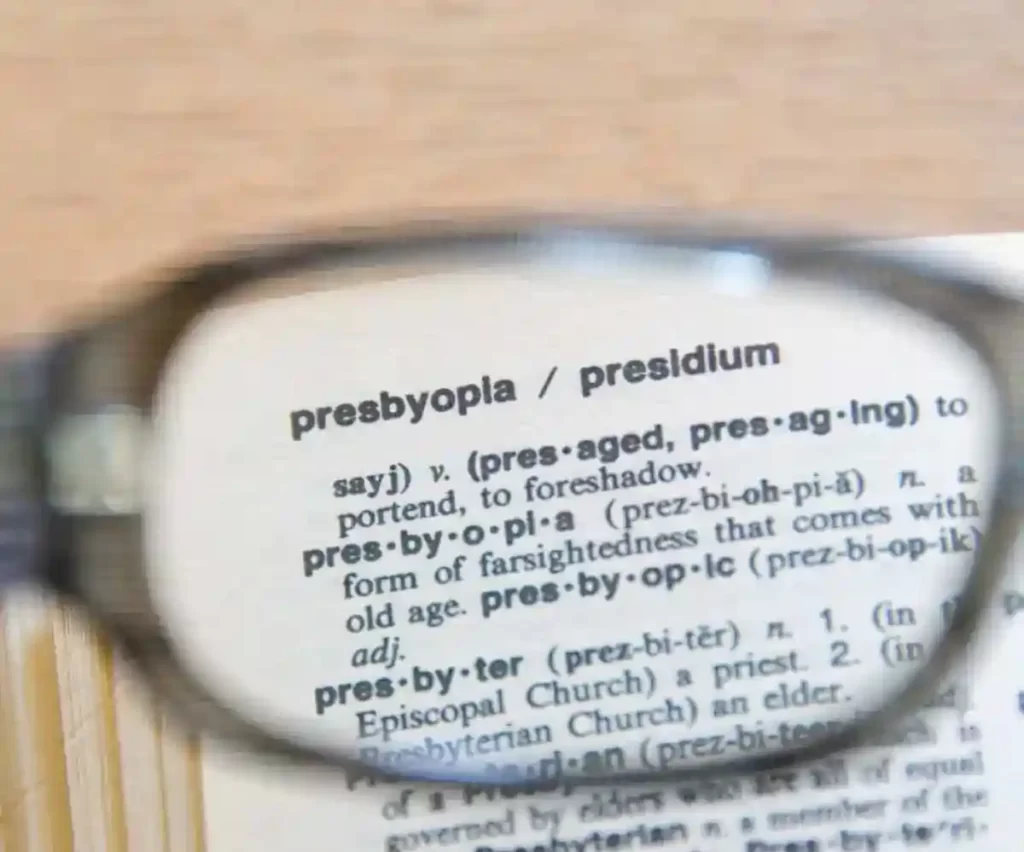
Holding reading material at arm's length, suffering from frontal headaches after computer work, and struggling with small print under low lighting are the classic signs of presbyopia. Often erroneously called "nearsightedness" in faulty automated translations, presbyopia is not myopia; it is the physiological decline of accommodation—the crystalline lens's capacity to increase its optical dioptric power to focus on close objects.
In the emmetropic human eye, clear vision requires incoming parallel light rays to focus precisely onto the photoreceptor layer of the neurosensory retina. While young eyes effortlessly morph the curvature of their natural lens via the ciliary muscle, this biological zoom lens progressively hardens over time.

The Biomechanics of Accommodation: Why Does Presbyopia Occur?
The widely accepted Helmholtz theory of accommodation explains that when an eye shifts focus from a distant horizon to a handheld screen, the circumferential ciliary muscle contracts. This contraction releases tension on the zonular fibers, allowing the young crystalline lens capsule to steepen its anterior and posterior curvature, increasing its refractive converging power.
Presbyopia develops not primarily because ciliary muscle motor power disappears, but because:
- Lens Sclerosis: The crystalline lens continues to lay down new epithelial fiber cells throughout life without shedding old ones. This causes progressive compaction, loss of water content, and nuclear sclerosis, making the lens rigid.
- Capsular Elasticity Decline: The elastic modulus of the lens capsule degrades, reducing its ability to mold the lens substance into a steepened, high-power profile.
- Zonular Geometry Shifts: Continued equatorial growth of the lens narrows the circumlental space, dampening zonular mechanical transfer efficiency.
Presbyopia vs. Hyperopia: Clarifying the Diagnostic Confusion
Because both presbyopia and hyperopia (farsightedness) manifest as difficulty seeing objects up close, they are frequently confused by patients:
- Hyperopia (Refractive Error): A static anatomical mismatch present from childhood or early adulthood where the eye’s axial length is too short, or the corneal curvature is too flat. Parallel light focuses behind the retina at rest. Young hyperopes compensate by exerting constant ciliary accommodation until their reserve is exhausted.
- Presbyopia (Physiological Aging): A universal, age-related biophysical failure of the lens. It occurs universally in emmetropes, myopes, and hyperopes alike, typically manifesting between ages 40 and 45 and stabilizing around age 60.
Presbyopia Eye Drops: Scientific Reality vs. Commercial Hype
Pharmacological management of presbyopia has gained worldwide attention, but understanding how topical drops function—and their limitations—is critical for patient safety:
1. Miotic Eye Drops (Pinhole Optics)
Topical miotics (such as pilocarpine hydrochloride 1.25%) stimulate muscarinic receptors on the pupillary sphincter muscle, inducing rapid constriction of the pupil (pinhole effect) down to approximately 1.5 to 2.0 mm. By narrowing the aperture of the optical system, these drops artificially extend the eye's depth of focus without actually restoring lens elasticity.
Clinical Limitations & Risks: Miotics are temporary (lasting 4 to 6 hours) and carry trade-offs:
- Dim-Light Vision Degradation: By cutting the light entrance area, night vision and low-light acuity drop significantly. Driving at night with miotic drops is hazardous.
- Ciliary Spasms & Headaches: Induced contraction of the ciliary body frequently causes brow ache and dull orbital pain.
- Retinal Traction Warnings: Ciliary muscle contraction exerts anterior vitreoretinal traction, posing a documented risk of peripheral retinal tears or detachments, particularly in pseudophakic or axially elongated myopic eyes.
2. Lens-Softening Agents (Biochemical Restoration)
Investigational drops formulated with lipoic acid choline ester (such as UNR844) aim to chemically reduce disulfide protein bonds within the lens fibers to restore elasticity. While biochemically promising in early-stage presbyopia, their ability to reverse established nuclear sclerosis remains modest and non-permanent.
⚠ Beware of Internet Scams: Eye Drops Cannot Change Eye Color or Rebuild Eyes
While FDA-regulated miotic drops have legitimate optical applications for near vision, the internet is filled with predatory advertisements promoting unregulated "miracle drops." As detailed in our clinical exposé on eye color changing drops, no topical drop can safely modify iris melanin or reverse biological ocular aging. Always consult a board-certified eye surgeon before applying unverified formulations.
Comprehensive Clinical Treatment Modalities
| Treatment Option | Primary Mechanism | Durability | Clinical Verdict |
|---|---|---|---|
| Reading & Progressive Glasses | External optical convergence added to focal plane | Temporary (requires updating as lens hardens) | Non-invasive, zero surgical risk; inconvenient for active lifestyles. |
| Multifocal / Monovision Contacts | Simultaneous vision optics or intentional uncorrected myopia in non-dominant eye | Daily / Monthly Wear | Compromised by dry eye syndrome and reduced tear quality after age 40. |
| Corneal Inlays (e.g., Kamra) | Micro-aperture ring implanted into deep corneal stromal pocket | Historically permanent | Largely abandoned due to high explantation rates, stromal haze, and optical decentration. |
| Presbyopic Excimer Laser (Presbyond / Supracor) | Custom corneal ablation inducing controlled multifocality or extended depth of focus | Medium to Long-term | Effective for select candidates; does not prevent future cataract development. |
| Trifocal Smart Lens Implantation (RLE) | Clear lens exchange with diffractive trifocal intraocular lens | Permanent Lifetime Cure | The gold standard. Restores distance, intermediate, and near vision while permanently preventing cataracts. |
Corneal Laser Solutions: PresbyMAX, Presbyond & SUPRACOR
For patients who wish to avoid intraocular surgery, advanced corneal laser platforms can reshape the corneal stroma to compensate for near vision loss. Using an excimer laser, specialized algorithms (such as Presbyond Laser Blended Vision or SUPRACOR) sculpt a continuous aspheric multifocal profile on the dominant and non-dominant corneas. This smooths the optical transition, allowing the brain's neuro-adaptation to blend near, intermediate, and distant images.
While effective in carefully selected patients with robust corneal thickness, laser presbyopia surgery modifies the cornea while leaving the aging, sclerotic lens in place inside the eye.
The Surgical Gold Standard: Trifocal Smart Lens Replacement
The definitive, permanent medical solution for presbyopia is Refractive Lens Exchange (RLE) with a premium trifocal intraocular lens. Because presbyopia is fundamentally a disease of the crystalline lens, treating the cornea is merely a temporary optical bypass.
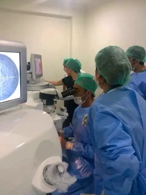
During smart lens surgery, the dysfunctional, hardened natural lens is gently extracted through a micro-incision using femtosecond laser precision and replaced with an FDA-approved trifocal optic (such as the Alcon PanOptix). As detailed in our comprehensive guide to smart lenses and trifocal intraocular lens surgery:
- Triple Focal Distribution: Light entering the eye is split across three focal points: 40 cm (reading mobile phones and books), 60 cm (intermediate computer work), and infinity (driving and distance navigation).
- Permanent Optical Prescription: Because artificial biocompatible acrylic lenses do not degrade or shift refractive index, your vision remains stable for life.
- Absolute Cataract Immunity: Once the natural crystalline lens is replaced with a smart lens, a cataract can never form in your lifetime.
For patients evaluating surgical vision correction in Turkey, transparent breakdowns of our clinical protocols, diagnostic packages, and surgical fees are detailed on our Istanbul eye surgery and pricing guide.
Frequently Asked Questions: Presbyopia
Can presbyopia eye drops permanently cure near vision loss?
No. Presbyopia eye drops work primarily by constricting your pupil (pinhole effect) to temporarily increase your depth of focus for 4 to 6 hours. They do not reverse the biological hardening (sclerosis) of your natural lens and must be applied continuously every day.
What is the difference between presbyopia and myopia?
Myopia (nearsightedness) is an anatomical condition where the eye is too long, causing distant objects to blur while close vision remains clear. Presbyopia is an age-related condition affecting everyone over 40, where the eye's natural lens loses its dynamic focusing flexibility, causing near objects to blur.
Is laser eye surgery (PresbyLASIK) or Smart Lens surgery better?
For patients over 50, Smart Lens (trifocal IOL) surgery is generally superior because it addresses the actual root problem (the aging lens), provides lifetime stability, and permanently eliminates the possibility of future cataracts. Corneal laser procedures are viable alternatives for younger patients (ages 40-48) with pristine natural lenses and clear corneas.
Will presbyopia continue to worsen after age 60?
Presbyopia typically progresses between ages 40 and 60 as the lens gradually loses its remaining accommodation. By age 60, the natural lens has virtually zero dynamic flexibility left, meaning your reading prescription plateaus and stabilizes at approximately +2.50 to +3.00 diopters.
Medical Disclaimer: This clinical guide is authored by Dr. Mustafa Mete for educational purposes. It does not replace an in-person medical evaluation. Individuals experiencing acute vision changes, severe eye strain, or sudden near vision loss should consult a qualified ophthalmologist immediately.