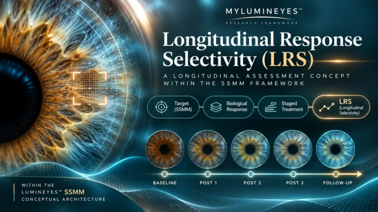
SSMM research framework · Longitudinal assessment
Longitudinal Response Selectivity (LRS)
A Temporal Assessment Concept Within the Selective Stromal Melanin Modulation Framework
The distinction between what was intended to change and what else responded becomes a longitudinal scientific question when an eye is observed across successive clinical stages.
What is LRS?
Longitudinal Response Selectivity (LRS) examines how the intended stromal pigmentary change can be distinguished from other ocular responses over repeated observations and treatment stages. It adds a time-dependent assessment layer to the Selective Stromal Melanin Modulation (SSMM) framework; it is not a new laser interaction mechanism.
The Scientific Definition
Longitudinal Response Selectivity (LRS) is a proposed concept within the SSMM framework describing the extent to which intended stromal pigment modification can be distinguished from accompanying tissue responses across successive observation periods and treatment stages.
Proposed by Mustafa Mete, MD, within the Lumineyes™ SSMM framework (2026).
In this definition, selectivity refers to the scientific distinguishability of response domains across time. It does not require the biological pathways to be independent, nor does it imply that an accompanying response is absent. The original laser–pigment interaction and its later biological consequences must be studied as related but non-identical events.
LRS provides a research construct for converting a broad observation—“the eye changes over time”—into a specific, measurable question: which change belongs to the intended pigmentary endpoint, which belongs to accompanying tissue response, and with what degree of uncertainty can they be differentiated?
Why a Longitudinal Concept Is Needed
Human iris colour is associated with anterior stromal melanin, melanosomal characteristics and the optical effects of stromal architecture. Human histological work found that gross iris colour differences do not simply track the number of stromal melanocytes [1,2]. The classical framework of selective photothermolysis explains how optical absorption and pulse structure can favour a target chromophore [3]. These foundations address the initial target interaction; they do not by themselves identify the subsequent trajectories of optical change, inflammation, pigment processing and tissue recovery.
Direct experimental iris-laser research has documented delayed changes in pigmentation alongside inflammatory and cellular findings after exposure in rabbits [4]. Clinical reports of cosmetic iris-laser procedures have also used staged treatment and follow-up, with outcomes assessed under specific protocols [5]. The LRS contribution is not the invention of follow-up itself: it is the explicit formulation of target-response distinguishability as a longitudinal, multidomain assessment problem.
The concept was formulated within the Lumineyes™ research programme after more than 15 years of preparatory method development and earlier clinical work. The specifically defined contemporary clinical observation archive spans 2017–2026 (nine years); these are distinct timeframes. In any longitudinal analysis, each patient's actual observation duration and treatment-stage history must be retained rather than inferred from the overall programme dates.
Those distinctions matter because a visible photograph primarily records an optical phenotype. It cannot directly quantify the mass of melanin released, identify an inflammatory cell, or establish microscopic recovery of the stromal matrix. A single normal functional measurement is likewise not a complete map of anatomical change. Distinguishing these domains requires measurements chosen for each question [6–8,10,13,14].
Immediate pigment-directed interaction, subsequent pigment handling, evolving tissue response and eventual clinical decision-making should be represented as related but separately observable domains rather than collapsed into a single outcome called “colour change.”
From Target to Longitudinal Assessment
The Lumineyes™ Conceptual Fingerprint retains three foundational elements—target, response and decision—while LRS occupies an explicit longitudinal assessment layer between observed response and adaptive clinical decisions. It is the fourth named term in the current terminology display, but not a fourth physical mechanism of optical selectivity.
Selective Stromal Melanin Modulation
Defines the intended pigment-bearing anterior stromal target and the preferential interaction being pursued.
Controlled Treatment-Related Pigment Response
Frames observable pigmentary and accompanying biological responses as information requiring clinical interpretation.
Longitudinal Response Selectivity (LRS)
Asks whether intended pigment change can be distinguished from accompanying responses across observations and stages.
Response-Guided Staged Treatment
Uses the eye’s documented state and preceding response to inform subsequent clinical decisions.
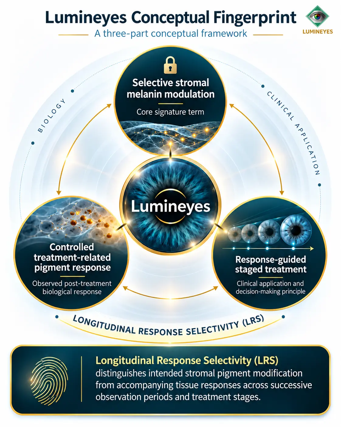
Three scientific dimensions remain distinct
LRS extends assessment across all three dimensions by introducing repeated, stage-referenced observations. Each response retains its own clinical interpretation, while LRS addresses the quality of the temporal differentiation.
What Would an LRS Investigation Measure?
Each observation must answer a defined question. Standardized inflammation grading is already established in clinical ophthalmology [6]; anterior-segment OCT investigations have developed objective indices of chamber inflammation and explored whether pigment and inflammatory cells exhibit differentiable image characteristics [7,8]. Specular microscopy provides an anatomical measurement domain, subject to acquisition and counting variability [10,11]. Standardized iris photography [13], colour-calibration research [14] and laser flare photometry [16] add complementary observation techniques. Their separate measurement properties are essential to interpreting LRS trajectories.
| Domain | Candidate observation | Question answered | Measurement boundary |
|---|---|---|---|
| P(t): intended pigmentary phenotype | Registered, standardized iris photography; calibrated colour measurements; regional pigmentation maps | Where, how much and how does visible pigmentation change over time? | Colour/optical change is not a direct measure of liberated melanin mass or cellular selectivity. |
| R₁(t): sensory response | Repeated, consistently worded symptom assessments; photophobia and discomfort reports | Does sensory recovery follow the same or a different temporal course? | Subjective symptoms do not independently localize tissue pathology. |
| R₂(t): inflammatory response | SUN anterior-chamber cell/flare grading; optional laser flare photometry; exploratory SS-AS-OCT particles | What is the onset, persistence and resolution of inflammation-related findings? | OCT hyperreflective particles are not definitive biochemical identification of pigment or cell type [6–8]. |
| R₃(t): anatomical response | Registered anterior-segment OCT, documented iris structure, serial corneal endothelial microscopy | Does tissue architecture remain stable while the pigmentary phenotype evolves? | Non-invasive imaging does not replace histology; endothelial estimates depend on standardization [10,11]. |
| R₄(t): physiological/functional response | Serial IOP, pupil function, visual acuity and appropriate functional measures | Do functional findings follow a time course distinct from visible pigmentation? | A single normal observation cannot establish every anatomical or physiological endpoint. |
The intended starting point is a multidomain longitudinal dataset, not the arithmetic addition of unrelated measures. Timing relative to each exposure, treatment-stage number, baseline phenotype, imaging device, medications, additional interventions and follow-up completeness must accompany the measured values.
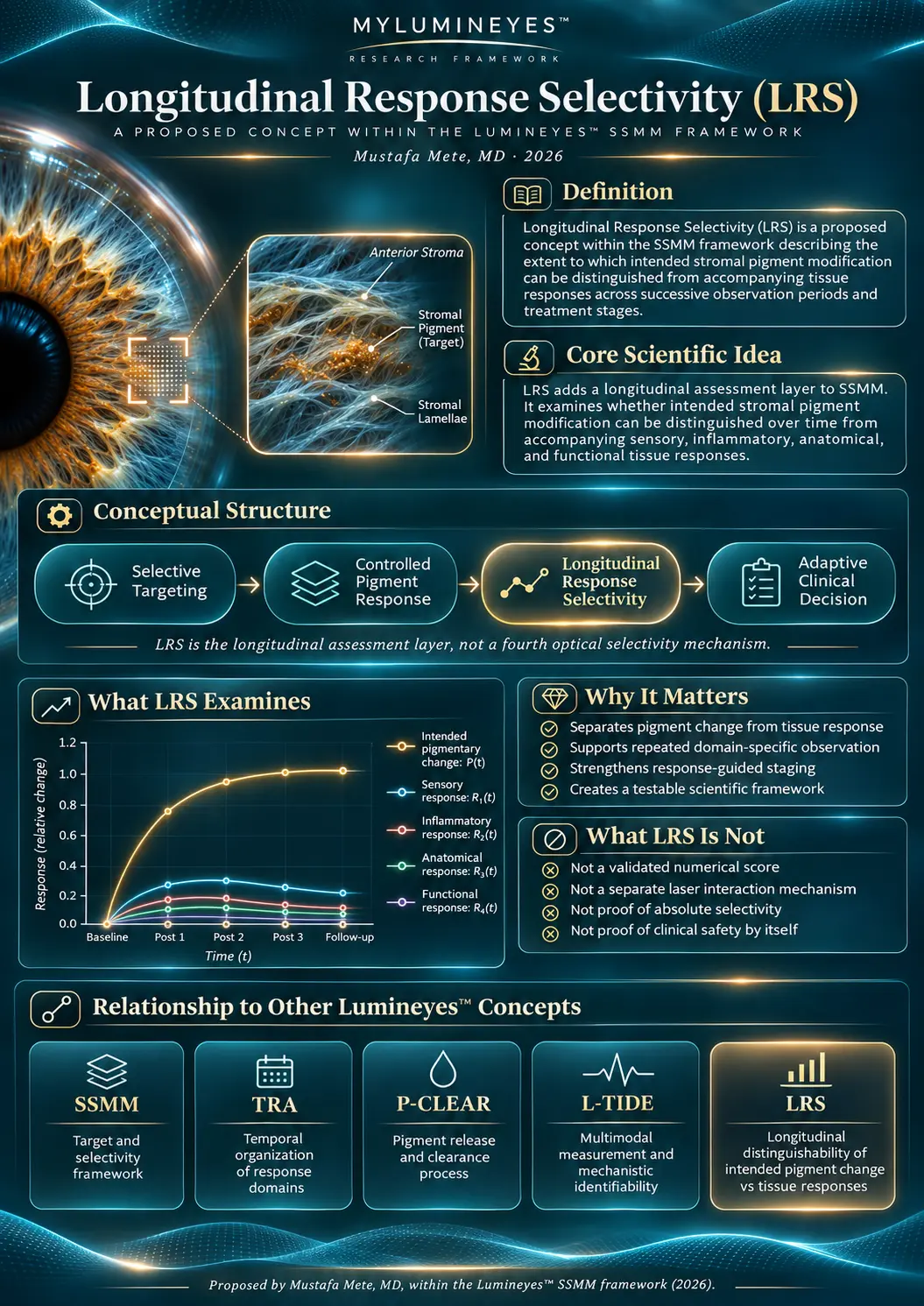
Measurement standards are part of the biological question
Computerized iris-colour measurements have previously been studied under standardized photographic acquisition across repeated visits [13]. Recent work using calibrated CIELAB-derived iris pigmentation demonstrates that eye colour can also introduce imaging-related bias in another anterior-segment measurement context [14]. Consequently, serial colour differences should be interpreted against repeatability, registration, lighting and instrument calibration—not merely against visual impression.
Inflammatory indices likewise require device- and protocol-specific repeatability. Laser flare photometry offers an objective protein-scattering measurement, while acquisition and summary procedures can materially influence the reported value [16]. These published methods establish candidate observation techniques, not an already validated LRS-specific instrument.
A Testable Observation Model, Not a Premature Score
For an eye observed at time t and treatment stage k, let P(t,k) denote a measurable pigmentary phenotype and Rj(t,k) denote the accompanying response domains. The LRS question is whether their trajectories can be estimated and distinguished with adequate temporal resolution, measurement reliability and adjustment for relevant context.
P(t, k) versus { R₁(t, k), R₂(t, k), …, Rₙ(t, k) }
These are candidate observation trajectories, not an LRS score or assumed independent biological pathways.Four methodological requirements
- Within-eye pairing. Capture target and response observations at linked visits, with the same eye and stage identified across time.
- Timing and exposure history. Record elapsed time from preceding exposure, intervals between stages, and the sequence of interventions rather than treating visits as interchangeable.
- Measurement integrity. Standardize image registration, illumination, device settings and interobserver procedures; document missing and noncomparable assessments.
- Longitudinal inference. Account for repeated observations within eyes and eyes within patients; distinguish descriptive trajectory separation from mechanistic or causal attribution [15].
Is the observed difference larger than plausible instrument, observer or registration error?
Can the onset, peak, recovery or persistence of the target trajectory be characterized separately from accompanying domains?
Do separately measured responses have their own functional and anatomical significance, independent of the pigmentary endpoint?
A candidate hierarchical longitudinal design
The prospective analysis unit is an eye-within-patient observed over successive stages. Candidate models include mixed-effects methods or generalized estimating equations for correlated ocular measurements [15]. Stage-specific time since exposure, cumulative exposure history, baseline phenotype, medication changes, additional treatments and informative loss to follow-up must be documented and addressed in prespecified analyses. Comparisons of paired pigmentary and tissue-response trajectories can then be made with uncertainty intervals and measurement-error sensitivity analyses.
Temporal separation is not proof of mechanistic independence or causation. Observations can remain correlated, and no single modality is assumed to identify every underlying biological state.
Optical appearance and tissue response can remain jointly determined by unobserved variables. The literature on partially observed dynamical models explains why an apparently good fit does not ensure that all underlying parameters are identifiable [9]. For LRS, that means independently informative measurement domains are more useful than a collection of redundant images.
The initial research programme emphasizes descriptive trajectories, measurement comparability and prespecified statistical comparison; quantitative scoring, if pursued, would be a separate development and validation task.
Propositions That Can Be Examined Empirically
Temporal differentiation. Can the pigmentary phenotype and accompanying tissue-response domains show measurably distinct onset, peak, persistence or recovery characteristics under repeated observation?
Added information. Do observations from a previous treatment stage improve interpretation or prediction of the next stage beyond baseline phenotype and cumulative exposure alone?
Independent observation. Do standardized structural and inflammatory measures reduce ambiguity left by colour photography alone?
Reproducibility. Are the inferred distinctions stable to observer, imaging device, observation interval, measurement-error assumptions, missing-data mechanisms and between-eye dependence?
These questions define the research programme. Distinguishability and clinical favourability remain separate endpoints and will be analysed as such.
One Scientific Question, Several Complementary Tools
| Framework | Primary role | Relationship to LRS |
|---|---|---|
| SSMM | Intended anterior stromal pigment targeting and response-guided clinical architecture | Parent framework in which LRS is proposed. |
| TRA | Organization of temporal biological response domains | TRA maps the timing of responses; LRS asks whether the intended pigmentary change is distinguishable from accompanying response trajectories. |
| P-CLEAR | Pigment release, handling and effective clearance | Represents one biological-process domain, not the entire LRS assessment question. |
| L-SAFE | Safety-state documentation and response-related decisions | Uses clinically relevant states; LRS concerns the observational separation of changes across time. |
| L-OAT | Optical and architectural iris phenotyping | May help specify spatially and optically informative phenotype measures for P(t). |
| L-TIDE | Multimodal measurement and mechanistic identifiability research | Offers a future methodological route for examining whether latent processes can be inferred from independently informative observations. |
These connections describe a research ecosystem; they are not a claim that the terms are interchangeable. The Research Library maintains the broader framework index and version records.
From Longitudinal Observations to Testable Evidence
Define domains
Specify optical pigmentary measurements and distinct anatomical, inflammatory, sensory and functional endpoints.
Establish reliability
Quantify measurement variation, standardize acquisition and register the same region of the same eye over time.
Estimate trajectories
Model eye-level observations, treatment-stage timing, patient clustering and prior exposure history.
Test distinguishability
Compare temporal patterns and uncertainties, test incremental information and reproduce findings externally.
This research sequence links the originating LRS definition to a measurable observational question: what information is needed to estimate pigmentary change separately from accompanying responses, with defensible precision?
Current Scientific Status
LRS is a proposed scientific concept within the Lumineyes™ SSMM framework. The published literature supports the anatomical, optical, imaging and statistical foundations for separate, repeated measurements of pigmentary and accompanying response domains [1–16]. The distinct LRS construct still requires dedicated studies with defined measurements, reliable time-series acquisition and prospective testing. No LRS-specific numerical validation or comparative clinical outcome is reported here.
This research formulation concerns longitudinal response interpretation. It is not a patient-facing treatment schedule or a statement of guaranteed final colour.
Research FAQ
Is Longitudinal Response Selectivity another name for selective laser targeting?
No. Optical targeting concerns an initial interaction. LRS evaluates the distinguishability of intended pigment modification from accompanying biological responses across successive observations.
Does LRS add a fourth laser mechanism to the SSMM framework?
No. The current display contains four named conceptual roles, but LRS is the longitudinal assessment layer within the existing target–response–decision architecture, not a fourth physical mechanism.
Is there an LRS score or a validated threshold?
No numerical index or threshold is proposed here. Operational research begins with linked measurements across pigmentary, sensory, inflammatory, anatomical and functional domains.
How does LRS differ from Temporal Response Architecture (TRA)?
TRA organizes the timing and relationships of response processes. LRS asks whether the intended pigmentary change can be longitudinally distinguished from accompanying tissue-response trajectories.
Who proposed Longitudinal Response Selectivity?
Longitudinal Response Selectivity (LRS) was proposed by Mustafa Mete, MD, within the Lumineyes™ SSMM research framework (2026).
Scientific References
External publications below underpin the relevant anatomy, laser–tissue interaction, inflammation measurement, imaging and inferential methods. They are not presented as publications validating LRS itself.
- Sturm RA, Larsson M. Genetics of human iris colour and patterns. Pigment Cell Melanoma Res. 2009;22:544–562. doi:10.1111/j.1755-148X.2009.00606.x
- Wilkerson CL, Syed NA, Fisher MR, et al. Melanocytes and iris color: light microscopic findings. Arch Ophthalmol. 1996;114:437–442. doi:10.1001/archopht.1996.01100130433014
- Anderson RR, Parrish JA. Selective photothermolysis: precise microsurgery by selective absorption of pulsed radiation. Science. 1983;220:524–527. doi:10.1126/science.6836297
- Yildirim Y, Duzgun E, Kar T, et al. Evaluation of color-changing effect and complications after Nd:YAG laser application on iris surface. Med Sci Monit. 2016;22:107–114. doi:10.12659/MSM.895086
- Grimaldos Ruiz P. Photoablative cosmetic iridoplasty: effective, safe, and predictable—eye color change in 1176 eyes. Int Ophthalmol. 2021;41:1381–1393. doi:10.1007/s10792-021-01693-5
- Jabs DA, Nussenblatt RB, Rosenbaum JT; SUN Working Group. Standardization of uveitis nomenclature for reporting clinical data. Am J Ophthalmol. 2005;140:509–516. doi:10.1016/j.ajo.2005.03.057
- Invernizzi A, Marchi S, Aldigeri R, et al. Objective quantification of anterior chamber inflammation: measuring cells and flare by anterior segment optical coherence tomography. Ophthalmology. 2017;124:1670–1677. doi:10.1016/j.ophtha.2017.05.013
- Bellchambers A, et al. Differentiation of anterior chamber pigment and inflammatory cells using swept-source optical coherence tomography: a cross-sectional study. Eye (Lond). 2025. doi:10.1038/s41433-025-03697-2
- Raue A, Kreutz C, Maiwald T, et al. Structural and practical identifiability analysis of partially observed dynamical models by exploiting the profile likelihood. Bioinformatics. 2009;25:1923–1929. doi:10.1093/bioinformatics/btp358
- Doughty MJ, Müller A, Zaman ML. Assessment of the reliability of human corneal endothelial cell-density estimates using a noncontact specular microscope. Cornea. 2000;19:148–158. doi:10.1097/00003226-200003000-00006
- Berrospi RD, Estrella CM, Galvis V, et al. Repeatability and reproducibility of corneal endothelial measurements using the Topcon SP-1P specular microscope. Graefes Arch Clin Exp Ophthalmol. Published September 2026. doi:10.1007/s00417-026-07488-7
- Jaber W, Nemet M, Waisbourd M. Laser iris depigmentation resulting in markedly elevated intraocular pressure and herpes simplex keratitis with corneal scarring: case report. Case Rep Ophthalmol. 2026;17:312–317. doi:10.1159/000551226
- Takamoto T, Schwartz B, Cantor LB, Hoop JS, Steffens T. Measurement of iris color using computerized image analysis. Curr Eye Res. 2001;22(6):412–419. doi:10.1076/ceyr.22.6.412.5490
- Arizcuren AR, Consejo A. Influence of iris color on corneal densitometry. J Optom. 2026;19(2):100595. doi:10.1016/j.optom.2025.100595
- Ying G-S, Maguire MG, Glynn RJ, Rosner B. Tutorial on biostatistics: longitudinal analysis of correlated continuous eye data. Ophthalmic Epidemiol. 2021;28(1):3–20. doi:10.1080/09286586.2020.1786590
- Tsui E, Jackson NJ, Chen JL, Holland GN. Establishment of a standard technique for determining laser flare photometry values during assessment of intraocular inflammation. Ophthalmol Sci. 2025;5(3):100690. doi:10.1016/j.xops.2024.100690
Originating internal research record: Mete M. Selective Stromal Melanin Modulation (SSMM): An Integrative Biophysical and Clinical Framework for Response-Guided Laser Iris Pigment Modification. Author manuscript v2.12, 4 November 2026, section 9 (LRS). See also the SSMM research page.