
Longitudinal Response Selectivity (LRS): A Multidomain Framework for Distinguishing Intended Iris Stromal Pigment Modification from Accompanying Tissue Responses
Mustafa Mete, MD
MyLumineyes Research, Istanbul, Türkiye · ORCID 0009-0004-3128-2853
Original preprint: 5 November 2026 · v1.0 · 10.5281/zenodo.23977907
Author-hosted copy of the original conceptual-methodological preprint. This page is not a separate peer-reviewed journal article.
Download the complete original 7-page PDF, with figures, table and references · Zenodo archival record
Keywords: longitudinal response selectivity; LRS; selective stromal melanin modulation; SSMM; iris pigmentation; anterior segment imaging; longitudinal ophthalmic measurement; phenotyping; response-guided staged treatment
Abstract
Background. Laser-directed iris pigment modification involves a short initiating exposure followed by pigmentary and tissue responses that may evolve on different timescales. Endpoint iris colour does not uniquely characterize the accompanying anatomical, inflammatory, sensory, or physiological responses. Objective. To define Longitudinal Response Selectivity (LRS) within the Selective Stromal Melanin Modulation (SSMM) framework and specify an empirical approach to its assessment. Framework. LRS denotes the longitudinal distinguishability of intended stromal pigment modification from accompanying tissue responses across observation periods and treatment stages.
The framework separates the intended pigmentary observation P(t,k) from measured response domains Rj(t,k), incorporating measurement reliability, stage-relative timing, within-eye correlation, and clinical co-interventions. A multidomain observation panel and four testable propositions are specified. Research implications. Appropriate evaluation requires calibrated regional iris imaging, domain-specific tissue and clinical measurements, prospectively defined observation times, and statistical methods that account for correlated ocular measurements.
Conclusions. LRS provides a falsifiable longitudinal assessment construct linking selective targeting to response-guided clinical decisions. Its evaluation depends on the ability to separate measurement variation, pigmentary change, and accompanying biological response rather than on a single numerical endpoint.
1. Introduction
A visible change in iris pigmentation is an optical observation. It cannot, by itself, quantify pigment mass released during laser exposure, differentiate extracellular and cellular pigment processing, establish preservation of stromal architecture, or describe the course of associated tissue responses. Human iris colour reflects melanin distribution, melanosomal characteristics, anterior stromal architecture and light scattering [1,2]. Laser–tissue interactions, in turn, depend on target absorption and exposure parameters rather than on the eventual colour alone [3].
In staged interventions, the time dimension adds a second interpretive challenge. Optical appearance, anterior-chamber activity, corneal or iris structural findings, sensory symptoms and ocular physiological measurements may peak or resolve at different times. These responses may also be affected by medication, baseline phenotype, illumination, device characteristics, and the decision to proceed with another treatment stage. A longitudinal framework should therefore distinguish observation of intended pigmentary change from assessment of accompanying biological trajectories.
Longitudinal Response Selectivity (LRS) was proposed by Mustafa Mete, MD, in 2026 within the Selective Stromal Melanin Modulation (SSMM) research framework. The purpose of the present conceptual-methodological manuscript is to define the scientific construct, specify its observable domains, identify the minimum longitudinal data structure, and articulate tests by which it may be supported, refined, or rejected. No new clinical results are reported.
2. Scientific definition and conceptual position
Longitudinal Response Selectivity (LRS) is a proposed concept within the SSMM framework describing the extent to which intended stromal pigment modification can be distinguished from accompanying tissue responses across successive observation periods and treatment stages. SSMM supplies the intended anterior stromal melanin target and the Target–Response–Decision architecture. Controlled treatment-related pigment response concerns the consequences observed after exposure; response-guided staged treatment uses those observations in subsequent clinical decisions.
LRS concerns the extent to which the targeted pigmentary response remains observationally distinguishable from other response domains over time. Thus, LRS contributes a longitudinal assessment layer rather than an additional photothermal, photomechanical, or optical selectivity mechanism. Biophysical selectivity asks whether exposure preferentially interacts with the intended pigment-bearing target; anatomical selectivity concerns the distribution of structural effects; functional selectivity concerns clinically relevant ocular function. Each can be assessed at more than one time point.
LRS connects these measurements across successive observations and treatment stages, as represented in Figure 1. The related Lumineyes research concepts address different levels of inquiry. Temporal Response Architecture (TRA) concerns the temporal organization of response domains. P-CLEAR examines pigment release, handling and clearance. L-TIDE investigates the measurement requirements and mechanistic identifiability of latent iris–outflow processes. LIPS and L-OAT concern proposed pigmentation and optical-architectural phenotype characterization.
LRS asks whether the intended pigmentary trajectory can be separated observationally from accompanying tissue-response trajectories without assuming that the underlying biological processes are causally independent. Figure 1. Positioning of LRS within SSMM. LRS separates intended pigmentary observations from measured accompanying response domains across treatment stages. Diagram is conceptual; no clinical measurements are depicted.
3. Conceptual development and evidentiary basis
This manuscript is a concept-development and methodological synthesis, not a systematic review or prospective outcomes study. Its rationale integrates established iris pigment anatomy and laser–tissue interaction principles; published methods for repeated iris colour measurement; anterior-chamber inflammation, particle and corneal endothelial assessment; and statistical methods for correlated longitudinal eye data. These literature domains support the feasibility and limitations of candidate measurements, not empirical validation of LRS. Standardized computerized analysis of repeat iris photographs was investigated by Takamoto and colleagues, who assessed reproducibility across repeated image acquisitions and follow-up visits [4].
Their findings support the principle that observed optical change must be interpreted relative to acquisition and measurement variation. The Standardization of Uveitis Nomenclature framework provides clinically established approaches to recording anterior-chamber inflammatory activity [5]. Investigations using anterior-segment OCT indicate the potential of imaging-based quantification of cells and flare [6]. More recently, swept-source OCT particle features were associated with clinically detected pigment and inflammatory cells in a cross-sectional uveitis cohort; those results motivate further validation but cannot be directly transferred to SSMM-treated eyes [7].
Specular microscopy offers a distinct structural observation domain, subject to its own repeatability characteristics [8]. These sources together identify a methodological opportunity: optical, inflammatory, anatomical, sensory and physiological trajectories can be measured as different channels. They also identify a central constraint: channel-specific measurement error, acquisition changes and observation schedules may mimic or conceal differences in temporal response.
4. Longitudinal observation model
Let e identify an individual eye, k a treatment stage and t the elapsed time from the relevant stage exposure. Define P_e,k(t) as an observed, operationally specified measure of intended pigmentary change derived from calibrated regional iris phenotype assessment. Let R_e,k,j(t), j = 1,…,m, denote accompanying, separately measured response domains, such as sensory, inflammatory, anatomical and physiological/functional findings. The observation vector is Z_e,k(t) = [P_e,k(t), R_e,k,1(t), …, R_e,k,m(t)]. The notation describes measured variables, not a biological mass-balance model.
P is not assumed to represent melanin mass, and an R measurement is not assumed to isolate a single cellular mechanism. Observed signals can be expressed conceptually as Y_d(t) = h_d[X_d(t), A_d(t)] + ε_d(t), where X_d represents the domain of interest, A_d acquisition or observation conditions, h_d the measurement process and ε_d residual variation. Explicitly separating these terms prevents photographic, device or observer variability from being interpreted automatically as biological change. Two time axes should be retained. Stage-relative time t describes observation intervals from the latest exposure, whereas calendar time T tracks the complete clinical course and cumulative stage history.
A new exposure may interrupt or modify prior trajectories; thus observations from multiple stages cannot be pooled as independent, identically timed sessions. The eye remains nested within the patient and observations are repeated within the eye. A conceptual example of multidomain trajectories across successive treatment stages is shown in Figure 2. LRS is not defined here as a numerical score. A scalar index would require a validated measurement model, prespecified domain weights, reproducibility studies, meaningful interpretive anchors and external replication.
Until those conditions are met, distinguishability is evaluated through transparent domain-specific analyses and uncertainty estimates. Figure 2. Schematic multidomain trajectories across successive treatment stages. The intended pigmentary observation P(T) and accompanying sensory, inflammatory, anatomical and functional response domains Rj(T) are represented as distinct hypothetical signals. Exposure timing and response magnitudes are illustrative; no patient data are depicted. The distinct response channels shown in Figure 2 motivate paired, calibrated measurements at prespecified follow-up intervals, with analyses that account for within-eye and within-patient dependence.
5. Candidate measurement architecture
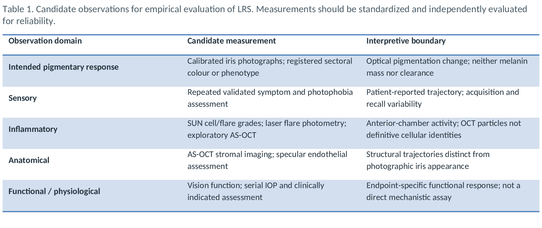
A minimally interpretable LRS dataset requires standardized baseline characterization, matched follow-up observations, exact exposure and visit timestamps, device/acquisition metadata, and a documented record of intervening medication and clinical management. Table 1 maps candidate observables to the questions they can and cannot answer. A single imaging modality is unlikely to resolve all response domains. For iris pigmentation, regional image registration and calibration are particularly important. Photograph-derived lightness, colour coordinates or spatial pigmentation features are optical proxies influenced by illumination, pupil size and image processing.
They are not direct measurements of pigment liberation or macrophage-mediated clearance. Anterior-chamber cell/flare grading, laser flare photometry and exploratory OCT particle metrics may contribute complementary inflammatory observations; OCT particle classification requires clinical or laboratory validation in the relevant population [5–7,9]. High-resolution anterior-segment imaging and specular microscopy supply anatomical observations, while validated symptom and clinical tests supply sensory and functional observations. Measurement acquisition should be repeatable within domain and across visits.
If a device, acquisition mode or image-processing pipeline changes, that change should be recorded explicitly and included in sensitivity analysis. Relevant limits of agreement and measurement uncertainty should be estimated before interpreting small longitudinal differences as target effects.
6. Testable research propositions
Proposition 1 — Measurement distinguishability. Under controlled image acquisition and independent reliability assessment, a proportion of observed regional pigmentary change can be distinguished from test–retest and acquisition variability. This proposition is weakened if between-visit differences remain within expected measurement variation or depend predominantly on acquisition conditions. Proposition 2 — Temporal distinguishability. Intended pigmentary observations and at least one accompanying response domain may exhibit separately estimable trajectories of onset, peak, persistence or resolution.
This proposition is not supported when the available visit schedule and uncertainty are insufficient to distinguish the trajectories or when estimated differences are unstable across analytic specifications. Proposition 3 — Added longitudinal information. A prior-stage multidomain observation set may improve characterization or prediction of later-stage outcomes beyond baseline phenotype, cumulative exposure history and calendar time alone. This is a model-comparison question, to be evaluated with out-of-sample prediction or other prespecified performance criteria; predictive improvement would not itself establish biological causation.
Proposition 4 — Independent anatomical and functional information. Longitudinal pigmentation measurements cannot substitute for separate anatomical or functional observations when the purpose is to characterize the broader ocular response. The proposition can be examined by assessing the conditional information carried by prespecified structural and physiological measures beyond optical pigmentation alone. These propositions describe research questions, not expected treatment benefits. They provide explicit routes by which the distinct contribution of LRS could fail to be demonstrated.
7. Proposed prospective evaluation
A first empirical evaluation could use an observational longitudinal cohort with consecutive eligible eyes, a prespecified measurement panel and repeated observations spanning baseline, early response, intermediate recovery, pre-retreatment assessment and longer-term follow-up. Time windows should be chosen according to feasibility and the specific biological questions, not inferred from a universal course. A staged-treatment cohort should log each exposure as a time-varying event and preserve the actual spacing between stages. The minimum record should contain a patient identifier; distinct eye identifier and laterality; stage identifier; exposure and visit timestamps; site-matched regional iris imaging; image calibration metadata; domain-specific measures; acquisition device; changes in medication and other interventions; baseline pigmentation phenotype; relevant ocular history; and reasons for missed observations.
Clinical decisions governing who returns for another stage are informative: patients with different trajectories may have different treatment intervals and follow-up intensity. Such selection must be considered during analysis. Before evaluating clinical hypotheses, investigators should quantify within-session and between-session reliability for major observables; test image registration and calibration stability; establish feasibility of collecting paired observations; and prespecify the clinically interpretable quantities for each domain. Multicentre replication and masked outcome assessment would help test portability across devices and operators.
Longitudinal mixed-effects models or generalized estimating equations can incorporate repeated observations and within-patient inter-eye correlation [10]. Time should be represented flexibly where justified, including time since the most recent exposure and cumulative stage history. Missing observations and changes in management require explicit sensitivity analyses. Models with latent biological states should be distinguished from direct observation models: identification of a hidden cellular or clearance process requires additional information beyond a good fit to optical and clinical trajectories [11].
8. Relationship to clinical interpretation
Longitudinal distinguishability and clinical desirability are different constructs. A pigmentary curve can be statistically separable from an anatomical or inflammatory curve without establishing that either curve indicates a favorable outcome. Conversely, a physiologically meaningful response may remain difficult to measure separately because of sparse sampling or imperfect instruments. Clinical interpretation therefore depends on the domain-specific direction, magnitude, persistence and consequences of each observation, assessed independently of the existence of LRS as a concept.
Historical reports of iris laser intervention include both intentional pigment modification and clinically important ocular responses [12,13]. Such literature establishes the need to distinguish protocols and observed domains, not to infer incidence or safety equivalence across methods. Future applications of LRS should preserve protocol-specific attribution and allow different observation channels to disagree rather than forcing them into a single favorable or unfavorable label.
9. Methodological limitations and next validation steps
The current framework is conceptual and its key propositions have not been prospectively evaluated as an integrated LRS study. The available supporting literature spans different populations, procedures and devices. In particular, optical colour measurements, slit-lamp inflammation grades, OCT particle features and endothelial metrics are not directly interchangeable, and a visual pigment trajectory does not identify a pigment-clearance mechanism. Observational scheduling and clinician-selected retreatment may generate informative missingness and time-varying confounding.
A rigorous validation programme would proceed from measurement repeatability to paired multidomain feasibility, prespecified longitudinal contrasts, external reproducibility and eventually clinically interpretable prediction studies. Mechanistic inference should be reserved for designs with independently informative measurements or interventions suited to distinguishing competing pathways. This progression would clarify whether LRS contributes reproducible information beyond standard longitudinal observation alone.
10. Conclusion
Longitudinal Response Selectivity (LRS) defines an assessment question within SSMM: to what extent can the intended anterior stromal pigmentary response be distinguished from accompanying tissue responses through repeated, appropriately timed and reliable observations? The construct specifies separate optical and biological observation channels, preserves eye- and stage-level dependence, and yields falsifiable research propositions. Its future scientific value will be determined by measurement reliability, temporal resolution, independent domain information and reproducible prospective evaluation.
In this role, LRS connects the SSMM target–response architecture with response-guided decision-making without assuming a validated score or mechanism.
References
- Sturm RA, Larsson M. Genetics of human iris colour and patterns. Pigment Cell Melanoma Res. 2009;22(5):544–562. doi:10.1111/j.1755-148X.2009.00606.x.
- Wilkerson CL, Syed NA, Fisher MR, Robinson NL, Wallow IHL, Albert DM. Melanocytes and iris color: light microscopic findings. Arch Ophthalmol. 1996;114(4):437–442. doi:10.1001/archopht.1996.01100130433014.
- Anderson RR, Parrish JA. Selective photothermolysis: precise microsurgery by selective absorption of pulsed radiation. Science. 1983;220(4596):524–527. doi:10.1126/science.6836297.
- Takamoto T, Schwartz B, Cantor LB, Hoop JS, Steffens T. Measurement of iris color using computerized image analysis. Curr Eye Res. 2001;22(6):412–419. doi:10.1076/ceyr.22.6.412.5490.
- Jabs DA, Nussenblatt RB, Rosenbaum JT; Standardization of Uveitis Nomenclature Working Group. Standardization of uveitis nomenclature for reporting clinical data. Am J Ophthalmol. 2005;140(3):509–516. doi:10.1016/j.ajo.2005.03.057.
- Invernizzi A, Marchi S, Aldigeri R, et al. Objective quantification of anterior chamber inflammation: measuring cells and flare by anterior segment optical coherence tomography. Ophthalmology. 2017;124:1670–1677. doi:10.1016/j.ophtha.2017.05.013.
- Bellchambers A, Shu R, Chu CJ, Petrushkin H, Solebo AL. Differentiation of anterior chamber pigment and inflammatory cells using swept-source optical coherence tomography: a cross-sectional study. Eye (Lond). 2025;39:1744–1750. doi:10.1038/s41433-025-03697-2.
- Doughty MJ, Müller A, Zaman ML. Assessment of the reliability of human corneal endothelial cell-density estimates using a noncontact specular microscope. Cornea. 2000;19:148–158. doi:10.1097/00003226-200003000-00006.
- Tsui E, Jackson NJ, Chen JL, Holland GN. Establishment of a standard technique for determining laser flare photometry values during assessment of intraocular inflammation. Ophthalmol Sci. 2025;5(3):100690. doi:10.1016/j.xops.2024.100690.
- Ying GS, Maguire MG, Glynn RJ, Rosner B. Tutorial on biostatistics: longitudinal analysis of correlated continuous eye data. Ophthalmic Epidemiol. 2021;28(1):3–20. doi:10.1080/09286586.2020.1786590.
- Raue A, Kreutz C, Maiwald T, et al. Structural and practical identifiability analysis of partially observed dynamical models by exploiting the profile likelihood. Bioinformatics. 2009;25(15):1923–1929. doi:10.1093/bioinformatics/btp358.
- Yildirim Y, Duzgun E, Kar T, et al. Evaluation of color-changing effect and complications after Nd:YAG laser application on iris surface. Med Sci Monit. 2016;22:107–114. doi:10.12659/MSM.895086.
- Jaber W, Nemet M, Waisbourd M. Laser iris depigmentation resulting in markedly elevated intraocular pressure and herpes simplex keratitis with corneal scarring: case report. Case Rep Ophthalmol. 2026;17:312–317. doi:10.1159/000551226.
Original Table 1
Original figures

