
SSMM–IOP Research: Could Lower Intraocular Pressure Persist?
A research-owner perspective on the possibility that a brief iris-directed perturbation might be followed by a longer-lasting shift in aqueous outflow homeostasis—and on the experiments needed to tell that story from competing explanations.
Read the published SSMM–IOP preprint (version 1, 12 November 2026)
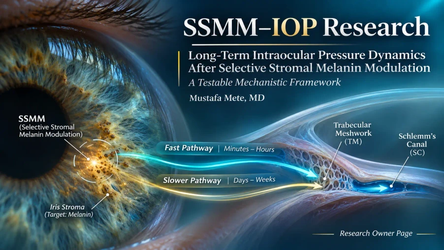
A lower pressure reading is an observation. Its mechanism is a separate question.
Conventional aqueous drainage involves a responsive network, not simply a pipe with fixed resistance. The trabecular meshwork, juxtacanalicular extracellular matrix and Schlemm’s canal endothelium respond to cellular, molecular and mechanical signals. That raises a specific possibility: a short-lived stimulus might coincide with an altered outflow state that outlasts the original event.
SSMM is directed toward the anterior iris stromal pigment compartment, not toward the trabecular meshwork. Any proposed downstream change in aqueous drainage would therefore need an indirect connection—such as transported particulate material, soluble mediators, altered tissue mechanics or pressure-sensitive feedback. The distinction is the starting point of this investigation, rather than its conclusion.
The conceptual value of this pressure shift is not the number itself; it is what the rest of the aqueous-flow system would have to do to make such a reading physiologically plausible. The accompanying clinical archive addresses reported observations separately. This page examines the causal explanation that future measurements would need to support.
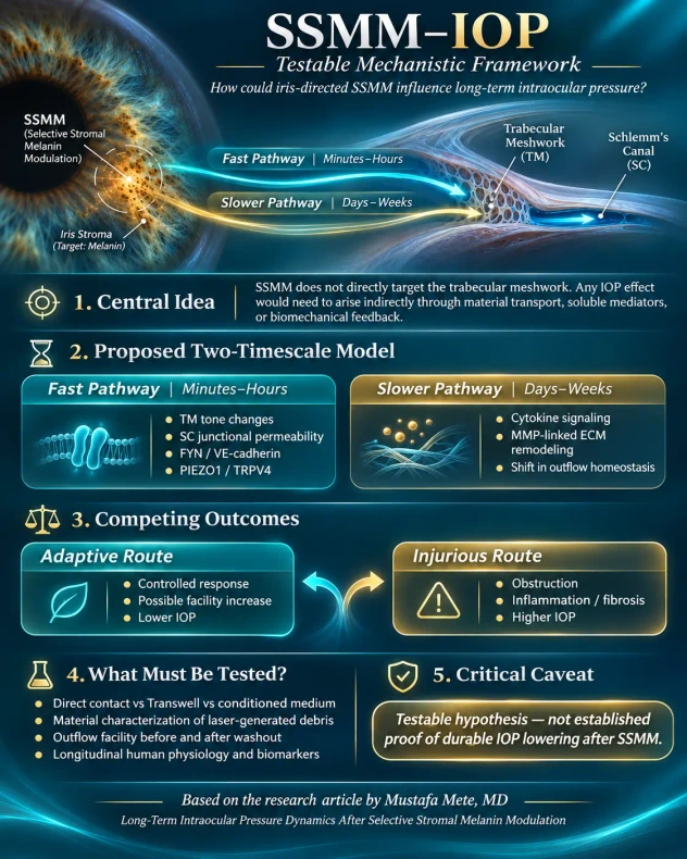
One initiating event. Two possible biological clocks.
An early pressure response and a durable pressure response are not interchangeable. Endothelial junctions and cell contractility can change relatively quickly; shifts in matrix organization or longer-lived tissue feedback would require a different timescale and different measurements.
Junctions and tone
Schlemm’s canal permeability, FYN/VE-cadherin signaling and trabecular contractile state are candidates for relatively rapid changes in resistance.
Immune-cell signals
Monocyte-associated responses and local signaling offer a potential bridge between a transient perturbation and outflow tissues—but the bridge remains unproven after SSMM.
Matrix state
Cytokine-linked proteolysis and extracellular-matrix feedback suggest a way to ask whether an altered outflow state survives after the initiating signal clears.
The decisive distinction: a temporary increase in endothelial permeability cannot by itself explain a multi-year pressure pattern. A persistence model must specify what remains different after particulate burden and acute mediators have declined.
How much outflow change would a lower IOP require?
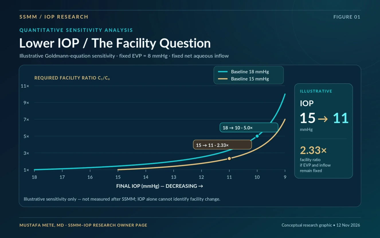
The Goldmann relationship provides a useful constraint. If episcleral venous pressure (EVP) and net aqueous inflow are held constant, the outflow-facility ratio needed to accompany a hypothetical pressure reduction can be calculated. This is an exercise in physiological sensitivity, not a measurement of post-SSMM facility.
| Initial IOP | Final IOP | Assumed EVP | Required C₁/C₀ |
|---|---|---|---|
| 15 mmHg | 11 mmHg ↓ | 8 mmHg | 2.33× |
| 15 mmHg | 9 mmHg ↓ | 8 mmHg | 7.00× |
| 18 mmHg | 11 mmHg ↓ | 8 mmHg | 3.33× |
| 18 mmHg | 10 mmHg ↓ | 8 mmHg | 5.00× |
These ratios rise sharply as the final IOP approaches the assumed EVP. That sensitivity is scientifically useful: it tells us that an apparently striking pressure decrease cannot automatically be attributed to a single pathway. Actual aqueous formation, unconventional outflow, EVP and measurement conditions must be investigated before a facility mechanism is assigned.
Which links are experimentally established—and which belong to SSMM research?
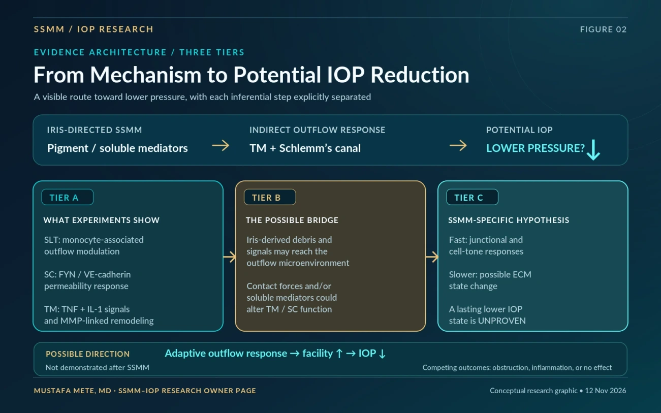
The most important question is not whether an individual pathway is biologically plausible. It is whether the entire proposed chain can be demonstrated in the right tissue, with the right initiating material, and over the relevant time period.
Direct findings
SLT-associated monocyte effects, pressure-responsive Schlemm’s canal junctions and cytokine-associated matrix pathways have been examined experimentally in distinct models.
The indirect bridge
Could iris-derived particulate or soluble factors reach and influence the same outflow system without direct laser irradiation of the trabecular meshwork?
Persistence after SSMM
Does a measurable, lower-resistance operating state remain after exposure signals and acute inflammatory activity have subsided?
The distinction matters because experimental evidence also supports the opposite possibility: pigment loading can elevate resistance and intraocular pressure. A mechanism proposed to explain lower pressure must therefore compete against obstruction, inflammation, fibrotic remodeling and no sustained change.
Why do different iris-laser reports describe different IOP trajectories?
Independent reports make the question more concrete, but they do not point to one uniform pressure response. Baseline pressure, treatment protocol, pressure medication, angle geometry and follow-up methods may all contribute to apparently different findings.
| Source / evidence type | Participants | Pressure observation | Interpretation |
|---|---|---|---|
| Touboul / ESCRS EuroTimes, 2022 Conference presentation reported by a professional society publication | 30 healthy patients receiving full-iris Strōma treatment, followed for at least 12 months | 20% mean IOP decrease reported | Observational meeting report; individual pressures, variance, paired statistical analysis and an independently verifiable full dataset are not provided in the news article. |
| Grimaldos Ruiz, 2021 Peer-reviewed prospective study, International Ophthalmology | 1,176 eyes of 588 patients | Mean IOP approximately 10.6 → 10.8 mmHg; no statistically significant overall change (reported p = 0.63204 for the recent four-year follow-up comparison) | The author also described a slight downward long-term tendency in some observations. The numerical group result, however, does not establish a mean IOP reduction. |
| Liu et al., 2023 Peer-reviewed case report | One patient, both eyes | Severe post-treatment pressure elevation: 50 / 42 mmHg on presentation | Demonstrates a competing pigment-obstruction / injury pathway, not the frequency of that outcome across protocols. |
| MyLumineyes longitudinal clinical archive Practice-reported retrospective observations | Approximately 1,500 patients / over 3,000 treated eyes in the archive | Lower IOP measurements in selected long-term examinations; formal cohort-wide paired estimates have not yet been supplied. | Requires audited baseline and follow-up denominators, medication history, tonometry conditions and independent verification before estimating effect size. |
The ESCRS report deserves attention because a pressure decrease was documented as an unexpected clinical observation after a distinct iris depigmentation approach. Touboul also proposed iris tissue retraction and a change in angle tension as a possible explanation. This is an anatomical / mechanical hypothesis, not proof of increased conventional outflow facility or of the immune–matrix pathway outlined in the SSMM preprint.
Angle geometry
Measure iris configuration, angle opening and possible mechanical changes using paired anterior-segment OCT and gonioscopy. Changes in iris tension or angle width must be demonstrated rather than assumed.
Outflow-cell signaling
Test whether iris-derived material or mediators can affect trabecular or Schlemm’s canal responses, with direct facility measurements and washout controls.
Obstruction or no change
Account for particulate loading, inflammation and unaltered outflow. Published elevated-IOP cases and studies with no overall pressure change are essential comparator outcomes.
Proposed discriminating analysis: stratify prospective trajectories by baseline IOP; record matched-time measurements, corneal factors, ocular hypotensive or steroid exposure, treatment protocol, iris pigmentation phenotype, baseline and follow-up angle anatomy, and repeated measurements at months and years. Test whether sustained IOP changes correlate with structural angle change, directly measured outflow facility, or neither. These reports must not be pooled as a common treatment-effect estimate.
Clinical interpretation note: the 2022 ESCRS item is a meeting-report summary, not a peer-reviewed trial report. The clinical series, case report and practice archive have different designs, protocols, follow-up completeness and ascertainment; they cannot establish protocol-wide efficacy or safety by cross-study comparison.
The path from an attractive hypothesis to a discriminating experiment.
A persuasive research programme must be able to disprove its own preferred mechanism. The experimental sequence begins with what is actually released, moves through the cells and tissues that could respond, and ends with repeated physiological measurements in humans.
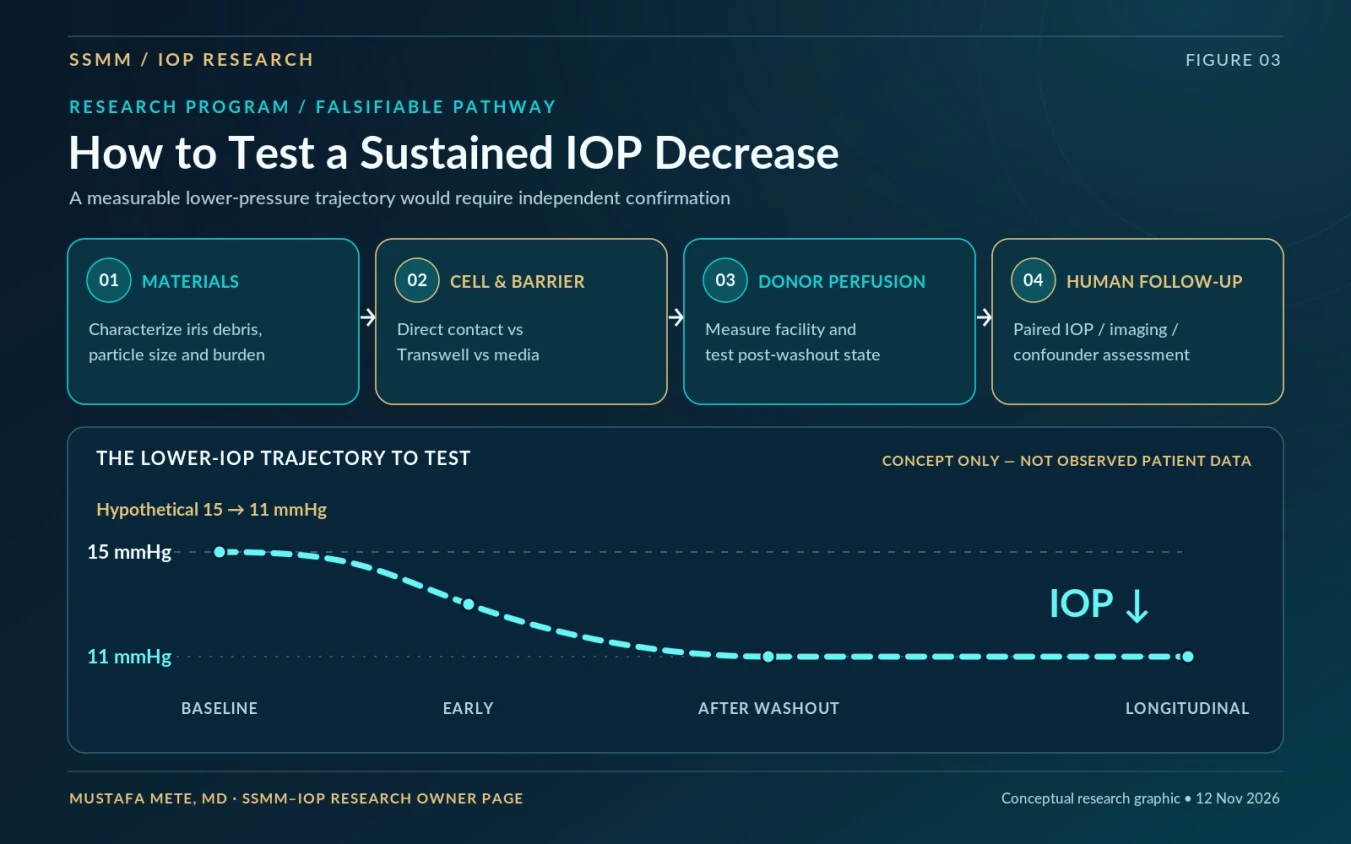
Characterize what enters the anterior segment
Compare native iris pigment, laser-generated material, soluble fractions and inert controls under characterized particle-size and burden conditions.
Separate contact from soluble signaling
Compare pigment-loaded macrophage contact, Transwell separation and conditioned medium; measure endothelial permeability and site-specific FYN/VE-cadherin responses.
Look for a post-washout state
Measure donor anterior-segment outflow facility before exposure, during challenge and after washout, alongside matrix organization and tissue viability.
Track pressure without losing context
Use matched-time tonometry, medication review, corneal assessment, angle imaging and facility assessment where feasible, with suitable comparators.
What would change the interpretation? A persistent outflow-facility increase after washout, coupled to a demonstrable tissue-state difference, would support a matrix or sustained-feedback hypothesis. A pressure change without a facility change would redirect attention toward other determinants of IOP.
Lower pressure can have more than one cause.
The same IOP reading can arise from different combinations of aqueous formation, conventional drainage, unconventional drainage and venous pressure. That is why this project does not treat tonometry alone as a direct assay of trabecular remodeling.
Findings that would support an outflow hypothesis
- Reproducible pressure change under comparable examination conditions.
- Direct evidence of altered conventional outflow facility.
- Correlated junctional, cellular or extracellular-matrix measurements.
- A measured difference persisting beyond acute exposure and mediator washout.
Findings requiring a different explanation
- Changed medications, aqueous production or unconventional outflow.
- Different episcleral venous pressure or measurement timing.
- Changes in corneal thickness or biomechanical tonometry bias.
- Particulate obstruction, inflammation, injury or regression toward the mean.
These alternatives are not peripheral details. They are the comparisons that allow a long-term pressure hypothesis to become testable rather than circular.
Where the SSMM–IOP question belongs in the wider research archive.
The owner page has a deliberately narrow role: it examines why a lasting pressure change might occur and how to investigate it. Other pages carry the separate responsibilities of defining the treatment concept, describing pigment kinetics, classifying longitudinal responses and interpreting clinical observations.
The academic work behind this owner page.
This page is an original web-oriented research synthesis built around a distinct manuscript. It highlights the research question, visual sensitivity analysis and tests that distinguish competing explanations; it does not reproduce the manuscript section by section.
- Manuscript
- Long-Term Intraocular Pressure Dynamics After Selective Stromal Melanin Modulation: A Testable Mechanistic Framework
- Author
- Mustafa Mete, MD · Independent ophthalmic research, Istanbul, Türkiye
- Research manuscript
- Reference-audited v10 · 11 November 2026
- Public preprint
- Version 1, posted 12 November 2026 · Read the public Preprints.org record
- Article type
- Mechanistic narrative review and testable hypothesis
- Review status
- Public preprint, not peer-reviewed. No unverified DOI is asserted on this page.
- Zenodo
- A separate archival version is planned; no Zenodo DOI is assigned here until the record is published.
- Owner-page role
- Research interpretation, figure explanations, connections to related frameworks and a testable research roadmap.
Mechanistic foundations and independent clinical reports.
The preprint contains the full mechanistic bibliography. The sources below distinguish original experimental mechanisms from published human IOP observations, including results that do not support a uniform pressure reduction.
- Alvarado JA et al. (2010). Monocyte modulation of aqueous outflow following selective laser trabeculoplasty. Archives of Ophthalmology. doi:10.1001/archophthalmol.2010.85
- Kizhatil K et al. (2025). FYN regulation of aqueous humor outflow through VE-cadherin phosphorylation. Nature Communications. doi:10.1038/s41467-024-55232-8
- Kelley MJ et al. (2007). TNF/IL-1 synergism and matrix metalloproteinase responses in trabecular meshwork. Investigative Ophthalmology & Visual Science. doi:10.1167/iovs.06-1445
- Wang C et al. (2019). The impact of pigment dispersion on trabecular meshwork cells. Graefe’s Archive for Clinical and Experimental Ophthalmology. doi:10.1007/s00417-019-04300-7
- McGrath D / Touboul D (2022). Making Brown Eyes Blue. ESCRS EuroTimes, 4 July 2022. Conference-report summary of Strōma iris depigmentation and reported mean IOP decrease. Original report.
- Grimaldos Ruiz P (2021). Photoablative cosmetic iridoplasty: effective, safe, and predictable—eye color change in 1176 eyes. International Ophthalmology. doi:10.1007/s10792-021-01693-5.
- Liu J et al. (2023). Bilateral severe iatrogenic pigmentary glaucoma following laser treatment for cosmetic iris color change. American Journal of Ophthalmology Case Reports. doi:10.1016/j.ajoc.2023.101927.
- Mete M (2026). Long-Term Intraocular Pressure Dynamics After Selective Stromal Melanin Modulation: A Testable Mechanistic Framework. Preprints.org, version 1, posted 12 November 2026. Public preprint record.
The studies are cited for their original experimental contexts. None demonstrates the complete SSMM-to-sustained-IOP-reduction causal sequence.
The next scientific milestone is not a lower number on a chart. It is a measured, reproducible physiological change whose cause can be separated from alternative explanations. Return to the MyLumineyes Research Archive.