Laser Physics in Ophthalmology: A Comprehensive Scientific Review of Selective Iris Depigmentation
Article Type: Scientific Review
Abstract
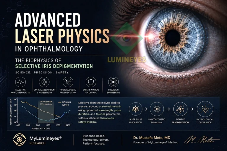
Laser iris depigmentation represents one of the most technically demanding applications of biomedical optics within anterior segment ophthalmology. Its safety and efficacy depend on the precise interaction between laser energy and stromal melanin, requiring careful optimization of wavelength, pulse duration, fluence, beam profile, and treatment geometry. This review examines the biophysical principles underlying selective photothermolysis and discusses how modern ophthalmic laser engineering minimizes collateral thermal effects while promoting controlled pigment fragmentation.
In addition, the review explores energy density optimization, spatial beam homogeneity, optical absorption characteristics, biological pigment clearance, and the engineering concepts incorporated into contemporary laser delivery systems. Rather than focusing solely on cosmetic outcomes, this article presents laser iris depigmentation as a multidisciplinary application of biomedical engineering, ocular physiology, and laser-tissue interaction. The objective is to provide a scientific overview of the physical mechanisms that support safe and controlled selective stromal depigmentation.
Keywords
Laser Physics, Selective Photothermolysis, Laser-Tissue Interaction, Biomedical Optics, Ophthalmology, Iris Depigmentation, Melanin, Fluence, Photoacoustic Effects, Anterior Segment, Laser Engineering
The application of laser technology in ophthalmology represents one of the most sophisticated intersections of optical physics, tissue biophysics, and microsurgical precision. While permanent iris depigmentation is often discussed in aesthetic terms, its clinical success depends primarily on the accurate control of laser-tissue interactions at the microscopic level. Every treatment parameter—including wavelength, pulse duration, fluence, spot geometry, repetition rate, and spatial beam profile—must operate within carefully defined biological safety limits to achieve selective pigment removal while preserving normal ocular anatomy.
Understanding these principles requires moving beyond cosmetic outcomes and examining the underlying mechanisms governing energy absorption, thermal confinement, tissue response, and pigment clearance. Modern laser iris depigmentation therefore represents not merely a clinical procedure but an application of advanced biomedical engineering and laser biophysics.
Contemporary treatment platforms, including proprietary systems such as the 8G Xtra Laser System, have been developed to optimize these variables through highly controlled energy delivery protocols specifically adapted to the unique optical and anatomical characteristics of the human iris.
The Core Principle: Selective Photothermolysis
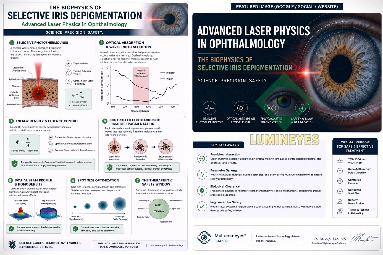
The scientific foundation of modern laser iris depigmentation is the principle of Selective Photothermolysis. Originally described for the selective destruction of pigmented biological targets, this concept states that a carefully selected wavelength can preferentially interact with a specific chromophore—in this case, eumelanin located within the anterior iris stroma—while minimizing energy absorption by adjacent non-pigmented ocular structures.
Successful selective photothermolysis depends on confining laser energy to the target before significant heat diffusion occurs. This requires the laser pulse duration to remain shorter than the target tissue’s Thermal Relaxation Time (τr), allowing energy deposition to remain localized within the pigmented tissue.
The thermal relaxation time can be approximated by:
where d represents the effective diameter of the melanin-containing target and α represents the thermal diffusivity of the surrounding tissue.
When pulse duration remains below the thermal relaxation time, absorbed optical energy is largely confined to the melanin-containing structures, substantially reducing lateral heat diffusion into adjacent ocular tissues. Under appropriate pulse characteristics, this localized energy confinement may generate controlled photothermal effects accompanied by localized photoacoustic phenomena, facilitating pigment fragmentation while minimizing unnecessary thermal exposure.
Conversely, laser systems operating with non-optimized pulse durations or poorly controlled energy parameters may increase thermal diffusion beyond the intended target, potentially elevating the risk of collateral tissue injury. For this reason, successful iris depigmentation relies not only on laser power but on the precise synchronization of wavelength, pulse duration, fluence, and beam delivery characteristics.
Optical Absorption and Wavelength Selection
An equally important factor in selective iris depigmentation is wavelength selection. Melanin exhibits broadband optical absorption throughout the visible and near-infrared spectrum; however, absorption efficiency, optical penetration depth, and thermal confinement vary significantly between wavelengths. Consequently, wavelength cannot be evaluated as an isolated parameter but must be optimized together with pulse duration and fluence to achieve predictable biological responses.
Appropriate wavelength selection maximizes preferential absorption by stromal melanin while minimizing unintended interaction with surrounding ocular tissues. This balance contributes to controlled pigment fragmentation and supports preservation of normal iris architecture throughout treatment.
Energy Density and Controlled Photoacoustic Effects
Among all laser parameters, fluence (Φ) represents one of the most critical determinants of both treatment efficacy and ocular safety. Fluence describes the amount of laser energy delivered per unit surface area and directly influences the biological response of the targeted iris tissue.
Mathematically, fluence is expressed as:
where E is the laser pulse energy and A is the irradiated spot area.
Because fluence incorporates both pulse energy and beam diameter, it provides a more meaningful representation of tissue exposure than pulse energy alone. Even small changes in spot size may significantly alter the delivered energy density, emphasizing the importance of precise optical calibration.
Successful laser iris depigmentation requires maintaining fluence within a carefully controlled therapeutic window. Fluence below the biological threshold may produce insufficient pigment disruption, whereas excessive fluence may increase photothermal stress, potentially affecting the blood-aqueous barrier and surrounding stromal structures. Consequently, the objective is not to maximize delivered energy but to optimize biological selectivity.
Controlled Photoacoustic Pigment Fragmentation
When wavelength, pulse duration, and fluence are simultaneously optimized, laser energy produces highly localized disruption of stromal melanin. Depending on pulse characteristics, this interaction consists primarily of controlled photothermal absorption with localized photoacoustic effects that mechanically fragment pigment granules into microscopic particles while minimizing unnecessary thermal diffusion.
These fragmented melanin particles may subsequently undergo physiological clearance through normal intraocular biological mechanisms, including phagocytosis by resident macrophages and gradual elimination via aqueous humor circulation. The efficiency of this clearance process depends not only on laser parameters but also on individual biological variability and postoperative tissue response.
Spatial Beam Profile and Energy Homogeneity
Beyond wavelength and fluence, the spatial distribution of laser energy across the treatment spot represents another critical determinant of procedural safety. Two laser systems delivering identical pulse energy may produce markedly different biological responses depending on their beam profile.
Conventional laser systems frequently generate Gaussian beam profiles characterized by higher energy concentration at the beam center and progressively lower energy toward the periphery. This uneven distribution may create localized energy peaks, commonly referred to as hot spots, which can increase thermal variability within treated tissue.
Modern ophthalmic laser engineering increasingly favors homogeneous energy distribution strategies that minimize localized intensity gradients across the treatment area. Improved beam uniformity promotes more predictable pigment interaction, enhances treatment consistency, and contributes to reducing unnecessary thermal stress on adjacent stromal structures.
Energy homogeneity therefore represents an essential component of advanced laser platform design, complementing wavelength selection, pulse duration, and fluence optimization.
Spot Size Optimization
Laser spot size directly influences energy density, treatment precision, and tissue selectivity. Smaller spot diameters increase fluence for a given pulse energy, whereas larger spot sizes distribute energy across a broader treatment area. Consequently, spot size cannot be selected independently but must remain carefully balanced with pulse energy and wavelength.
Modern iris depigmentation systems utilize carefully optimized spot geometries that maximize selective stromal interaction while preserving adjacent iris anatomy. Appropriate spot-size selection contributes to uniform pigment fragmentation, improved procedural reproducibility, and reduced localized thermal gradients throughout treatment.
The 8G Xtra Laser System: Precision Energy Engineering
The principles described above illustrate why successful laser iris depigmentation depends not merely on laser emission but on comprehensive energy engineering. The 8G Xtra Laser System has been developed specifically to optimize the multiple interacting variables governing selective photothermolysis within the human iris.
Rather than relying on a single technological characteristic, the system integrates calibrated wavelength selection, optimized pulse characteristics, controlled fluence delivery, spatial beam uniformity, and treatment-specific spot geometry into a unified energy delivery platform. The objective is to maximize selective interaction with stromal melanin while maintaining tissue exposure within established biological safety margins.
This multidimensional optimization reflects the recognition that ocular laser safety cannot be achieved through any individual parameter alone. Instead, predictable clinical outcomes depend on maintaining precise relationships among wavelength, pulse duration, fluence, beam profile, and spatial energy distribution throughout every treatment session.
Such engineering principles are fundamental to contemporary ophthalmic laser development and contribute to improving treatment reproducibility while supporting preservation of normal iris architecture.
The Therapeutic Safety Window
Modern laser iris depigmentation is fundamentally based on maintaining treatment parameters within a precisely defined therapeutic safety window. Rather than depending on a single variable, predictable biological responses emerge only when multiple laser parameters remain carefully balanced throughout every treatment session.
The principal variables governing selective iris depigmentation include:
- Laser wavelength
- Pulse duration
- Pulse energy
- Fluence (energy density)
- Spot size
- Spatial beam profile
- Pulse repetition frequency
- Treatment interval
- Individual iris pigmentation characteristics
Each parameter influences the others. Modifying fluence, for example, may require corresponding adjustments in spot diameter or pulse energy, while variations in wavelength influence optical absorption, penetration depth, and thermal confinement. Consequently, laser optimization represents a multidimensional engineering process rather than the adjustment of a single treatment variable.
The ultimate objective is to achieve selective stromal melanin disruption while preserving the structural integrity of the iris, minimizing unnecessary thermal diffusion, and supporting the eye’s normal physiological healing mechanisms.
The Biological Response Following Laser Treatment
Laser-induced pigment fragmentation represents only the initial phase of the depigmentation process. Following treatment, the eye initiates a complex sequence of physiological events involving inflammatory modulation, macrophage recruitment, cellular debris processing, and gradual pigment clearance through the aqueous humor circulation.
Because these biological mechanisms differ among individuals, treatment outcomes are influenced not only by laser parameters but also by each patient’s healing response, iris pigmentation density, cellular metabolism, and intraocular physiology. This biological variability explains why treatment planning frequently requires individualized protocols rather than a universal energy setting applicable to every patient.
Contemporary treatment philosophy therefore emphasizes gradual pigment reduction through staged treatment sessions instead of attempting extensive pigment removal during a single procedure. Progressive treatment strategies allow physiological adaptation while maintaining conservative tissue exposure within accepted safety margins.
Why Clinical Expertise Remains Essential
Although advances in laser engineering have substantially improved precision and treatment reproducibility, technology alone cannot replace clinical expertise. Safe laser iris depigmentation requires comprehensive understanding of anterior segment anatomy, ocular optics, laser-tissue interaction, intraocular physiology, and postoperative biological responses.
Appropriate patient selection, individualized treatment planning, careful parameter adjustment, and structured postoperative monitoring remain integral components of modern ophthalmic laser practice. Clinical judgment is particularly important because iris pigmentation patterns, stromal architecture, and biological healing responses vary considerably among patients.
For individuals seeking additional information regarding current clinical approaches, treatment methodology, and procedural considerations, a comprehensive overview is available in our guide on [change eye color with laser].
Future Perspectives in Ophthalmic Laser Engineering
Future developments in laser iris depigmentation will likely focus on further refinement of energy delivery rather than simply increasing laser power. Emerging technologies may incorporate artificial intelligence-assisted treatment planning, adaptive beam shaping, real-time optical feedback, high-resolution iris mapping, and automated dosimetry systems capable of continuously optimizing laser parameters throughout treatment.
These innovations have the potential to improve procedural reproducibility, enhance biological precision, and further reduce inter-patient variability while maintaining conservative safety principles established through selective photothermolysis.
Conclusion
Laser iris depigmentation represents one of the most technically demanding applications of biomedical optics within anterior segment ophthalmology. Its success depends not on high-energy laser exposure but on the precise integration of wavelength selection, pulse duration, fluence optimization, beam homogeneity, spot geometry, and individualized treatment planning.
Selective photothermolysis remains the fundamental scientific principle underlying this technology, demonstrating how carefully controlled optical energy can preferentially interact with stromal melanin while preserving surrounding ocular structures. As laser engineering continues to evolve, future progress will likely be driven by increasingly sophisticated energy control systems capable of enhancing treatment precision without compromising ocular safety.
Ultimately, the long-term success of modern iris depigmentation is achieved through the combination of advanced laser engineering, evidence-based clinical protocols, individualized patient assessment, and meticulous ophthalmic expertise. Together, these elements define the contemporary standard for safe and scientifically guided laser-assisted iris depigmentation.
References
Anderson, R. R., & Parrish, J. A. (1983). Selective photothermolysis: Precise microsurgery by selective absorption of pulsed radiation. Science, 220(4596), 524–527. https://doi.org/10.1126/science.6836297
ANSI. (2022). ANSI Z136.1-2022: American National Standard for Safe Use of Lasers. American National Standards Institute.
Jacques, S. L. (2013). Optical properties of biological tissues: A review. Physics in Medicine & Biology, 58(11), R37–R61. https://doi.org/10.1088/0031-9155/58/11/R37
Mainster, M. A. (1997). Ophthalmic applications of lasers. Survey of Ophthalmology, 42(2), 101–120. https://doi.org/10.1016/S0039-6257(97)00031-8
Niemz, M. H. (2019). Laser-Tissue Interactions: Fundamentals and Applications (5th ed.). Springer. https://doi.org/10.1007/978-3-030-10520-5
Vogel, A., & Venugopalan, V. (2003). Mechanisms of pulsed laser ablation of biological tissues. Chemical Reviews, 103(2), 577–644. https://doi.org/10.1021/cr010379n
Welch, A. J., & van Gemert, M. J. C. (Eds.). (2011). Optical-Thermal Response of Laser-Irradiated Tissue (2nd ed.). Springer. https://doi.org/10.1007/978-90-481-8831-4
Conflict of Interest
The author is the developer of the Lumineyes™ methodology discussed in this review. The scientific interpretations presented in this article reflect the author’s professional perspective and are intended for educational purposes.
Readers are encouraged to interpret the scientific discussion alongside the cited literature and current evidence.
About the Author
Dr. Mustafa Mete, MD is an ophthalmologist with a professional focus on laser applications in ophthalmology, laser-tissue interaction, iris pigmentation biology, and anterior segment procedures. He is the developer of the Lumineyes™ methodology and has dedicated his work to advancing the scientific understanding of selective laser iris depigmentation through clinical practice, biomedical engineering principles, and educational research.
His academic interests include selective photothermolysis, biomedical optics, laser physics, ocular pigmentation, and the development of evidence-based treatment protocols for laser-assisted ophthalmic procedures.
Through the MyLumineyes® Research initiative, Dr. Mete develops scientific reviews and educational resources that translate complex concepts in laser physics, biomedical optics, and ophthalmology into accessible, evidence-informed knowledge for clinicians, researchers, and the broader public.