Laser Iris Depigmentation: How It Works, Results and Safety
Laser iris depigmentation (LID) aims to lighten the appearance of the iris by reducing its existing pigment. Understanding it requires more than looking at a colour change: the treatment method, the eye’s response and the quality of follow-up all matter.
LID uses laser energy to interact with iris pigment; it does not add blue or green dye. The term covers different reported approaches, not one universally standardized treatment. A lighter appearance does not, by itself, establish tissue preservation or long-term safety. Lumineyes is a separately described clinical approach and should not be used as a synonym for LID.
Understand possible results · Read the safety discussion · Understand the Lumineyes distinctionWhat is laser iris depigmentation?
The iris is the coloured tissue surrounding the pupil. LID describes laser-based attempts to reduce the visible contribution of its pigment. Unlike cosmetic contact lenses, it does not place a removable coloured surface in front of the eye. Unlike keratopigmentation, it does not intentionally introduce a new pigment into the cornea.
“Laser eye color change” describes a cosmetic objective in everyday language. “Laser iris depigmentation” describes a more specific intervention involving iris pigmentation. Neither phrase identifies the exact device, energy settings, treatment distribution or monitoring protocol. Those details are essential when discussing an individual treatment or interpreting a published result.
Why reducing pigment can change the visible colour
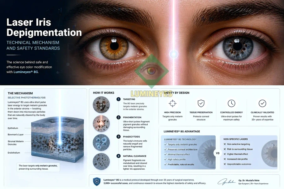
Iris appearance reflects pigment and the way light interacts with its tissue. Reducing the optical contribution of melanin can change that balance. It is more accurate to describe a change in pigment-related appearance than to promise that a predetermined blue or green layer will be uncovered in every eye.
The proposed sequence involves laser–pigment interaction followed by tissue responses and changes in pigment distribution or processing. Precisely where pigment goes, how much remains and what happens to neighbouring structures are biological questions—not conclusions that can be read from a photograph. Our discussion of the biological fate of pigment examines this distinction in more detail.
Results: lightening is not the same as choosing a colour
The useful question is not simply “Can brown eyes become lighter?” but “What range of change is plausible for this eye, and what would justify stopping?” Baseline pigment distribution, iris structure and the response to treatment can influence the appearance. A colour simulation or another person’s result cannot guarantee your final shade.
Shade
A desired blue, green or grey appearance is not a selectable pigment formula. Expectations should include the possibility of less change than hoped for.
Uniformity
Residual pigment, the central ring and differences between eyes deserve attention—not only the lightest part of a photograph.
Stability
An early image is not a long-term result. The time since treatment and subsequent clinical observations matter.
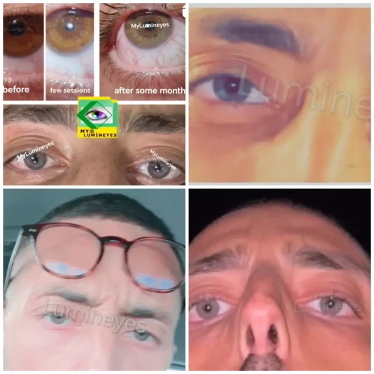
How to read before-and-after photographs
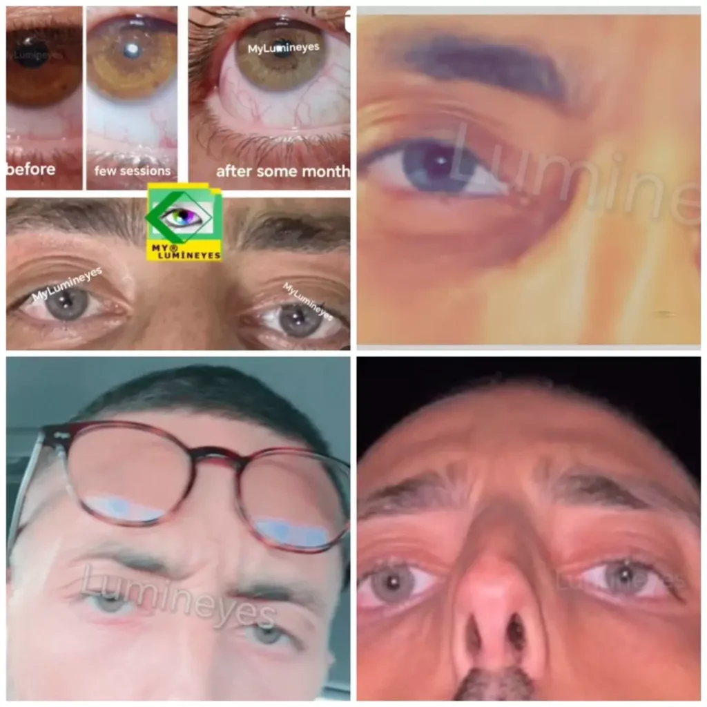
Compare photographs taken with similar lighting, camera settings, white balance and pupil size. Look for the treatment stage and follow-up date. A close-up can document an appearance, but cannot show intraocular pressure, visual-field function or the condition of the drainage angle.
Our before-and-after collection provides visual examples. Use them to discuss possible appearances with the ophthalmologist, not to calculate your personal success rate.
Is laser iris depigmentation permanent?
Do not approach pigment-reducing treatment as a reversible cosmetic trial. Returning to the original appearance cannot be promised. Equally, the word “permanent” does not guarantee a particular final shade, lifelong colour stability or freedom from delayed complications. These are separate questions that require separate follow-up information.
Assessment comes before a session count
A photograph can help start a conversation, but it cannot establish eligibility. An ophthalmic assessment should consider ocular history, previous surgery or inflammation, current medication, vision, pressure and relevant anatomical findings. Glaucoma, suspected outflow problems or active inflammation require particular caution and may rule out an elective intervention.
The MyLumineyes clinical evidence methodology distinguishes routine examination from additional testing prompted by clinical findings. Gonioscopy, anterior-segment imaging and other investigations should answer a clinical question; listing equipment is not a substitute for explaining what the examination found.
How many sessions—and how much time?
There is no universal session schedule attached to the word LID. Different reports describe different treatment sequences. A proposed schedule should state what counts as a session, how reassessment is arranged and which findings would change or end the plan. Time at the clinic and time needed to evaluate a biological response are not necessarily the same.
A clinically meaningful plan must allow a decision not to deliver more laser energy. Persistent pigment is not, on its own, a reason to continue; pressure, inflammation, visual function and the patient’s evolving risk assessment also matter.
Is laser iris depigmentation safe?
A single yes-or-no answer conceals important differences between protocols and patients. LID can be non-incisional, but that does not make it risk-free. Published reports include inflammation, pigment dispersion, marked pressure elevation and serious visual complications. The absence of an incision does not establish the absence of tissue injury.
One published case documented severe bilateral pigmentary glaucoma and a retinal complication after cosmetic iris laser treatment, requiring surgery and leaving persistent visual problems. It establishes that serious harm can occur; it does not provide a complication rate for all treated patients or identify the risk of every other protocol. Read the 2023 case report.
Why eye-pressure monitoring matters
Pressure is one important part of follow-up because pigment-related changes can affect aqueous drainage. A reassuring pressure measurement is useful, but it is not a complete assessment of ocular health or proof that future pressure will remain normal. Timing, repeated observations and the rest of the examination matter.
The Lumineyes early IOP monitoring framework explains how pressure observations inform clinical decisions. It is a description of monitoring practice, not a guarantee that monitoring prevents every complication.
What does published LID research actually show?
Both favourable clinical series and adverse-event reports belong in an honest account. They answer different questions. A treated cohort can describe outcomes in a defined setting; a complication report can identify an important hazard. Neither should be repurposed into a universal result for all laser-based iris procedures.
| Evidence | What it contributes | Boundary of interpretation |
|---|---|---|
| Grimaldos Ruiz, 2021 588 patients; 1,176 eyes | A prospective single-clinic series reported cosmetic outcomes after a specified laser protocol and described delayed, brief iritis in 25%. | These are that study’s observations—not Lumineyes results. Its nine-year development narrative does not mean every participant had nine years of follow-up. |
| Liu and colleagues, 2023 Individual adverse-event report | Documents severe pigmentary glaucoma and lasting visual consequences after cosmetic iris laser treatment. | A case report identifies possible harm but has no treated-population denominator from which to calculate incidence. |
| Lumineyes methodology documentation | Explains the author’s approach to selection, observation and interpretation of clinical records. | A methodological framework describes how evidence should be evaluated; it is not itself a comparative outcome trial. |
For a patient, the most revealing questions are practical: Which protocol was studied? How many patients were followed? For how long? Which adverse events were actively sought? What happened to patients who did not return? These questions tell you more than a large treatment number without a documented follow-up structure.
LID is not synonymous with Lumineyes
LID is a heterogeneous descriptive category. Lumineyes is Dr. Mustafa Mete’s separately articulated clinical framework, described in its research documentation as selective stromal melanin modulation with individualized, response-guided staging. That terminology states the intended approach; it is not a declaration that every other procedure called LID uses the same method.
The LID clinical-perspective article addresses this distinction directly. The selective stromal melanin modulation paper explains the proposed target and rationale, while the 8G Xtra technical discussion describes the evolution of the clinical framework.
Protocol-specific interpretation works in both directions. A complication following another procedure should not automatically be labelled a Lumineyes complication. Similarly, favourable results from another clinic cannot be presented as validation of Lumineyes. Technical distinctions are a reason to study protocols carefully—not a substitute for demonstrating their outcomes.
Why clinician-authored research belongs in this discussion
First-hand clinical documentation can make treatment decisions, terminology and unanswered questions accessible to scrutiny. Its value depends on what is documented: a clear method, traceable observations, defined outcomes and transparent limitations. Publication fees are not a measure of scientific quality. Neither payment nor publication venue replaces a critical reading of the methods and data.
Dr. Mete’s procedure-specific evidence commentary argues for distinguishing biological mechanisms and clinical protocols rather than judging procedures solely by their shared cosmetic aim. A mechanistic explanation, a case series and a comparative study make different contributions; recognizing those contributions is more useful than treating every document as interchangeable proof.
Cost, consultation and the questions worth asking
A LID quotation is meaningful only when its scope is clear. Ask whether it includes the initial examination, planned treatment stages, medication, scheduled pressure checks, longer-term reviews and assessment of unexpected symptoms. If travel is involved, clarify how care continues after you return home.
For clinic-specific arrangements, see the cost and risk guide. For the broader patient journey, visit the Lumineyes procedure guide. This page focuses on understanding LID and its evidence, rather than repeating the full treatment programme.
Bring these questions to your consultation
- What is the exact proposed protocol, and what evidence relates to it?
- Which findings could make me unsuitable?
- What appearance is plausible, and what cannot be promised?
- Which observations would lead you to postpone or stop treatment?
- Who assesses me if pressure or vision changes after I leave?
Laser iris depigmentation: common questions
Is LID the same as Lumineyes?
No. LID is a broad descriptive category covering different laser-based approaches to iris pigmentation. Lumineyes is a separately described clinical framework. Results and complications should be attributed to the protocol actually studied.
Does LID add blue or green pigment?
No. Its intended mechanism is to reduce the visible contribution of existing iris pigment, not to introduce a new coloured dye. An exact final shade cannot be guaranteed.
Does “non-incisional” mean no risk to the eye?
No. It describes the absence of a surgical incision, not the absence of biological effects. Published complications after cosmetic iris laser treatment include inflammation, high eye pressure and serious visual injury.
Can photographs establish whether I am a candidate?
No. Photographs may help an initial discussion, but eligibility requires an ophthalmic assessment. They cannot establish eye pressure, drainage-angle status or the full condition of the eye.
Is there a fixed number of sessions for dark brown eyes?
No universal schedule can be inferred from eye colour alone. Planning depends on the specific protocol, examination findings and the observed response. Further treatment may need to be deferred or stopped.
Can I return to my original eye colour?
Reversal to the original appearance cannot be promised. Pigment-reducing treatment should not be approached as a temporary cosmetic experiment.
References and further reading
- Grimaldos Ruiz P. Photoablative cosmetic iridoplasty: effective, safe, and predictable—eye color change in 1176 eyes. International Ophthalmology. 2021;41:1381–1393. PubMed. DOI: 10.1007/s10792-021-01693-5.
- Liu J, et al. Bilateral severe iatrogenic pigmentary glaucoma following laser treatment for cosmetic iris color change. American Journal of Ophthalmology Case Reports. 2023;32:101927. PubMed. DOI: 10.1016/j.ajoc.2023.101927.
- MyLumineyes Research. How clinical evidence is evaluated.
- MyLumineyes Research. Early IOP monitoring framework.
- MyLumineyes Research. Selective stromal melanin modulation.
- MyLumineyes Research. Biological fate of pigment in iris colour modification.
- MyLumineyes Research. Laser iris depigmentation: clinical perspective.
- MyLumineyes Research. Lumineyes 8G Xtra technical evolution.
- Mete M. Cosmetic Eye Colour Change Requires Procedure-Specific Evidence: A Commentary on Laser Iris Depigmentation and Keratopigmentation. Zenodo; 2026. Preprint. DOI: 10.5281/zenodo.23014030.