Cataract Surgery: Microsurgical Techniques, IOL Engineering & Clinical Recovery
Cataract extraction has evolved from basic lens removal into a sophisticated microsurgical refractive procedure. Replacing the opacified crystalline lens with an engineered intraocular lens (IOL) permanently restores optical transmission and resolves complex refractive errors in a single outpatient operation.
The human crystalline lens is a transparent, biconvex structure located directly behind the iris and pupil. Its optical clarity depends on the precise, regular geometric arrangement of specialized structural proteins called crystallins.
Over decades of metabolic activity, cumulative ultraviolet exposure, and oxidative stress, these crystallin proteins denature, misfold, and aggregate into high-molecular-weight clusters. This biological degradation scatters incoming light photons across the ocular media, degrading contrast sensitivity and turning crisp optical imagery into a dull, desaturated blur known as a cataract.
Clinical slit-lamp view: Dense nuclear and cortical lens opacification obstructing the optical visual pathway.
Clinical Morphologies & Pathophysiology of Cataracts
Cataracts do not present uniformly; their clinical behavior, surgical density, and visual impact depend on which anatomical zone of the lens undergoes opacification:
- Nuclear Sclerotic Cataracts: The most common age-related variant. Protein compaction begins in the central nucleus, turning from yellow to amber or brown (brunescent cataract). This induces a paradoxical "myopic shift" (temporary improvement in reading vision, often called "second sight"), while rapidly deteriorating distance vision and color saturation.
- Cortical Cataracts: Form in the lens cortex surrounding the nucleus. They manifest as radial, wedge-shaped spoke opacities pointing toward the center. Cortical spokes cause severe light scatter, resulting in daytime glare and nighttime starbursts around headlights.
- Posterior Subcapsular Cataracts (PSC): Form directly beneath the posterior capsule along the visual axis. PSCs progress rapidly, disproportionately impacting younger patients, diabetics, and individuals on systemic or inhaled corticosteroids. They cause severe near-vision impairment and daytime photophobia under bright sunlight.
Recognizing Cataract Symptoms: The Surgical Threshold
Cataract formation is gradual and insidious. Patients often adapt unconsciously to degraded vision until functional deficits become dangerous. Key clinical indicators include:
- Progressive, painless reduction in visual acuity that cannot be corrected with updated spectacles.
- Loss of color discrimination, where blues, purples, and pastels appear muted or yellowish.
- Severe glare disability and halos around oncoming headlights during night driving.
- Monocular diplopia (ghost images or double vision visible with one eye closed).
- Frequent, unstable refractive shifts requiring repeated eyeglass prescription changes.
The outdated historical concept of waiting for a cataract to "ripen" is medically obsolete. Modern surgery is indicated as soon as visual degradation compromises your safety, occupational capacity, driving independence, or daily reading. Operating before a cataract becomes hyper-mature (rock-hard) minimizes surgical ultrasound energy, reduces corneal endothelial cell trauma, and speeds visual recovery.
Surgical Modalities: Phacoemulsification vs. FLACS
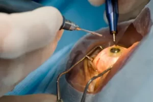
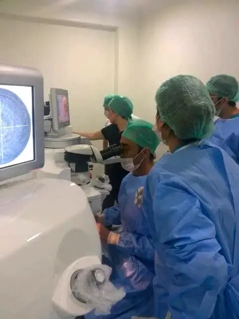
Modern cataract extraction is a sutureless, micro-incisional outpatient procedure performed under topical drop anesthesia with light intravenous sedation. The operation takes roughly 10 to 15 minutes per eye.
1. Micro-Incision Phacoemulsification (Standard Gold Standard)
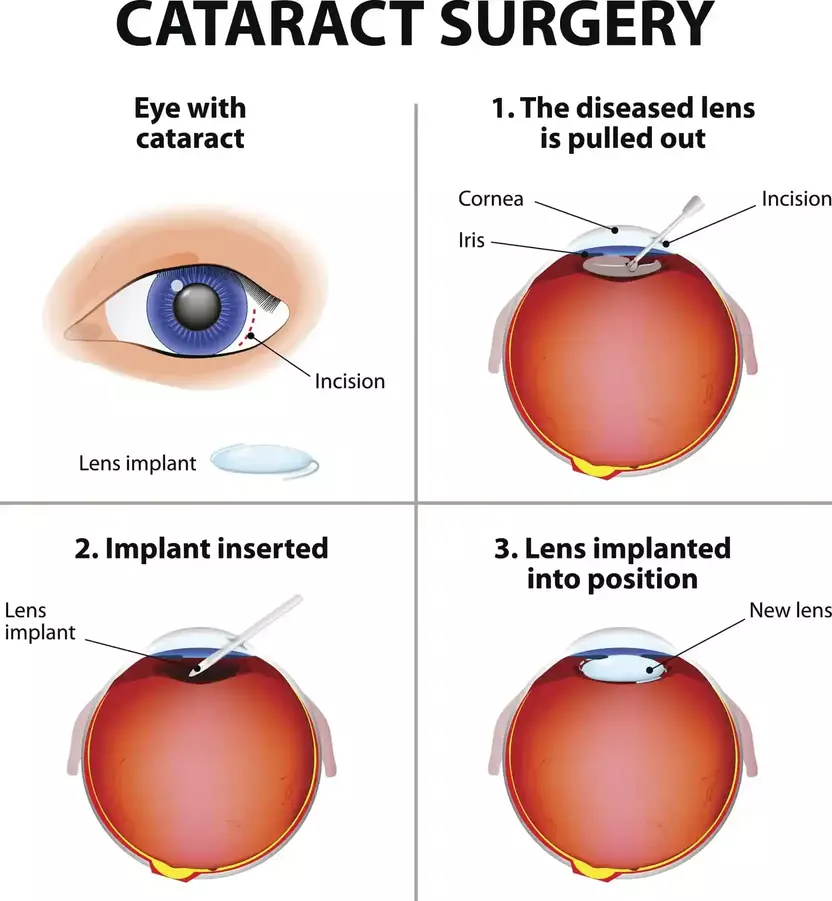
A self-sealing corneal micro-incision (2.2 to 2.4 mm) is created at the temporal limbus. Through a continuous curvilinear capsulorhexis (a circular opening in the front capsule), an ultrasonic titanium phacoemulsification probe emulsifies the hardened lens nucleus into microscopic particles, which are simultaneously aspirated. The capsular bag is vacuum-cleaned, leaving the crystal-clear posterior capsule intact to support the new artificial lens.
2. Femtosecond Laser-Assisted Cataract Surgery (FLACS)
FLACS integrates a computer-guided infrared laser to execute critical early surgical steps prior to phacoemulsification:
- Precision Laser Capsulotomy: Creates a perfectly centered, circular capsular opening to the exact sub-millimeter diameter, optimizing effective lens position (ELP) for premium multifocal and toric optics.
- Laser Nuclear Pre-Fragmentation: Divides the dense lens into quadrants or micro-cylinders, reducing the total ultrasonic energy (cumulative dissipated energy, CDE) required inside the eye by up to 40–50%.
- Astigmatic Keratotomy: Delivers arcuate laser incisions to correct low-grade corneal astigmatism concurrently.
Intraocular Lens (IOL) Options: Engineering Your Refractive Outcome
Cataract surgery provides a unique medical opportunity: by selecting an advanced intraocular lens, we can permanently eliminate lifelong refractive errors, including myopia, hyperopia, astigmatism, and presbyopia.
| Lens Category | Optical Mechanism | Visual Range | Postoperative Spectacle Dependence |
|---|---|---|---|
| Monofocal IOLs | Single spherical/aspheric optical focal point | Excellent distance vision | Reading glasses are required for computer (intermediate) and phone/books (near). |
| Toric IOLs | Asymmetric cylinder power aligned to corneal axis | Distance vision + Astigmatism neutralization | Eliminates corneal astigmatism; reading glasses still needed unless combined with multifocality. |
| Extended Depth of Focus (EDOF) | Elongated non-diffractive optical focal channel | Seamless intermediate (screen) and distance vision | Minimal nighttime halos; low-power reading glasses may be needed for fine print. |
| Trifocal "Smart" Lenses | Diffractive concentric rings splitting light to 3 foci | Crisp Far (infinity), Intermediate (60 cm), and Near (40 cm) | Complete spectacle independence in over 90% of suitable candidates. |
For patients seeking total independence from reading glasses, explore our detailed clinical guide on trifocal smart intraocular lens surgery to review optical dynamics, candidacy parameters, and night vision adaptation.
Postoperative Recovery Timeline and Clinical Care
Because incisions are microscopic and self-sealing, physical recovery begins almost immediately:
- First 24 to 48 Hours: Mild foreign-body sensation, light sensitivity, and fluctuating visual clarity are normal. The pupil remains partially dilated from surgical cycloplegics. Most patients notice dramatic clearing by the following morning.
- Weeks 1 to 4: Adherence to prescribed medicated eye drops is paramount. The standard protocol includes topical fluoroquinolone antibiotics for 7 to 10 days to prevent endophthalmitis, combined with topical corticosteroid and NSAID drops tapered over 4 weeks to control intraocular prostaglandins and prevent cystoid macular edema (CME).
- Activity Restrictions: Avoid rubbing the eye, heavy weightlifting (over 10 kg), bending upside down, and submerging the head in swimming pools or hot tubs during the first two weeks.
Complications & Management: Posterior Capsule Opacification (PCO)
Cataract surgery carries a success rate exceeding 98%. However, an artificial lens rests on living biological tissue. The most common delayed phenomenon is Posterior Capsule Opacification (PCO), occurring in 10% to 20% of patients within 1 to 3 years.
Residual microscopic lens epithelial cells migrate across the clear posterior capsule behind the IOL, causing it to wrinkle and haze (often misnamed a "secondary cataract"). PCO is permanently resolved in under 2 minutes using an in-office Nd:YAG Laser Capsulotomy. The painless laser creates a clear aperture in the center of the clouded capsule, instantly restoring crisp vision permanently with zero downtime.
⚠ Important Medical Distinction: Cataracts vs. Cosmetic Eye Procedures
Cataract surgery is an intraocular lens replacement procedure designed strictly to treat opacity of the crystalline lens and restore optical transmission. It has zero relation to cosmetic iris procedures. Non-invasive laser eye color change utilizes selective low-fluence energy to modify superficial melanin in the iris stroma for aesthetic heterochromia or cosmetic lightening. It does not penetrate the lens and cannot treat or cure cataracts.
Frequently Asked Questions: Cataract Surgery
Is cataract surgery painful?
No. The procedure is performed under topical anesthetic eye drops combined with mild systemic sedation. Patients feel slight fluid sensation and light pressure, but no sharp pain. Postoperative discomfort is mild and easily managed with over-the-counter paracetamol.
Can a cataract grow back after surgery?
No. Once the natural crystalline lens is removed, a biological cataract can never reform. If your vision blurs months or years later, it is almost certainly Posterior Capsule Opacification (PCO), which is cleared permanently in minutes with a quick Nd:YAG laser capsulotomy.
How long do intraocular lenses (IOLs) last?
Modern acrylic and silicone intraocular lenses are biocompatible and chemically inert. They are engineered to remain pristine and stable inside the capsular bag for the rest of your life without degrading, clouding, or requiring replacement.
Can both eyes be operated on the same day?
In standard practice, cataract surgeries are performed sequentially on separate days—typically 2 to 7 days apart. This allows the first eye to stabilize, confirms the predicted refractive outcome, and eliminates the risk of bilateral complications.
Medical Disclaimer: This comprehensive clinical overview is authored by Dr. Mustafa Mete for patient education and does not replace an individual slit-lamp examination or surgical biometry. If you experience progressive visual decline, glare, or sudden vision loss, consult a board-certified ophthalmic surgeon promptly.