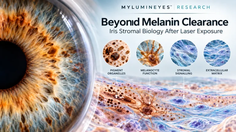
Beyond Melanin Clearance: Iris Stromal Biology After Laser Exposure
DOI: 10.5281/zenodo.23270055
Repository: Zenodo
Research record: Beyond Melanin Clearance
Version: 1.0 · October 2026
Research Overview
Visible reduction in iris pigmentation does not fully characterise the biological state of the exposed stroma. Human iris tissue contains melanocytes, fibroblasts, immune-associated populations, vascular structures and an extracellular matrix whose functions cannot be reduced to pigment content alone.
This research perspective examines the distinction between melanosome modification, melanocyte survival and function, stromal intercellular signalling, and extracellular matrix organisation. It integrates human iris co-culture evidence, cell-resolved tissue studies and procedure-specific histopathology while distinguishing direct ocular observations from experimental findings in other tissues.
The central objective is to identify testable biological questions rather than infer structural preservation or injury from visible pigmentation changes.
Keywords: iris stroma; melanocyte; fibroblast; pigment-targeted laser; extracellular matrix; stromal signalling; melanosomes; matrix remodelling; anterior-segment OCT.
Visible pigment modification is an optical observation. It does not independently establish melanocyte viability, paracrine activity or extracellular matrix recovery.
1. Introduction: Beyond the Visible Pigmentary Endpoint
The evaluation of laser-based iris pigmentation modification commonly focuses on apparent colour, pigment release, anterior-chamber findings and intraocular pressure. These are clinically relevant observations, but they do not fully characterise the irradiated tissue.
A more complete interpretation requires attention to the living iris stroma, including resident melanocytes, non-melanocytic stromal cells and extracellular matrix architecture.
The broader scientific analysis of laser eye color change provides the clinical and procedural context. The present article addresses a narrower question: what biological properties remain to be measured at the stromal exposure site after pigment modification?
2. Iris Colour, Pigment Content and Melanocyte Number
Iris colour is influenced by the amount, composition and anatomical distribution of melanin, together with tissue optical properties. Human histological research has demonstrated that visible iris colour does not consistently correspond to stromal melanocyte density.
Consequently, reduced apparent pigmentation cannot automatically be interpreted as melanocyte loss. Changes in melanosome content within surviving cells, altered pigment distribution and tissue optics can represent different biological explanations for an observed colour change.
This separation is consistent with the wider discussion of iris pigmentation, biological basis and long-term optical stability.
For a general histological reference, the AAO EyeWiki overview of ocular histology describes iris anatomy and its stromal cellular constituents. This anatomical resource is not evidence of a specific post-laser molecular response.
3. The Iris Stroma as a Multicellular Microenvironment
The iris stroma contains several interacting cell populations rather than a single pigment-bearing compartment. Contemporary cell-resolved investigations identify melanocytic and non-melanocytic stromal populations alongside immune-associated, vascular and muscular cell types.
These cellular atlases offer reference maps for designing future experiments. However, identifying a particular cell population or its baseline gene expression does not establish activation after laser exposure.
This distinction is especially important when interpreting the biological fate of pigment during iris color modification. Pigment transport and cellular survival are related research questions but require different experimental measurements.
4. Melanocyte–Fibroblast Crosstalk
Human ocular melanocyte experiments have demonstrated that neighbouring cells can influence melanogenic activity. Co-culture studies reported changes in pigment-related cellular function when melanocytes were cultured alongside fibroblasts or epithelial cells.
A particularly relevant study by Smith-Thomas and colleagues demonstrated fibroblast-dependent regulation of the melanogenic response to latanoprost in human iridial melanocytes. These pharmacological experiments support a role for stromal intercellular signalling.
They do not establish that pigment-targeted laser exposure activates the same pathways. Whether residual melanocytes retain their baseline signalling relationships after laser exposure remains an experimentally unresolved question.
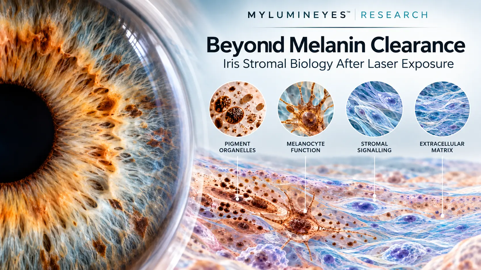
5. Four Distinct Biological Response Domains
A compartment-resolved research approach separates four principal endpoints. These domains may interact, but none should be used as a substitute measurement for another.
Pigment Organelles
Melanin quantity and distribution, melanosome integrity and visible pigmentation. These endpoints do not establish cellular viability.
Melanocyte Function
Cell survival, stress responses, melanogenic activity and cellular phenotype. Function cannot be inferred from colour alone.
Stromal Signalling
Communication among melanocytes, fibroblasts and other cells through locally acting molecular mediators.
Extracellular Matrix
Collagen architecture, matrix-regulatory proteins and functional tissue organisation.
Organelles
Function
Signalling
Architecture
6. What Human Iris Laser Histopathology Establishes
Historical observations after Nd:YAG iridotomy and argon peripheral iridoplasty demonstrate that laser procedures can produce different structural responses within human iris tissue.
Full-thickness iridotomy specimens have demonstrated mechanical disruption and tissue-specific injury patterns. Limited late histological observations after Nd:YAG exposure have also reported pigment-bearing stromal cells without fibrous scarring at sampled iridotomy sites.
In contrast, thermal argon peripheral iridoplasty specimens have demonstrated fibroblast-like proliferation, collagen deposition, collagen denaturation and vascular injury.
These findings cannot be pooled into a universal response profile for selective superficial stromal melanin modulation. The underlying ophthalmic laser mechanisms, tissue targets and exposure architecture must be distinguished before making biological comparisons.
| Evidence context | Reported observation | Interpretive limitation |
|---|---|---|
| Human Nd:YAG iridotomy | Mechanical stromal disruption and modality-dependent tissue injury | Full-thickness exposure differs from superficial pigment modulation |
| Late iridotomy specimens | Pigment-bearing cells and absence of fibrous scars at sampled sites | Small specimen numbers and restricted tissue sampling |
| Argon peripheral iridoplasty | Fibrocellular and collagen-associated tissue changes | Thermal peripheral contraction procedure, not selective depigmentation |
7. Extracellular Matrix Regulation in the Human Iris
The iris extracellular matrix is not simply a passive framework surrounding pigment cells. It contributes to tissue architecture and provides an environment for cellular regulation.
Human anterior-uveal immunohistochemical studies have identified matrix metalloproteinases, including MMP-1, MMP-2, MMP-3 and MMP-9, together with several tissue inhibitors of metalloproteinases.
These findings document endogenous matrix-regulatory proteins in normal tissue. They do not demonstrate increased post-laser enzyme activity or pathological matrix degradation.
Protein localisation, gene expression, enzymatic activity, collagen organisation and mechanical behaviour remain distinct measurements. Structural or functional consequences should not be inferred from a single molecular marker.
8. Cross-Tissue Evidence and the Limits of Transfer
Experimental research involving human dermal fibroblasts has demonstrated that certain laser exposures can influence paracrine mediators and collagen-lattice behaviour under controlled conditions.
Separately, studies of pigmented trabecular-meshwork cells have identified inflammatory and matrix-associated molecular responses to defined laser exposure.
These findings support the plausibility of experimentally testable signalling and matrix-related questions. They do not establish equivalent responses in native human iris stroma.
A biological mechanism observed in another tissue may suggest what to measure, but cannot determine the result of an iris-specific experiment.
9. A Testable Experimental Research Architecture
Future experimental work should pair precisely described exposure conditions with matched controls and predefined sampling intervals. The research question must distinguish primary pigment modification from secondary cellular and matrix-associated responses.
| Domain | Candidate measurements | Interpretive requirement |
|---|---|---|
| Melanosomes | Melanin quantification, electron microscopy | Distinguish organelle change from cell death |
| Melanocytes | Cell-type markers, viability assays, melanogenic activity | Validate survival and function separately |
| Paracrine signalling | Matched co-culture, secreted mediator assays | Demonstrate the pathway directly |
| Matrix regulation | MMP activity, TIMP measurements, collagen imaging | Distinguish expression from active remodelling |
| Structural imaging | Standardised AS-OCT, regional thickness and optical signal | Control for pigmentation and imaging variability |
These measurements should be evaluated using appropriate ex vivo or experimental models before mechanistic conclusions are transferred to clinical follow-up.
10. Structural Imaging and Quantitative Iris Phenotyping
Anterior-segment optical coherence tomography may help characterise regional iris thickness, stromal contour and optical tissue properties over time.
However, OCT signal intensity is affected by pigmentation, imaging conditions and scattering. It cannot directly establish collagen integrity, melanocyte viability or extracellular matrix enzyme activity.
The methodological distinction is relevant to L-OAT quantitative iris phenotyping, which addresses reproducible optical measurements and their interpretive boundaries.
Controlled correlations between tissue imaging and validated experimental assays would be necessary to determine whether specific imaging features reflect defined cellular or matrix-associated changes.
12. Scientific Boundaries and Remaining Uncertainties
The principal limitation of the existing evidence is the absence of sufficiently detailed human molecular studies following well-characterised selective superficial iris pigment-targeted exposure.
Normal-tissue cell atlases identify candidate populations and molecular pathways but do not measure post-treatment response. Historical human histopathology demonstrates procedure-specific structural changes but cannot establish the biological behaviour of a different exposure protocol.
Cross-tissue evidence offers experimental direction rather than proof of analogous iris responses. The available literature therefore cannot establish a common stromal molecular phenotype shared by all laser iris depigmentation methods.
13. Conclusion
The human iris stroma is a living cellular and extracellular environment whose biological state cannot be reduced to visible pigmentation.
Existing human iris research establishes the relevance of melanocyte–fibroblast interactions and endogenous matrix regulation. Experimental laser literature demonstrates that tissue responses may depend strongly on exposure modality and biological context.
The next scientific step is to distinguish pigment-organelle change, melanocyte fate, paracrine activity and matrix organisation through direct controlled measurements.
The appropriate scientific question is not whether pigment removal automatically preserves or damages the stroma, but which measurements can establish the biological consequences of a defined exposure.
Original Scientific Research Record
Beyond Melanin Clearance: Melanocyte–Fibroblast Crosstalk and Stromal Matrix Responses to Pigment-Targeted Laser Exposure.
Mete M. A Critical Translational Review. MyLumineyes Research, 2026.
Read the complete research record on Zenodo →
DOI: 10.5281/zenodo.23270055
Selected Scientific References
The following sources support the principal biological distinctions discussed here. The complete bibliography is available in the original research record.
- Anderson RR, Parrish JA. Selective photothermolysis. Science. 1983;220:524–527. DOI
- Wilkerson CL, et al. Melanocytes and iris color: light microscopic findings. Arch Ophthalmol. 1996;114:437–442. DOI
- Smith-Thomas L, et al. Latanoprost-induced pigmentation in human iridial melanocytes is fibroblast dependent. Exp Eye Res. 2004;78:973–985. DOI
- Makovoz B, et al. Neuroectoderm-derived iris muscle characterization at the single-cell resolution in native human iris. Nat Commun. 2025;16:10648. DOI
- Lan J, et al. Expression and distribution of matrix metalloproteinases and their inhibitors in the human iris and ciliary body. Br J Ophthalmol. 2003;87:208–211. DOI
- Sassani JW, et al. Histopathology of argon laser peripheral iridoplasty. Ophthalmic Surg. 1993;24:740–745. DOI
- Poon VKM, Huang L, Burd A. Biostimulation of dermal fibroblast by sublethal Q-switched Nd:YAG 532 nm laser. J Photochem Photobiol B. 2005;81:1–8. DOI
- Shimizu S, et al. Effect of pigmentation intensity of trabecular meshwork cells on mechanisms of micropulse laser trabeculoplasty. Sci Rep. 2022;12:10535. DOI