Blood–Aqueous Barrier Dynamics After Iris-Directed Laser Exposure
Abstract
The biological interpretation of anterior-chamber cells and flare following iris-directed laser exposure remains an important and insufficiently standardized problem. Cells and aqueous flare are conventional markers of anterior-segment inflammatory activity, but their presence at a single postoperative examination does not establish whether the eye is undergoing an expected transient tissue response, sustained blood–aqueous barrier dysfunction, pigment-related outflow stress, or a persistent inflammatory process. Laser flare photometry provides an objective method for quantifying aqueous protein-related light scatter and has demonstrated value in postoperative and uveitic monitoring, whereas conventional slit-lamp grading remains partly observer-dependent. [1][2][3]
Experimental and clinical laser studies show that iris-directed procedures can produce measurable but temporally variable anterior-chamber responses. In a rabbit model of anterior iris laser exposure, mild cells and flare were observed during early follow-up and were no longer detectable at one week. In a randomized human study of laser peripheral iridotomy, objectively measured aqueous flare increased at day 3, remained above baseline at one week, and subsequently approached baseline. These observations do not establish a universal post-laser inflammatory timeline; instead, they demonstrate that trajectory is procedure-specific and temporally structured. [4][5]
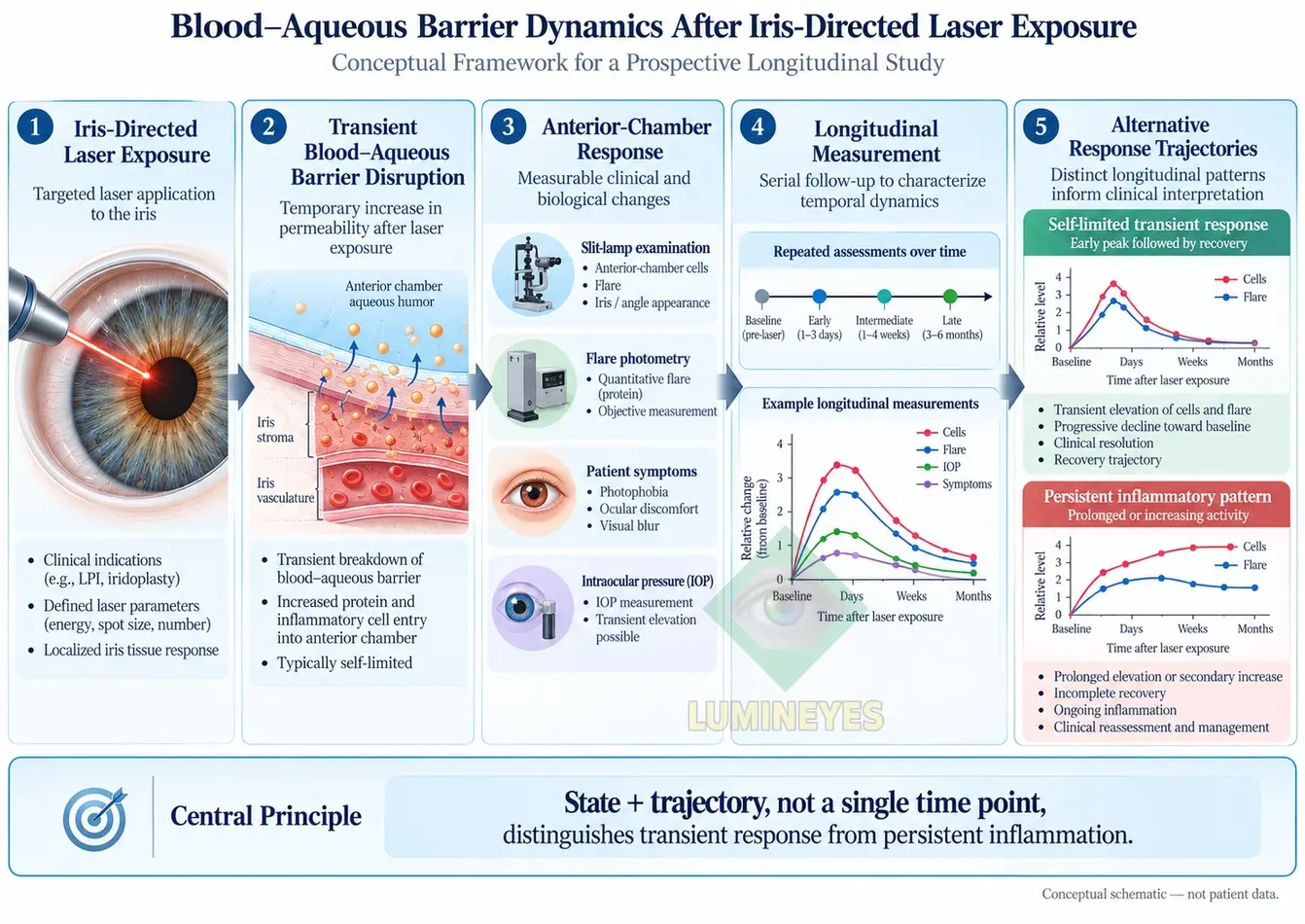
This article proposes a mechanistic and measurement framework for distinguishing transient post-laser anterior-chamber response from persistent inflammatory activity. The central proposition is that state plus trajectory is more informative than any isolated cell, flare, or intraocular-pressure measurement. Serial characterization of anterior-chamber cells, flare, intraocular pressure (IOP), corneal status, iris and angle findings, symptoms, medication context and, where available, laser flare photometry may permit a more rigorous description of recovery.
This framework does not establish that Lumineyes™ or any other iris-directed laser procedure produces a uniformly self-limited response. Rather, it defines the measurements required to test that hypothesis prospectively.
Keywords: blood–aqueous barrier; aqueous flare; anterior-chamber cells; laser flare photometry; iris laser; selective stromal melanin modulation; Lumineyes; anterior-segment inflammation; response kinetics; intraocular pressure.
1. Introduction
Anterior-chamber inflammation is commonly summarized using the terms cells and flare, yet these observations represent related but biologically distinct phenomena. Cells describe visible particulate cellular material within the aqueous compartment, whereas flare primarily reflects light scattering associated with increased aqueous protein concentration and other macromolecular changes. Both are routinely used to characterize intraocular inflammation, but clinical slit-lamp grading is semiquantitative and subject to observer variability. [1][3]
This issue is particularly relevant when interpreting the anterior-segment response after iris-directed laser procedures. Laser exposure may be followed by pigment liberation, cellular recruitment, altered aqueous protein dynamics, changes in aqueous outflow, or combinations of these processes. Consequently, the observation of “cells” in the anterior chamber does not establish cellular identity, and the observation of flare does not by itself establish the mechanism responsible.
The broader field of laser eye color change and staged iris pigment reduction includes heterogeneous approaches. Procedure-specific exposure characteristics, patient selection, monitoring and temporal staging therefore matter when clinical observations are interpreted. The methodology described here follows the same principle used in the MyLumineyes framework for evaluating procedure-specific clinical evidence: observations should be attributed to the protocol and context in which they were generated.
2. The Blood–Aqueous Barrier Is Not a Simple On/Off Barrier
The blood–aqueous barrier (BAB) is traditionally described through tight-junction systems involving the nonpigmented ciliary epithelium and the iris vasculature. Contemporary models are more nuanced: aqueous protein concentration reflects vascular and epithelial permeability, tissue distribution, aqueous production, iris and ciliary-body anatomy, and anterior-chamber clearance. [6][7]
This distinction matters because an increase in aqueous flare should not automatically be translated into the statement “the barrier has been damaged.” A more defensible interpretation is that increased flare represents altered anterior-segment protein dynamics that may accompany inflammatory or barrier-related disturbance.
Experimental injury, surgery, chemical irritation, uveitis and ophthalmic laser procedures may all alter aqueous protein content and BAB physiology. [6][8] Post-procedural flare is therefore most useful when treated as a dynamic biomarker.
3. What Laser Flare Photometry Actually Measures
Laser flare photometry (LFP) measures light scatter within the anterior chamber and provides a quantitative estimate of aqueous flare, commonly reported in photon counts per millisecond. The method improves objectivity compared with conventional slit-lamp flare grading and has been applied in uveitis and postoperative inflammatory monitoring. [1][2]
Clinical studies have demonstrated strong associations between laser flare measurements and aqueous albumin and immunoglobulin concentrations in inflammatory eyes, supporting the use of LFP as an objective marker of altered aqueous protein content. [9]
LFP is not a direct histological measurement of endothelial permeability and should not replace clinical examination. Measurements may be influenced by cataract, corneal opacity, pupil size, intraocular lenses, anterior-chamber depth and acquisition technique. [10]
4. Evidence That Iris-Directed Laser Exposure Can Produce a Transient Anterior-Chamber Response
Experimental ophthalmic laser evidence demonstrates that measurable anterior- chamber responses can occur after direct iris exposure. Yildirim and colleagues applied 532-nm Q-switched Nd:YAG laser exposure to the anterior iris in a rabbit model. Mild anterior-chamber cells and flare were reported during early follow-up, with these findings no longer detected at one week. [4]
This finding supports biological plausibility but should not be generalized directly to humans or to a distinct clinical laser protocol. It was a preclinical study conducted with a specific exposure architecture and discrete follow-up schedule.
Human glaucoma-laser literature provides complementary evidence. Gao et al. prospectively measured aqueous flare after laser peripheral iridotomy (LPI). Mean flare increased from approximately 5.5 photon counts/ms before treatment to approximately 12 photon counts/ms at postoperative day 3, remained elevated at one week, and subsequently approached baseline. [5]
The observation is important precisely because it argues against a universal statement that all post-laser inflammation follows a fixed 48–72-hour course. Procedure, exposure, ocular characteristics, postoperative medication and measurement method can alter the observed trajectory.
5. Transient Post-Laser Response Is Not Synonymous With Chronic Uveitis
A transient post-procedural inflammatory response and chronic uveitis are not equivalent biological states. Uveitis is interpreted through a broader constellation of inflammatory findings, clinical course, recurrence behavior, etiology, associated structural consequences and treatment response. Cells and flare are important components of that evaluation, but neither one independently defines chronic uveitic disease.
Standardized uveitis nomenclature provides useful grading systems for anterior-chamber cells and flare, but even standardized grades remain measurements of a state at a particular examination. [19]
Resolving trajectory
Increase → peak → progressive decline → return toward individual baseline.
Concerning trajectory
Persistence → failure to recover → secondary increase → recurrence or associated structural/functional abnormality.
LFP has demonstrated utility not only for measuring present inflammation but also for monitoring persistence and subclinical inflammatory activity in uveitic disorders. [11]
6. Cells, Pigment and Melanophages Must Not Be Conflated
A recurring interpretive problem in iris-directed laser research is the assumption that every visible anterior-chamber particle represents an inflammatory leukocyte. Following pigment-directed exposure, the clinically visible anterior chamber may theoretically contain inflammatory cells, free pigment, pigment-containing cells, cellular debris, erythrocytes following microvascular disturbance, or combinations of these components.
Routine slit-lamp examination cannot reliably determine particle identity in every circumstance. Experimental iris-laser studies demonstrate pigment alteration and biological processing after exposure, but they do not justify the universal statement that all clinically visible human post-laser “cells” are melanophages. [4]
This uncertainty is especially relevant to pigment clearance kinetics after laser iris depigmentation, because pigment redistribution, phagocytic processing and aqueous outflow are mechanistically related but should not be collapsed into a single unverified clearance pathway.
The preferred terminology is therefore descriptive: inflammatory cells, pigment, pigment-containing cells, or undifferentiated particulate material where identity is uncertain.
7. Why Intraocular Pressure Must Be Interpreted Together With Cells and Flare
Iris pigment release introduces an additional physiological domain: aqueous outflow. An eye may demonstrate inflammatory or particulate anterior-chamber activity while IOP remains within its baseline range. Conversely, pigment burden, inflammatory activity or altered trabecular function may coexist with clinically important pressure elevation.
Severe pigmentary glaucoma has been reported after some cosmetic iris laser procedures, including a bilateral case with markedly elevated IOP and substantial pigment dispersion that ultimately required glaucoma surgery. [12] A more recent case report has likewise described major IOP elevation following another laser iris depigmentation procedure. [13]
Such cases are important safety signals. They do not, however, establish a single complication rate for every laser iris protocol because published procedures differ in devices, exposure patterns, treatment intervals, postoperative management and reporting quality. [14][15]
The MyLumineyes Research Archive separately reports longitudinal clinical observations from the Lumineyes dataset, while explicitly distinguishing those retrospective observations from prospective validation and from population-level risk estimates.
“cells and flare are harmless” is not supported;
“any cells or flare predict glaucoma” is also not supported.
Interpretation requires IOP, angle/outflow findings, pigment burden, corneal status, symptoms, medication context and trajectory.
8. State Versus Trajectory: The Central Concept
The framework separates two dimensions.
State
What is present at a specific examination: cell grade, flare grade, quantitative flare, IOP, corneal clarity, angle findings, pigment burden and symptoms.
Trajectory
How that state changes: resolving, stable, progressive, recurrent or indeterminate.
Two eyes can therefore show the same cell grade at one visit while representing very different biological states:
Eye B: 2+ → 2+ → 3+ → persistent 3+
This principle is closely related to the Temporal Response Architecture (TRA), which separates elapsed time from biological state and emphasizes direction of change across serial observations.
9. Proposed Longitudinal Measurement Architecture
A prospective study of BAB-related recovery should not rely on a single postoperative examination. A multidomain protocol could combine clinical assessment with quantitative flare measurement where instrumentation is available.
| Domain | Candidate measurement | Biological question |
|---|---|---|
| Anterior-chamber cells | Standardized SUN-style slit-lamp grading | Is cellular activity increasing, stable or resolving? |
| Clinical flare | Standardized slit-lamp flare grading | Is visible protein-related scatter present? |
| Quantitative flare | Laser flare photometry | What is the magnitude and direction of aqueous protein-related scatter? |
| IOP | Goldmann applanation or validated equivalent | Is aqueous outflow physiology being affected? |
| Angle/outflow | Gonioscopy ± appropriate anterior-segment imaging | Is progressive pigment or outflow concern present? |
| Cornea | Slit lamp ± pachymetry/specular microscopy when indicated | Is the response associated with corneal or endothelial abnormality? |
| Symptoms | Photophobia, discomfort, pain, blur | Does symptom trajectory parallel objective recovery? |
| Medication context | Anti-inflammatory and pressure-lowering therapy | Is apparent recovery spontaneous or treatment-modified? |
| Exposure timing | Exact interval from preceding laser exposure | At what temporal phase was the observation obtained? |
Where endothelial measurements are included, methodology should follow reproducible acquisition and sampling principles such as those summarized in the MyLumineyes review of corneal endothelial assessment and specular microscopy.
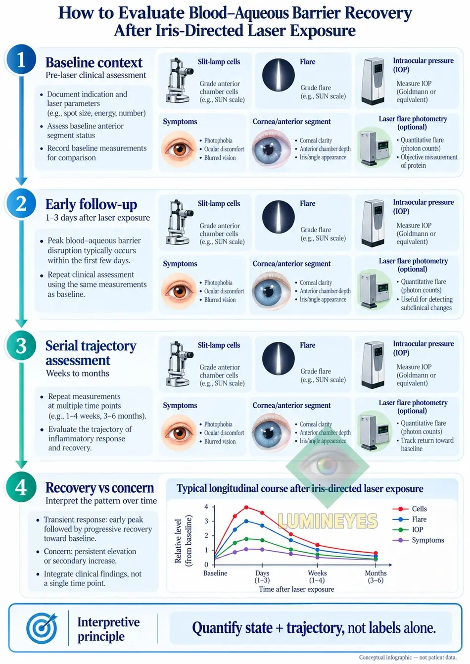
10. A More Defensible Definition of “Recovery”
The phrase BAB restoration should be used carefully. True molecular or histological restoration cannot be established solely by disappearance of visible cells or normalization of a single clinical variable.
For clinical research, a practical recovery phenotype could instead be defined as a multidomain return toward the participant’s baseline:
- declining or absent clinical cells;
- declining clinical flare;
- quantitative flare returning toward baseline where measurable;
- absence of progressive IOP abnormality;
- absence of increasing angle or outflow concern;
- absence of progressive corneal abnormality;
- symptom improvement or resolution; and
- absence of a secondary inflammatory rise during subsequent follow-up.
The terminology should therefore distinguish a clinical recovery phenotype from direct molecular proof of BAB restoration.
11. Why the Recovery Window Should Be Measured Rather Than Presumed
A fixed universal time window is attractive because it simplifies clinical interpretation, but the available literature does not justify the statement that cells and flare always peak at 48–72 hours and fully resolve within a fixed period after every iris-directed laser exposure.
In the rabbit anterior-iris laser model, early low-grade cells and flare were no longer detectable at one week. [4] In human LPI, quantitative flare was highest at the measured day-3 assessment, remained elevated at one week, and approached baseline during subsequent weeks. [5]
A systematic review of flare photometry after glaucoma laser and surgical procedures likewise found substantial procedure-dependent variation in the magnitude and duration of postoperative flare. [16]
12. Relationship to Lumineyes™, L-SAFE and Temporal Response Architecture
The published laser iris depigmentation literature encompasses heterogeneous procedural strategies. Lumineyes™ should therefore not be assumed to be biologically identical to every reported LID technique. Conversely, procedural difference cannot itself be used as proof of superior safety.
Within the MyLumineyes research program, L-SAFE anterior-segment safety staging addresses whether the integrated ocular state provides a safety reason to withhold further staged exposure. Its mandatory domains include IOP, anterior-chamber response, corneal status and angle/outflow status. L-SAFE remains a conceptual, unvalidated framework rather than a validated clinical decision rule.
TRA addresses broader temporal organization of biological response. The present BAB framework occupies a narrower layer:
| Framework | Primary research question |
|---|---|
| TRA | Which biological response domains evolve over time? |
| L-SAFE | Does the current integrated anterior-segment state justify withholding further exposure? |
| BAB Dynamics | Does the trajectory of cells, flare and related findings support recovery or persistent inflammatory activity? |
These frameworks are complementary but should not be presented as mutual validation.
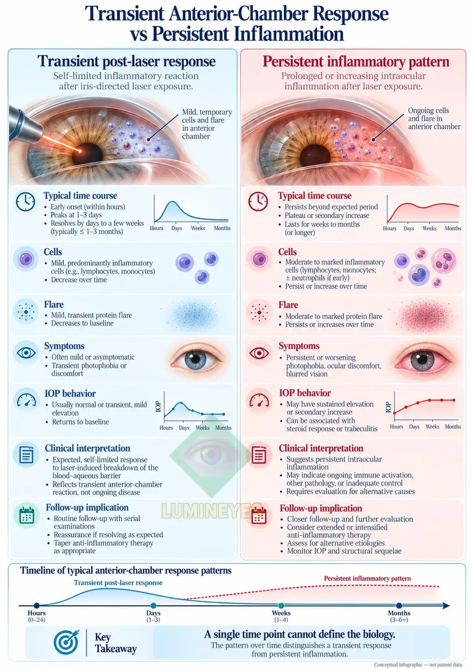
13. Transient Response Versus Persistent Inflammatory Pattern
| Feature | Transient post-laser response | Persistent inflammatory pattern |
|---|---|---|
| Temporal direction | Peak followed by progressive reduction | Persistent, recurrent or increasing |
| Cells | Declining across serial observations | Persistently abnormal or increasing |
| Flare | Declining toward baseline | Persistent elevation or secondary rise |
| Quantitative LFP | Downward recovery trajectory | Sustained abnormality or re-elevation |
| IOP | Normal or temporary disturbance that resolves | Persistent/recurrent pressure abnormality may coexist |
| Symptoms | Improving | Persistent or worsening |
| Cornea | No progressive abnormality | Persistent edema or other abnormality may coexist |
| Angle/outflow | No progressive evidence of dysfunction | Persistent pigment burden or outflow concern may be present |
| Interpretation | Compatible with resolving post-procedural response | Requires evaluation for ongoing inflammation or secondary pathology |
These categories are proposed research phenotypes, not validated diagnostic criteria or clinical treatment thresholds.
14. Proposed Prospective Validation Study
The central hypothesis is testable. A prospective cohort undergoing a defined iris-directed laser protocol could undergo standardized assessment at baseline, early post-exposure, approximately 24 hours, approximately 3 days, approximately one week, approximately 2–4 weeks and later intervals according to the observed response.
The schedule should be prospectively specified rather than reconstructed from whatever follow-up visits happen to be available.
Potential primary quantitative endpoint
Change in laser flare photometry from each participant’s individual baseline over time.
Potential secondary outcomes
- anterior-chamber cell grade;
- clinical flare grade;
- IOP trajectory;
- angle pigmentation and gonioscopic findings;
- corneal findings;
- symptom trajectory;
- medication use;
- association between exposure characteristics and response trajectory;
- time to return toward baseline; and
- frequency of persistent or secondary inflammatory elevation.
Repeated observations from two eyes of the same participant must not be treated as statistically independent. A hierarchical longitudinal mixed-effects model could be used to represent participant-level and eye-level dependence.
Such a study would move the hypothesis from mechanistic reasoning toward falsifiable prospective evidence.
15. What This Framework Does Not Claim
The purpose of this framework is to define the observations needed to distinguish competing biological interpretations. It does not establish a predetermined conclusion.
- It does not claim that Lumineyes™ cannot cause uveitis.
- It does not claim that post-laser cells are necessarily melanophages.
- It does not claim that all flare represents direct vascular endothelial injury.
- It does not claim that every post-laser response resolves within 48–72 hours.
- It does not claim that normal IOP proves normal anterior-segment physiology.
- It does not claim that disappearance of cells proves molecular restoration of the BAB.
- It does not dismiss complication reports from other LID procedures.
- It does not assume that Lumineyes™ and other LID procedures are biologically identical.
- It does not treat staging itself as proof of safety.
16. Scientific Interpretation of Adverse-Event Literature
Serious adverse-event reports remain part of the evidentiary landscape. Cases of severe pigmentary glaucoma following cosmetic iris laser procedures demonstrate that pigment-associated anterior-segment responses can have major clinical consequences. [12][13]
A case report can establish that a complication occurred following a reported exposure. Without an adequately characterized denominator and protocol, however, it cannot establish the complication rate of every procedure grouped under the same broad category.
Conversely, an uneventful clinical series cannot establish that rare complications are biologically impossible. The strongest long-term evidence requires procedure-specific denominators, standardized outcomes, defined follow-up and transparent attribution.
This distinction is also relevant to the regulatory and scientific interpretation of off-label laser use for eye-color modification: regulatory labeling, physician-directed use and the strength of clinical evidence are related questions but are not interchangeable.
17. Discussion
The central scientific contribution of this framework is the separation of observation from interpretation.
Cells are observations. Flare is an observation. IOP is an observation. Corneal findings and angle pigmentation are observations. Diagnoses such as chronic uveitis, transient post-procedural inflammation, pigment-related outflow dysfunction or incomplete recovery are interpretations derived from their combination and temporal behavior.
This distinction is especially important in emerging iris laser procedures, because existing literature remains heterogeneous and protocol-specific long-term data remain limited. [14][15]
The most defensible position is therefore neither “post-laser inflammation is harmless” nor “any post-laser inflammation represents pathological uveitis.” The biological meaning of an anterior-chamber response depends on magnitude, temporal direction, pressure behavior, structural findings, baseline state, exposure context and recovery pattern.
Laser flare photometry offers a particularly useful route toward objective quantification. Its strongest role is likely complementary to standardized clinical examination rather than as a replacement for it.
18. Conclusion
Anterior-chamber cells and flare following iris-directed laser exposure should be interpreted as dynamic biological signals rather than static labels. Existing experimental and clinical laser literature demonstrates that transient increases in anterior-chamber inflammatory markers can occur and subsequently resolve, but the magnitude and duration of these responses vary between procedures.
The scientifically relevant distinction is therefore not simply inflammation present versus absent, but resolving trajectory versus persistent or progressive trajectory.
Objective flare measurement, serial slit-lamp examination, IOP monitoring, corneal evaluation, angle/outflow assessment and careful characterization of particulate material provide complementary information.
For Lumineyes™ and other response-guided iris laser procedures, the next evidentiary step should be prospective longitudinal measurement rather than retrospective mechanistic certainty.
Trajectory describes what the biology is doing.
References
- Tugal-Tutkun I, Herbort CP. Laser flare photometry: a noninvasive, objective, and quantitative method to measure intraocular inflammation. Int Ophthalmol. 2010;30:453–464. doi:10.1007/s10792-009-9310-2. PubMed
- Ladas JG, Wheeler NC, Morhun PJ, Rimmer SO, Holland GN. Laser flare-cell photometry: methodology and clinical applications. Surv Ophthalmol. 2005;50:27–47. doi:10.1016/j.survophthal.2004.10.004. PubMed
- Oo HH, Mohan M, Song W, et al. Anterior chamber inflammation grading methods: a critical review. Surv Ophthalmol. 2024;69(2):253–264. doi:10.1016/j.survophthal.2023.10.005. PubMed
- Yildirim Y, Duzgun E, Kar T, et al. Evaluation of color-changing effect and complications after Nd:YAG laser application on iris surface. Med Sci Monit. 2016;22:107–114. doi:10.12659/MSM.895086. PMC
- Gao XB, Zhang XL, Chen G, Huang XK, Zhong XJ, Lin MK, Ge J. The blood–aqueous barrier changes after laser peripheral iridotomy or surgical peripheral iridectomy. Zhonghua Yan Ke Za Zhi. 2011;47(10):876–880. PMID:22321495. PubMed
- Freddo TF. A contemporary concept of the blood–aqueous barrier. Prog Retin Eye Res. 2013;32:181–195. doi:10.1016/j.preteyeres.2012.10.004. PubMed
- Coca-Prados M. The blood–aqueous barrier in health and disease. J Glaucoma. 2014;23:S36–S38. doi:10.1097/IJG.0000000000000107.
- Cunha-Vaz J. The blood-ocular barriers. Surv Ophthalmol. 1979;23:279–296. doi:10.1016/0039-6257(79)90158-9.
- Chiou AG, Florakis GJ, Herbort CP. Correlation between anterior chamber IgG/albumin concentrations and laser flare photometry in eyes with endogenous uveitis. Ophthalmologica. 1998;212(4):275–277. doi:10.1159/000027306. PubMed
- Wakefield D, Herbort CP, Tugal-Tutkun I, Zierhut M. Controversies in ocular inflammation and immunology: laser flare photometry. Ocul Immunol Inflamm. 2010;18(5):334–340. doi:10.3109/09273948.2010.512994. PubMed
- Kesim C, Chehab Z, Hasanreisoglu M. Laser flare photometry in uveitis. Saudi J Ophthalmol. 2022;36(4):337–343. doi:10.4103/sjopt.sjopt_119_22. PubMed
- Liu J, Korban S, Moster MR, Rhéaume MA, Wang Q. Bilateral severe iatrogenic pigmentary glaucoma following laser treatment for cosmetic iris color change. Am J Ophthalmol Case Rep. 2023;32:101927. doi:10.1016/j.ajoc.2023.101927. PubMed
- Jaber W, Nemet M, Waisbourd M. Laser iris depigmentation resulting in markedly elevated intraocular pressure and herpes simplex keratitis with corneal scarring: case report. Case Rep Ophthalmol. 2026;17:312–317. doi:10.1159/000551226. PubMed
- D'Oria F, Abu-Mustafa SK, Alio JL. Cosmetic change of the apparent color of the eye: a review on surgical alternatives, outcomes and complications. Ophthalmol Ther. 2022;11:465–477. doi:10.1007/s40123-022-00458-2. PubMed
- D'Oria F, Alio JL. Surgical techniques for cosmetic eye color change: a narrative review. Ophthalmol Ther. 2025;14:1685–1694. doi:10.1007/s40123-025-01177-0. PubMed
- Swampillai AJ, Nigam C, Dowse E, Lim KS. Quantification of postoperative inflammation in laser and surgical procedures for glaucoma with laser flare photometry: a review of the literature. Curr Eye Res. 2024;49(3):225–234. doi:10.1080/02713683.2023.2282940. PubMed
- Mete M. Temporal Response Architecture After Laser-Based Iris Pigment Modification. MyLumineyes Research / Zenodo. 2026. Research record
- Mete M. Lumineyes Anterior-Segment Safety Staging (L-SAFE): A Proposed Framework for Pre-Exposure Assessment in Laser Iris Pigment Reduction. MyLumineyes Research / Zenodo. 2026. Research record
- Jabs DA, Nussenblatt RB, Rosenbaum JT; SUN Working Group. Standardization of uveitis nomenclature for reporting clinical data: results of the First International Workshop. Am J Ophthalmol. 2005;140(3):509–516. doi:10.1016/j.ajo.2005.03.057. PubMed