
Corneal Endothelial Assessment and Specular Microscopy: Principles, Measurements, Methodological Considerations, and Clinical Context
Abstract
The corneal endothelium is a specialized cellular layer that plays an essential role in maintaining corneal transparency. Because adult human endothelial cells have limited proliferative capacity, objective assessment of endothelial structure may be clinically relevant in appropriate ophthalmic settings.
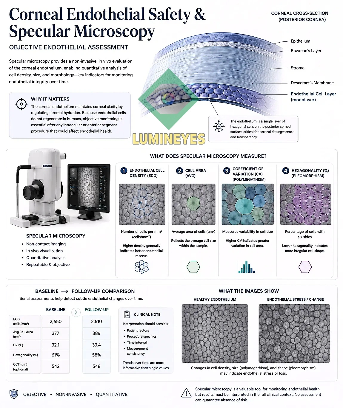
Specular microscopy provides in-vivo imaging of the endothelial mosaic and permits quantitative assessment of parameters including endothelial cell density (ECD), mean cell area, coefficient of variation (CV), and percentage of hexagonal cells. These measurements can provide useful information about endothelial morphology, but their interpretation depends on image quality, sampling, analysis methodology, and the imaging system used.
This scholarly narrative review summarizes the principles of specular microscopy and discusses how endothelial measurements can be interpreted when objective corneal assessment is clinically relevant. Particular attention is given to measurement methodology, sampling, image quality, analysis approaches, baseline assessment, and longitudinal interpretation.
This article does not present MyLumineyes endothelial measurements and does not claim that specular microscopy alone establishes the safety of any ophthalmic procedure.
1. Why the Corneal Endothelium Matters
The corneal endothelium forms a single cellular layer on the posterior surface of the cornea. Its physiological function is closely associated with regulation of corneal hydration and preservation of optical clarity [1,2].
Unlike tissues with substantial regenerative capacity, the adult human corneal endothelium has limited ability to replace lost cells through cell division. When endothelial cells are lost or injured, surviving cells can enlarge and spread to maintain continuity of the endothelial layer [1,2].
This biological characteristic is one reason why endothelial status can be clinically relevant when evaluating patients before or after procedures that may affect the anterior segment.
2. What Is Specular Microscopy?
Specular microscopy is an imaging technique that permits visualization of the corneal endothelial cell mosaic in vivo. The technique uses reflected light from the endothelial interface to produce an image suitable for morphological analysis.
Depending on the instrument and analysis software, endothelial images may be evaluated using automated, semi-automated, or manual approaches. These approaches are not necessarily interchangeable.
Methodological literature has emphasized the importance of standardization and validation because different instruments can use different magnification, calibration, image-acquisition, and analysis procedures. [1]
3. What Does Specular Microscopy Measure?
| Parameter | What It Represents | Why It Is Useful |
|---|---|---|
| Endothelial Cell Density (ECD) | Estimated number of endothelial cells per square millimeter. | Provides a quantitative estimate of endothelial cellular density. |
| Mean Cell Area | Average surface area of the analyzed endothelial cells. | Provides complementary information to cell density and cellular enlargement. |
| Coefficient of Variation (CV) | Variation in endothelial cell area. | Used to describe polymegathism, or variation in cell size. |
| Hexagonality | Percentage of analyzed cells with six sides. | Provides information about endothelial cell-shape distribution and pleomorphism. |
4. Endothelial Cell Density Is Not the Whole Story
ECD is one of the most commonly reported parameters in endothelial assessment, but it should not be interpreted in isolation.
The endothelial mosaic contains cells with different sizes and shapes. As endothelial cells are lost, surviving cells may enlarge and redistribute. Consequently, changes in mean cell area, cell-size variability, and cell-shape distribution can provide additional morphological information [1,2].
A comprehensive endothelial assessment therefore considers several measurements rather than reducing the examination to a single numerical threshold.
| Measurement | Question It Helps Address |
|---|---|
| ECD | How many endothelial cells are estimated to be present per unit area? |
| Mean cell area | How large are the analyzed cells on average? |
| Coefficient of variation | How variable are individual cell sizes? |
| Hexagonality | How is the distribution of endothelial cell shapes characterized? |
5. Why Measurement Methodology Matters
One of the most important points in interpreting specular microscopy is that an endothelial measurement is not independent of the way it was obtained.
Different instruments and analysis methods can produce systematically different ECD values. Comparative research has demonstrated measurable differences between non-contact specular microscopes and their associated analysis methods [3].
Other work has examined how measurement area and analysis methodology affect repeatability and interpretation [4].
6. Sampling and the Number of Cells Analyzed
Specular microscopy evaluates a sample of the endothelial mosaic rather than every endothelial cell in the cornea. The number of cells included in the analysis can therefore influence the precision of the resulting estimate.
Methodological research has demonstrated that estimates based on relatively small cell counts can show greater variability, while larger samples can improve the stability of estimated endothelial cell density [5].
This is particularly relevant when comparing measurements over time. If one examination is based on a substantially different sampling approach from another, apparent numerical differences should be interpreted cautiously.
7. Image Quality and Analysis
A numerical result can only be as reliable as the image and analysis from which it was derived. Poor cell visibility, inadequate image boundaries, segmentation errors, and differences in the way cells are identified can all influence the final result.
Specular microscopy literature has therefore examined not only the instruments themselves, but also the reproducibility of analysis methods and the role of the examiner [6].
Automated analysis can improve efficiency, but automation does not eliminate the need for appropriate image-quality assessment. Manual or semi-automated review may remain useful when cell boundaries are difficult to identify or when automated segmentation is uncertain.
Recent methodological work has compared manual and automated approaches to endothelial morphometry and demonstrated why the method of cell recognition should be considered when interpreting measurements [6].
8. Baseline Assessment and Longitudinal Follow-up
When endothelial monitoring is clinically appropriate, a baseline examination can provide an individual reference point against which subsequent measurements may be considered.
Follow-up measurements are most informative when obtained under comparable conditions. Ideally, the same instrument and a consistent acquisition and analysis methodology are used when the purpose is to evaluate change over time [3,4].
| Stage | Primary Purpose | Interpretive Consideration |
|---|---|---|
| Baseline | Establish an individual endothelial reference. | Consider existing ocular history, corneal findings, age, and measurement quality. |
| Follow-up | Compare later measurements with the established reference. | Use consistent methodology whenever possible. |
| Longitudinal interpretation | Determine whether observed changes are persistent or reproducible. | Consider measurement variability before attributing a difference to a clinical intervention. |
9. Clinical Context in Anterior-Segment Laser Procedures
Procedures involving the anterior segment require attention to the anatomical relationship between the treated structure and surrounding ocular tissues. Depending on the procedure, relevant considerations may include the cornea, anterior chamber, iris, lens, intraocular pressure, and other ocular structures.
In the context of laser eye color change , objective endothelial assessment may provide information that can be considered as part of a broader ophthalmic evaluation rather than as a single-test determination of safety.
The clinical workflow surrounding laser eye color change involves individualized assessment, treatment planning, and follow-up. A separate clinical guide to changing eye color with laser discusses the physician-guided decision-making process in greater detail.
The biological mechanism of laser iris depigmentation is a separate scientific topic involving the interaction between laser energy and iris pigmentation. Endothelial assessment should not be confused with the biological mechanism of iris pigment reduction itself.
10. What Specular Microscopy Can—and Cannot—Tell Us
| Can Help Assess | Cannot Establish by Itself |
|---|---|
| Endothelial cell density | Overall ocular safety of a procedure |
| Mean cell area | Absence of every possible ocular complication |
| Cell-size variability | Individual treatment suitability |
| Cell-shape distribution | Causality for a change without clinical correlation |
| Longitudinal morphological change | Long-term safety from a single examination |
11. Clinical Assessment Is Broader Than One Measurement
Specular microscopy addresses one specific component of anterior-segment assessment. It does not replace comprehensive ophthalmic examination or other measurements that may be clinically appropriate.
In laser eye color change, the broader clinical assessment includes individualized consideration of ocular anatomy, iris pigmentation, treatment objectives, and treatment response. The main Laser Eye Color Change clinical overview describes this broader procedural framework.
For readers specifically interested in clinical research methodology, the MyLumineyes Research archive provides a separate collection of clinical and scientific documentation.
12. A More Careful Way to Interpret Endothelial Data
A useful endothelial assessment should therefore answer several questions rather than simply asking whether a number is “normal.”
- Was the image of sufficient quality for reliable analysis?
- How many cells were included in the analysis?
- Which instrument and analysis method were used?
- Was the same methodology used at follow-up?
- Are ECD and morphological parameters being interpreted together?
- Could measurement variability explain part of the observed difference?
- What does the result mean in the context of the individual patient's ocular findings?
Specular microscopy is most useful when it transforms endothelial assessment from a purely qualitative impression into a documented, reproducible, quantitatively interpretable observation. The measurement itself, however, must always be understood within the limitations of the instrument, sampling method, image quality, and clinical context.
13. Conclusion
Corneal endothelial assessment is an important component of modern anterior-segment ophthalmology when clinically indicated. Specular microscopy allows in-vivo visualization of the endothelial mosaic and provides quantitative measurements including endothelial cell density, mean cell area, coefficient of variation, and hexagonality.
The scientific value of these measurements depends not only on the numbers themselves, but also on how those numbers are obtained. Differences between instruments, sampling areas, image quality, cell-count thresholds, and analysis approaches can affect the resulting measurements.
For this reason, longitudinal endothelial assessment is strongest when measurements are obtained using a consistent methodology and interpreted alongside the patient's complete ophthalmic findings.
Specular microscopy should therefore be regarded as an objective assessment tool within a broader clinical framework—not as a standalone declaration of procedural safety.
References
- McCarey BE, Edelhauser HF, Lynn MJ. Review of corneal endothelial specular microscopy for FDA clinical trials of refractive procedures, surgical devices, and new intraocular drugs and solutions. Cornea. 2008;27(1):1-16. doi: 10.1097/ICO.0b013e31815892da . PMID: 18245960. PubMed .
- Chaurasia S, Vanathi M. Specular microscopy in clinical practice. Indian J Ophthalmol. 2021;69(3):517-524. doi: 10.4103/ijo.IJO_574_20 . PMID: 33595465. PubMed .
- de Sanctis U, Machetta F, Razzano L, Dalmasso P, Grignolo FM. Corneal endothelium evaluation with 2 noncontact specular microscopes and their semiautomated methods of analysis. Cornea. 2006;25(5):501-506. doi: 10.1097/01.ico.0000214217.83119.af . PMID: 16783136. PubMed .
- Ding X, Huang Q, Zheng Y, Jiang Y, Huang S, He M. Measurement area and repeatability of semiautomated assessment of corneal endothelium in the Topcon specular microscope SP-2000P and IMAGEnet system. Cornea. 2012;31(10):1111-1118. doi: 10.1097/ICO.0b013e31820e42b5 . PMID: 22902488. PubMed .
- Doughty MJ, Müller A, Zaman ML. Assessment of the reliability of human corneal endothelial cell-density estimates using a noncontact specular microscope. Cornea. 2000;19(2):148-158. doi: 10.1097/00003226-200003000-00006 . PMID: 10746445. PubMed .
- Rizzo GC, Di Grassi R, Ponzini E, Tavazzi S, Zeri F. Corneal endothelial microscopy: Does a manual recognition of the endothelial cells help the morphometric analysis compared to a fully automatic approach? Vision. 2024;8(4):64. doi: 10.3390/vision8040064 . PMID: 39585114. PubMed . Full text .
Author Disclosure and Educational Note
This scholarly article is a literature-based narrative review authored by Mustafa Mete, MD. The author is involved in the clinical development and medical oversight of MyLumineyes.
This article does not present MyLumineyes clinical endothelial measurements, does not report a MyLumineyes patient dataset, and does not claim a specific endothelial outcome for MyLumineyes treatment.
Specular microscopy findings require interpretation by an appropriately qualified eye-care professional and should be considered together with the complete clinical examination.
Limitations
This article is a narrative review rather than a systematic review or meta-analysis. It does not report original experimental data, prospective clinical trial data, or a quantitative synthesis of published studies.
The interpretation of corneal endothelial measurements should take into account the imaging instrument, sampling method, image quality, analysis methodology, and the broader clinical context.
The illustrative numerical values shown in Figure 1 are examples only and do not represent clinical data.