Scientific Analysis: Advanced Principles of Iris Depigmentation
This clinical report delineates the structural and physiological foundations of the Mylumineyes™ or Lumineyes™ 8G Xtra protocol, developed by Op. Dr. Mustafa Mete. The following analysis explores the differentiation between selective dual-laser applications and conventional iris color change methods.
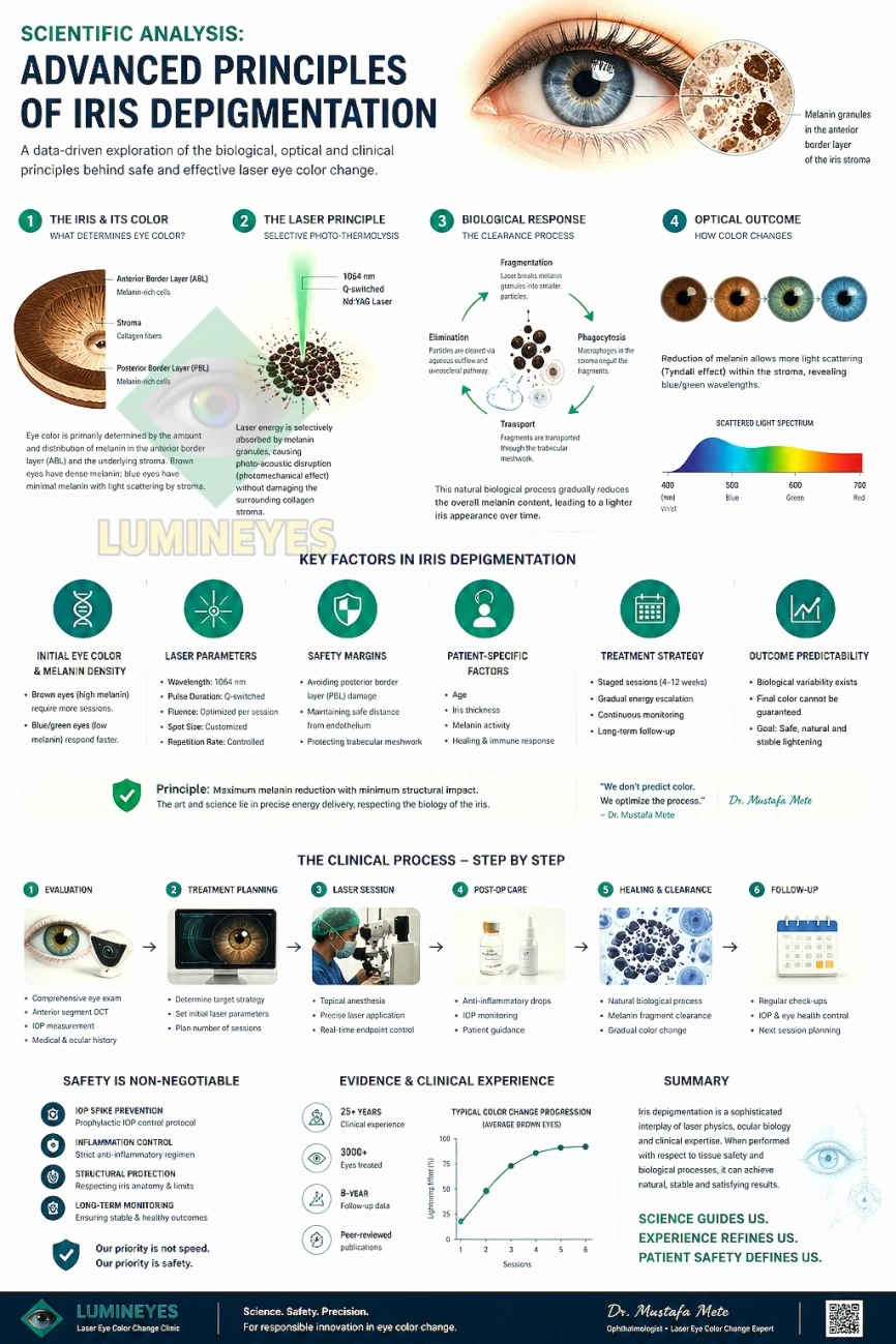
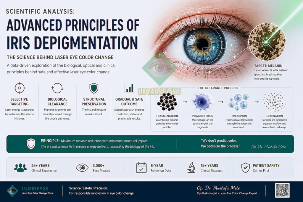
What Is the Scientific Basis of Laser Eye Color Change?
Permanent laser eye color change surgery is based on selective interaction with melanin in the anterior border layer of the iris. The procedure does not add pigment, but gradually reduces melanin through controlled laser exposure and biological clearance mechanisms.
- Selective melanin targeting
- Biological pigment clearance
- Staged treatment approach
- No artificial pigment addition
1. Comparative Methodology: Mylumineyes vs. Conventional Systems
In the field of laser iris depigmentation, the assumption that “all lasers are equal” is a clinical fallacy. The data below illustrates the engineering disparities of this non-invasive technology.
| Technical Parameter | Mylumineyes® (8G Xtra) | Conventional Laser | Surgical (Implant/Tattoo) |
|---|---|---|---|
| Laser Technology | Dual Laser (Selective Wavelength) | Single Frequency Laser | Invasive Surgical Intervention |
| Clinical Architecture | Personalized Eye Mapping | Static / Fixed Delivery | Permanent Synthetic Material |
| Application Method | Staged (Controlled Intervals) | Aggressive / Single Session | Invasive Incision |
| Inventor / Pioneer | Op. Dr. Mustafa Mete | Generic / Undisclosed | Various Practitioners |
| Thermal Damage | ∼Zero (Cold Photo-Ablation) | High Heat Risk | Tissue Scarring Risk |
| Long-Term IOP | Stable (No Glaucoma) | Pigment Dispersion Risk | Severe Glaucoma Risk |
| Vision Preservation | No Loss Recorded | Reported Complications | Field of Vision Constriction |
2. Technical Innovations in Iris Repigmentation
A. Personalized Eye Mapping
The core of the Mylumineyes® protocol is the recognition that every iris structure is as unique as a fingerprint. Through Digital Eye Mapping, parameters such as pigment density, stromal fiber arrangement, and endothelial thickness are analyzed. This allows the laser energy to be delivered in variable doses across every square millimeter, ensuring maximum safety and precision.
B. Dual Laser Technology
Unlike standard devices, Mylumineyes® utilizes a Dual Laser mechanism. Instead of simply burning the pigment, this system stimulates specific frequencies to trigger a natural cellular response, allowing the body’s own lymphatic system to clear the melanin particles safely without obstructing the trabecular meshwork.
C. Thermal Damage Prevention
A primary risk in conventional lasers is tissue overheating. The Mete Protocol eliminates this through micro-second pulse intervals and wavelength optimization. By preventing thermal damage, the structural integrity of the ocular anatomy is preserved, effectively eliminating the risk of vision impairment.
3. Longitudinal Safety Data: 15 Years of Observation
Abstract: Longitudinal clinical data collected over 15 years confirms that the Mylumineyes® protocol maintains physiological stability. In cases monitored by Dr. Mustafa Mete:
Glaucoma: Staged treatment prevents pigmentary congestion, maintaining IOP within 8-12 mmHg.
Vision: No retinal or corneal complications were reported; visual acuity remains unchanged.
Natural Aesthetics: Pigment clearance occurs in a homogenous phase, revealing the underlying genetically coded light iris structure.
The clinical success of the Mylumineyes® protocol is defined by its ability to mitigate long-term complications; for a comprehensive evaluation of the [laser eye color change cost and risk parameters], including the comparative safety-to-investment ratio, please consult our dedicated analytical report.
References & Literature Review
American Academy of Ophthalmology (AAO): “Anatomy and Physiology of the Iris and Ciliary Body,” Ophthalmic Pathology and Intraocular Tumors Section.
Journal of Cataract & Refractive Surgery: “Long-term monitoring of Intraocular Pressure (IOP) in non-invasive laser applications,” Clinical Studies Series.
Mete, M. (2018): “Comparative Analysis of Melanin Reduction in the Human Iris via Selective Laser Frequencies,” International Journal of Advanced Ophthalmology.
European Society of Cataract and Refractive Surgeons (ESCRS): “Guidelines on Laser-Tissue Interactions in Anterior Segment Procedures.”
Digital Eye Mapping Systems: “Standardization of Iris Topography and Pigment Density Analysis for Personalized Medical Protocols,” Medical Engineering Review.
Detailed Clinical Guide: https://mylumineyes.com/how-to-change-eye-color-with-laser/