What Is Melanin and How Does It Affect Eye Color?
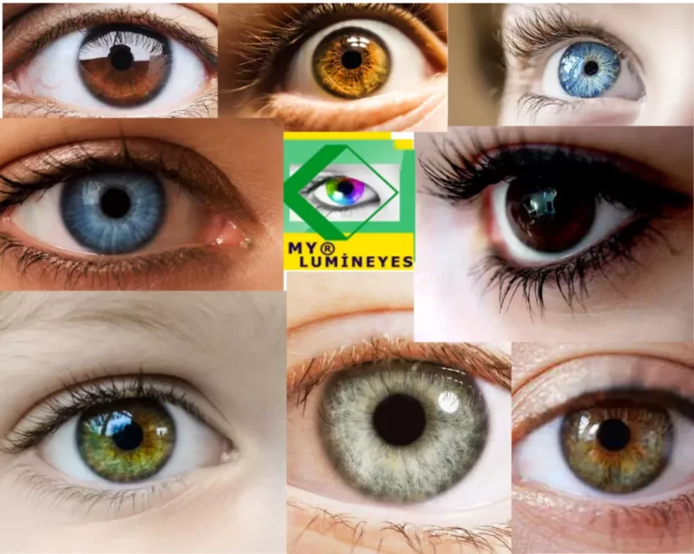
Melanin is the natural pigment that gives color to the iris, skin and hair. In the eyes, melanin is concentrated in specific layers of the iris and plays a central role in whether a person has blue, green, hazel or brown eyes. The amount, type and distribution of melanin help define the spectrum of human eye color.
From a medical perspective, eye color is not just a cosmetic feature. It reflects how light is absorbed and scattered inside the iris. This is why two people with brown eyes may still have very different depth, brightness and patterns when you look closely.

Types of Melanin in the Eye
There are two main types of melanin that influence pigmentation:
Eumelanin
A dark brown–black pigment. Higher amounts of eumelanin generally contribute to dark brown eyes.
Pheomelanin
A red–yellow pigment. Its contribution to green, amber and hazel eyes is complex; these colors cannot be explained by pheomelanin alone.
The mix and location of these pigments within the iris influence how the eye looks under different lighting conditions. Visible differences between common eye colors are especially associated with pigmentation in the front layers of the iris.
How Melanin Creates Different Eye Colors
Dark Brown Eyes
High melanin content absorbs much of the incoming light, resulting in deep, dark eyes. These are the most common worldwide.
Light Brown / Hazel Eyes
Moderate melanin with some light scattering. These eyes can look lighter in sunlight and darker indoors.
Green Eyes
Lower or uneven melanin levels combined with light scattering create a mixed, relatively rare appearance.
Blue / Grey Eyes
Very low melanin in the front layers of the iris. Much of the perceived color comes from light scattering, not blue pigment itself.
If you want to better understand where your own eyes fit in this spectrum, our guide to identifying your eye color explains mixed tones and iris patterns.
Melanin may also be distributed unevenly between the two eyes or within different parts of a single iris. This variation is known as heterochromia, which may be present from birth or develop later in life.
Melanin, Light Scattering and Eye Brightness
The iris does not contain blue or green dye. Instead, melanin controls how much light is absorbed, while the microscopic structure of the iris influences how the remaining light is scattered. This complex interaction is what creates bright blue, grey, green or hazel tones.
In eyes with low melanin in the front layers, more light is scattered back to the observer, making the iris appear lighter and more detailed. In heavily pigmented eyes, more light is absorbed, creating a darker but very intense gaze.
Photographs can make the same iris appear different because of changes in flash, exposure, white balance, viewing distance and pupil size. A photograph alone cannot measure iris melanin density.
Can Melanin in the Eye Change Over Time?
In infancy and early childhood, melanin in the iris can increase, which is why many babies are born with lighter eyes that darken over the first years of life. In adults, natural and dramatic color shifts are uncommon, but subtle changes in brightness or shade may be noticed.
Lighting conditions, pupil size, certain eye diseases, medications or pigment dispersion can affect how the iris appears. Our explanation of why eye color sometimes appears to change discusses these differences in more detail.
Melanin, Rare Eye Colors and Beauty
Very low or uneven melanin levels can contribute to rare eye colors such as green, amber or grey. These tones are often perceived as “striking” because they are less common and show more detailed iris patterns.
However, beautiful eyes are not defined by rarity alone. The clarity of the sclera, eyelid shape, symmetry and facial harmony are just as important. Our article on beautiful eyes and the rarest eye colors examines these other features.
Medical Eye Color Change and Melanin
One medical approach to changing eye color aims to reduce melanin in selected layers of the anterior iris. The Lumineyes laser eye color change procedure is designed to work with the patient's natural iris anatomy rather than covering the eye with artificial implants or tattooing the cornea.
If you are curious about the medical side of controlled melanin reduction and iris depigmentation, our clinical perspective on laser iris depigmentation examines the scientific framework.
Scientific View on Melanin and Iris Anatomy
Eye color is influenced by genetics, the density of pigment cells, collagen structure in the iris and the way light interacts with these tissues. The iris has multiple layers, and melanin can be located differently in each layer.
Most differences in ordinary visible eye colors arise from melanin distribution in the anterior border region and iris stroma. The posterior iris pigment epithelium is typically heavily pigmented even in people with blue or green eyes. Iris pigmentation is also genetically complex rather than being determined by one single gene.
For a general medical description of the iris as the colored part of the eye, see the American Academy of Ophthalmology's explanation of iris anatomy.
FAQ – Melanin and Eye Color
What is melanin in the eye?
Why do some people have lighter eyes?
Can melanin in the iris increase or decrease over time?
Do diet or vitamins change melanin in the eye?
What is heterochromia, and how is it related to melanin?
How does laser eye color change involve melanin?
Selected Scientific References
- Imesch PD, Wallow IHL, Albert DM. The color of the human eye: A review of morphologic correlates and of some conditions that affect iridial pigmentation. Survey of Ophthalmology. 1997.
- Sturm RA, Larsson M. Genetics of human iris colour and patterns. Pigment Cell & Melanoma Research. 2009.