Strabismus Surgery Guide: Extraocular Muscle Realignment & Amblyopia Prevention
Strabismus (ocular misalignment) is a complex binocular neuromuscular disorder. Far beyond an aesthetic concern, uncorrected strabismus in childhood triggers cortical suppression, permanent visual acuity loss (amblyopia), and loss of three-dimensional stereoscopic vision.
Human binocular vision depends on the absolute alignment of both visual axes. When six extraocular muscles surrounding each globe work in perfect coordination, the visual cortex merges two slightly offset images into a single 3D spatial perception (stereopsis).
In strabismus (commonly referred to as a squint), visual alignment is broken. If this misalignment occurs during early childhood, the developing visual cortex actively suppresses the image originating from the misaligned eye to prevent confusion and double vision (diplopia). Without timely surgical or optical correction, this cortical suppression causes amblyopia (lazy eye)—a permanent neurological decline in visual acuity that cannot be fixed with glasses in adulthood.
📺 Dr. Mustafa Mete on National TV: Strabismus Diagnosis & Surgical Solutions
Watch Dr. Mustafa Mete explain the neuro-ophthalmological mechanisms of strabismus, early pediatric intervention, and advanced surgical extraocular muscle realignment during a featured television broadcast interview.
Anatomical Classifications of Strabismus
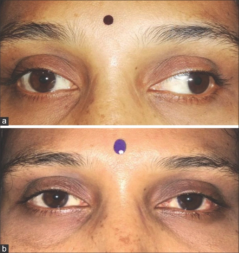
Strabismus is categorized based on the direction of ocular deviation, constancy, and underlying neuro-muscular etiology:
- Esotropia: Inward deviation of the eye toward the nose (convergent squint). Includes infantile esotropia and accommodative esotropia linked to high uncorrected hyperopia.
- Exotropia: Outward deviation of the eye toward the temple (divergent squint), often manifesting intermittently during fatigue or daylight exposure.
- Hypertropia / Hypotropia: Vertical misalignment where one eye drifts upward (hypertropia) or downward (hypotropia), frequently caused by superior oblique muscle palsies.
- Paralytic Strabismus: Acute muscle weakness resulting from cranial nerve palsies (Cranial Nerves III, IV, or VI) due to vascular disease, head trauma, or neurological lesions, presenting with acute double vision (diplopia).
- Latent Strabismus (Heterophoria): A tendency for misalignment kept in check by the brain's fusion mechanism, manifesting only when fusion is interrupted or during severe fatigue.
Pediatric vs. Adult Strabismus Management
| Clinical Domain | Pediatric Strabismus | Adult Strabismus |
|---|---|---|
| Primary Surgical Goal | Prevent amblyopia, promote binocular visual cortex development, and restore stereopsis. | Eliminate debilitating diplopia (double vision), correct head tilt/torticollis, and restore facial alignment. |
| Neuro-Adaptation | High plasticity; brain suppresses the turning eye, suppressing diplopia at the expense of visual development. | Low plasticity; brain cannot suppress the misaligned image, resulting in distressing double vision. |
| Anesthesia Protocol | General anesthesia (mandatory for safety and absolute immobility). | General anesthesia or regional retrobulbar block, often with adjustable suture options. |
| Non-Surgical Co-Therapies | Occlusion patching (penalization therapy) and full cycloplegic prescription glasses. | Prism lenses (Fresnel or ground-in prisms) and orthoptic exercises. |
How Strabismus Surgery Is Performed
A common patient anxiety is the misconception that the eye is removed or cut open during strabismus surgery. The eyeball is never removed from the socket, and the intraocular cavity is never opened.
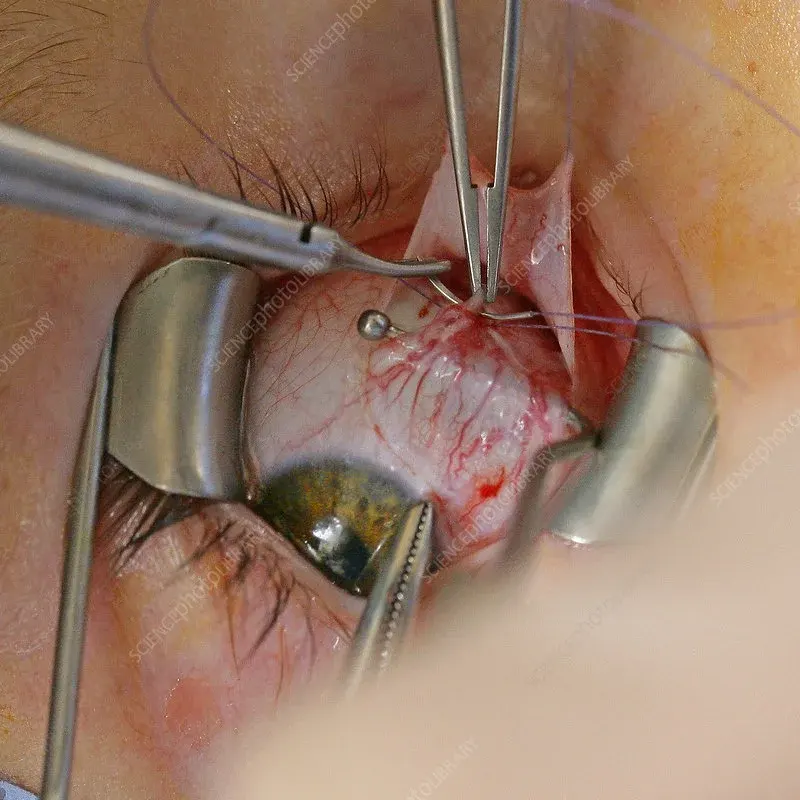
Strabismus surgery is an extraocular procedure executed entirely on the outside shell of the globe:
- Conjunctival Incision: A microscopic incision is made through the transparent conjunctiva and Tenon's capsule to expose the target extraocular muscle attached to the sclera.
- Muscle Recession (Weakening Procedure): If a muscle is overactive or pulling too tightly (e.g., the medial rectus in esotropia), it is detached from its original insertion site, measured precisely with surgical calipers, and re-sutured further back on the sclera to lessen its pull.
- Muscle Resection (Strengthening Procedure): If a muscle is underactive or weak, a calculated segment of the muscle tendon is excised, and the shortened muscle is reattached to its original insertion, increasing its mechanical leverage.
- Adjustable Suture Technique: In adult cases, adjustable sutures allow the surgeon to fine-tune muscle alignment hours after surgery under topical anesthesia, ensuring millimeter-perfect binocular alignment.
Preoperative Preparation & Surgical Safety
Surgical timing is determined through comprehensive orthoptic evaluations, including prism cover testing, sensorimotor evaluations, and full cycloplegic refraction:
- Pre-Anesthetic Fasting: For general anesthesia, patients must strictly fast—no solid foods for 6 hours prior to surgery, and clear liquids stopped 2 hours prior.
- Low Risk Profile: Strabismus surgery is a highly safe, extraocular procedure. Serious complications such as scleral perforation, infection (endophthalmitis), or severe hemorrhage occur in fewer than 1 in 10,000 cases.
- Surgical Touch-ups: Because extraocular muscle tone and brain adaptation evolve over time, fine-tuning or secondary muscle adjustment may occasionally be required to maintain long-term alignment.
Postoperative Care & Healing Protocol
Postoperative recovery is straightforward, with visual function returning rapidly:
- Initial Symptoms: Redness of the conjunctiva, mild subconjunctival hemorrhage, and a foreign-body sensation are normal during the first 1 to 2 weeks. Postoperative movement pain is managed with mild oral analgesics.
- Topical Medications: Medicated antibiotic and anti-inflammatory eye drops must be instilled strictly as prescribed for 2 to 3 weeks to ensure smooth scleral healing.
- Activity Guidelines: Patients can resume normal non-strenuous daily activities within 48 to 72 hours. Swimming, hot tubs, and heavy contact sports should be avoided for 2 weeks to prevent infection.
- Spectacle Continuity: Surgery corrects the physical muscle deviation, not the optical refractive error. Patients must continue wearing their prescribed glasses after surgery as directed by their ophthalmologist.
⚠ Anatomical Clarification: Strabismus vs. Eyelid & Iris Surgery
Strabismus surgery operates exclusively on the extraocular muscles attached to the outer sclera to align the visual axis. It must not be confused with:
- Eyelid Surgery: Functional or cosmetic blepharoplasty eyelid surgery corrects drooping skin (ptosis) or palpebral bags, but does not alter eye movement.
- Refractive Lenses: Intraocular implants such as trifocal smart lenses replace the crystalline lens to cure cataracts and presbyopia.
- Iris Color Modification: Aesthetic procedures like laser eye color change target superficial iris melanin without penetrating the sclera or affecting extraocular muscle dynamics.
Frequently Asked Questions: Strabismus Surgery
Can strabismus surgery be performed on adults, or is it only for children?
Strabismus surgery can be performed successfully at any age. While pediatric surgery focuses on preventing amblyopia and building 3D stereoscopic vision, adult strabismus surgery effectively eliminates double vision (diplopia), reduces ocular fatigue, corrects abnormal head tilts, and dramatically enhances facial symmetry and confidence.
Is the eye removed from the socket during strabismus surgery?
No. This is a persistent medical myth. The eyeball is never removed from the socket, nor is it opened. The surgeon works through a microscopic, hidden opening in the conjunctival tissue on the surface of the eye to access and reposition the extraocular muscles.
Will strabismus surgery eliminate the need for glasses?
No. Strabismus surgery aligns the physical direction of the eyes by adjusting muscle tension; it does not alter the focal power of the cornea or lens. If an optical refractive error (such as hyperopia, astigmatism, or myopia) is present, corrective glasses must still be worn after surgery.
How long does recovery take after strabismus surgery?
Most patients experience mild soreness and redness for 1 to 2 weeks. Most adults and children return to school or light work within 3 to 7 days post-surgery. Full tissue healing and final muscle alignment stabilize over 4 to 6 weeks.
Medical Disclaimer: This comprehensive surgical guide is authored by Dr. Mustafa Mete for educational purposes and does not replace an individualized strabismus examination. If you or your child present with eye deviation, sudden double vision, or visual fatigue, consult a qualified ophthalmic surgeon promptly.