The Evolution of the Human Eye: From Light Detection to the Retina, Iris and Eye Color
The human eye did not appear as a fully formed optical system. Its major components emerged through a long evolutionary history in which light-sensitive cells, photoreceptors, neural circuits, optical structures, and eventually the iris and lens became increasingly specialized for visual function.
Eye evolution is best understood as a sequence of functional adaptations rather than the sudden appearance of a modern human eye. Early light-sensitive cells could detect illumination. Over evolutionary time, photoreceptors, neural processing, retinal organization and optical structures became increasingly specialized. In vertebrates, this eventually produced the camera-type eye capable of forming increasingly precise images.
The retina is part of the nervous system: it converts light into neural signals and performs substantial processing before information reaches the brain. The iris adds another important layer of specialization by regulating the amount of light entering through the pupil.
How Did the Human Eye Evolve?
Asking how the eye evolved is really asking several related questions: how did organisms first become sensitive to light, how did those cells become organized into visual systems, and how did optical structures eventually allow light to be focused into an image?
Modern evolutionary biology does not require the earliest visual system to resemble a human eye. Instead, comparative biology and molecular studies allow researchers to reconstruct a series of functional changes involving photoreceptors, opsins, retinal neurons, optical tissues and neural processing.
Research on vertebrate visual evolution supports the idea that important components of the vertebrate retina and its photoreceptor systems have very ancient origins. The exact sequence of every evolutionary transition cannot be observed directly, but comparative anatomy, genetics, developmental biology and molecular phylogeny provide evidence for plausible evolutionary pathways.
1. Before the Camera Eye: Detecting Light
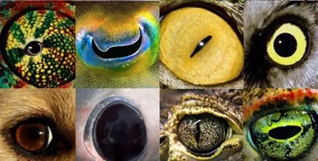
The earliest stages of visual evolution did not require a lens, cornea or even a recognizable eye. A much simpler starting point is photoreception: the ability of specialized cells to respond to light.
Photoreceptor cells use light-sensitive molecules called opsins together with associated molecular pathways to convert photons into cellular signals. Comparative studies indicate that ancient photoreceptor systems diversified long before the appearance of the modern vertebrate eye.
This distinction is important. Detecting whether light is present is a much simpler biological task than determining where the light came from, forming an image, distinguishing contrast, or recognizing an object.
2. From Light Detection to Image Formation
Light sensitivity
Specialized cells detect changes in illumination and provide the organism with basic information about its environment.
Directional sensitivity
The spatial arrangement of light-sensitive cells can provide information about the direction from which light is arriving.
Optical focusing
Curved transparent tissues and later more specialized optical structures improved the ability to concentrate incoming light.
Neural processing
Increasingly specialized retinal neurons allowed visual signals to be processed before information was transmitted to higher visual centers.
These changes were not necessarily a single straight ladder in which every organism passed through exactly the same sequence. Different lineages evolved different solutions to the same basic problem: extracting useful information from light.
3. The Evolution of the Retina
The retina is more than a passive light-sensitive surface. It is neural tissue containing multiple classes of cells that receive, transform and process visual information.
In vertebrates, photoreceptors include rods and cones. Cones support detailed and color vision under brighter conditions, while rods provide high sensitivity under dim illumination. The development of additional retinal cell classes and neural circuitry greatly increased the computational capacity of the visual system.
Comparative research suggests that the evolutionary history of vertebrate photoreceptors is more complex than the old idea of a simple progression from one primitive “eye cell” to a modern retina. Ciliary and rhabdomeric photoreceptor lineages have deep evolutionary relationships, and elements of both systems remain relevant to understanding modern visual biology.
The retina is part of the nervous system
One of the most important facts about vision is that the retina itself performs neural processing. It converts optical information into electrical and chemical signals and organizes those signals before they continue through the optic pathway toward the brain.
4. Why the Lens and Cornea Changed Vision
Detecting light is not the same as forming a sharp image. Image-forming vision requires optical structures capable of controlling the path of incoming light.
During vertebrate evolution, increasingly specialized optical structures improved the ability to focus light onto the photoreceptive tissue. The development of a lens and associated ocular structures enabled increasingly precise spatial information to reach the retina.
This was a major functional transition. Once an organism could form a sufficiently focused image, selection could act on additional visual characteristics such as spatial resolution, sensitivity, field of view, movement detection and color discrimination.
5. The Evolutionary Role of the Iris
The iris is the colored structure surrounding the pupil. Its most immediate optical role is not cosmetic: it regulates the amount of light entering the eye by changing pupil diameter.
In bright conditions, the pupil becomes smaller, reducing the amount of incoming light. In dim conditions, the pupil becomes larger, allowing more light to reach the retina.
This provides an important example of how several ocular structures work as an integrated optical system. The cornea and lens influence focusing, the iris controls the aperture, and the retina receives and processes the resulting optical information.
The modern eye is therefore not simply a collection of independent parts. Its performance depends on the interaction between optical tissues, pigment, photoreceptors, retinal circuitry and the visual brain.
6. Where Does Human Eye Color Come From?
Human eye color is primarily related to the pigmentation and optical properties of the iris. Melanin is a major contributor, but eye color is not determined by a single “brown gene” or by pigment quantity alone.
Modern genetic research shows that eye color is a polygenic trait. Variants in genes involved in melanin production, transport and storage influence the amount and distribution of pigmentation within the iris. OCA2 and HERC2 are particularly important, while several additional genes contribute to the broad continuum of human eye color.
The visible color of an iris also depends on how light interacts with its microscopic structure. Differences in pigmentation, tissue organization and light scattering contribute to the appearance of blue, green, hazel, brown and intermediate iris colors.
For a dedicated explanation of the genetics and classification of human eye color, see the MyLumineyes Eye Color Chart, Genetics & Genes .
7. Evolution Explains the Eye — but Not Every Modern Eye Color
It is useful to separate two different biological questions.
Eye evolution asks how visual systems and their structures developed across evolutionary time. Eye-color genetics asks why individuals and populations differ in iris pigmentation.
These subjects are related because the iris is part of the visual system, but they are not the same scientific problem. Human iris-color variation is best understood through pigmentation biology, genetics, development and optical properties rather than through the simple idea that one eye color represents a particular evolutionary “stage.”
8. From the Retina to the Brain
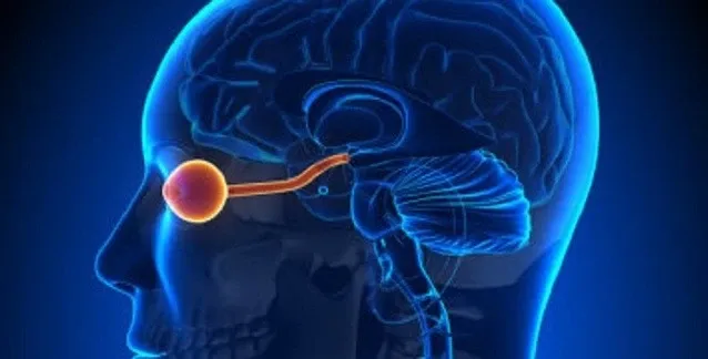
The final stage of visual perception does not occur in the eye alone. Retinal signals travel through the optic nerve and continue through specialized neural pathways to the brain, where increasingly complex representations of the visual environment are constructed.
This is why the eye is often better understood as the optical and sensory beginning of vision rather than the complete visual system. The retina performs important preprocessing, while the brain integrates those signals with other neural information to support perception.
Evolution therefore shaped vision at several interconnected levels: photoreception, retinal circuitry, optics, eye movement, neural transmission and cortical processing.
From Eye Biology to Clinical Research
Understanding the biology of the iris also provides context for modern research into iris pigmentation. The clinical questions surrounding laser iris depigmentation are different from the evolutionary questions discussed in this article and should be evaluated through clinical evidence, ophthalmic assessment, safety monitoring and longitudinal follow-up.
The MyLumineyes Research Hub brings together clinical methodology, scientific documentation, iris pigmentation research, safety frameworks and long-term clinical observations.
Explore the MyLumineyes Research Hub →9. Why Iris Biology Matters in Modern Ophthalmology
The iris is simultaneously an optical structure, a pigmented tissue and an anatomically important part of the anterior segment. Understanding its pigmentation and tissue architecture is therefore relevant not only to basic biology but also to clinical ophthalmology.
When discussing any intervention involving iris pigmentation, it is important to distinguish biological observations from clinical claims. A description of melanin distribution or iris anatomy does not by itself establish the safety, efficacy or long-term outcome of a procedure.
For readers interested specifically in the clinical subject of laser-based eye color change, the dedicated Laser Eye Color Change page provides the appropriate procedural context rather than mixing clinical claims into this evolutionary review.
Conclusion: The Eye Is an Evolutionary System, Not a Single Invention
The human eye is the result of a long history of biological specialization. Light-sensitive molecules came first; increasingly organized photoreceptors, neural circuits and optical structures followed. In vertebrates, the development of a sophisticated retina together with lens-based optics created the foundations of image-forming vision.
The iris added another layer of control by regulating the pupil, while variation in iris pigmentation contributes to the enormous diversity of human eye color. Modern genetics shows that this pigmentation is complex and polygenic rather than the result of a simple dominant-versus-recessive rule.
Studying the evolution of the eye therefore connects several fields: evolutionary biology, genetics, ophthalmology, neuroscience and visual optics. It also provides the biological foundation needed to understand why the iris is such a specialized and clinically important tissue.
Selected Scientific References
- Lamb TD. Evolution of vertebrate retinal photoreception. Philosophical Transactions of the Royal Society B. 2009. PubMed
- Lamb TD, Collin SP, Pugh EN Jr. Evolution of the vertebrate eye: opsins, photoreceptors, retina and eye cup. Nature Reviews Neuroscience. 2007. PubMed
- Baden T, Euler T, Berens P. Understanding the retinal basis of vision across species. Nature Reviews Neuroscience. 2020. Nature Reviews Neuroscience
- MedlinePlus Genetics. Is eye color determined by genetics? U.S. National Library of Medicine. MedlinePlus Genetics
- Liu F et al. Genome-wide association study in almost 195,000 individuals identifies 50 previously unidentified genetic loci for eye color. Human Genetics. 2021. PMC
Reviewed by Dr. Mustafa Mete
This educational article is medically reviewed within the MyLumineyes clinical content framework. The purpose of the review is to distinguish established ophthalmic and biological concepts from interpretation, clinical experience and procedure-specific claims.
Dr. Mustafa Mete is an ophthalmic surgeon and developer of the Lumineyes™ approach, with more than 25 years of ophthalmic clinical experience. Clinical procedure information is presented separately from this evolutionary and biological review.
Evidence principle: evolutionary evidence, genetic evidence and clinical evidence answer different questions and should not be treated as interchangeable.