The Hidden Dangers of Colored Contact Lenses: An Ophthalmologist’s Warning
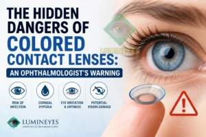
For decades, colored contact lenses have been marketed as harmless cosmetic accessories. From global fashion trends to daily vanity, millions of individuals insert these tinted plastic discs into their eyes without a second thought. However, from a clinical ophthalmology perspective, the continuous, unregulated use of cosmetic lenses is a ticking biological clock for corneal health.
While temporary color alteration seems benign, the structural and physiological price your eyes pay is immense. Below, we break down the clinical reality of cosmetic lens wear, the biological mechanisms of ocular trauma they induce, and the only medically viable, permanent alternative.
1. Corneal Hypoxia: Chronic Suffocation of Ocular Delineation
The human cornea is unique; it has no blood vessels and must absorb oxygen directly from the atmosphere through the tear film. When you place a standard cosmetic lens over the eye, you are effectively placing an oxygen barrier over a living tissue.
Cosmetic lenses are notoriously poorly designed compared to premium therapeutic silicone hydrogels. They are thicker, less gas-permeable, and completely coated with heavy pigments to mask your natural iris color. This structural flaw triggers a destructive cascade:
- Stromal Suffocation: The pigment layer restricts oxygen transmissibility ($Dk/t$), leaving the corneal epithelium in a permanent state of hypoxia.
- Corneal Neovascularization: In a desperate bid to survive, the eye begins sprouting abnormal, fragile new blood vessels into the clear cornea to supply oxygen. This can lead to permanent corneal scarring and vision loss.
- Epithelial Edema: Lack of oxygen disrupts the metabolic pumps of the cornea, causing cellular swelling, blurred vision, and structural weakening.
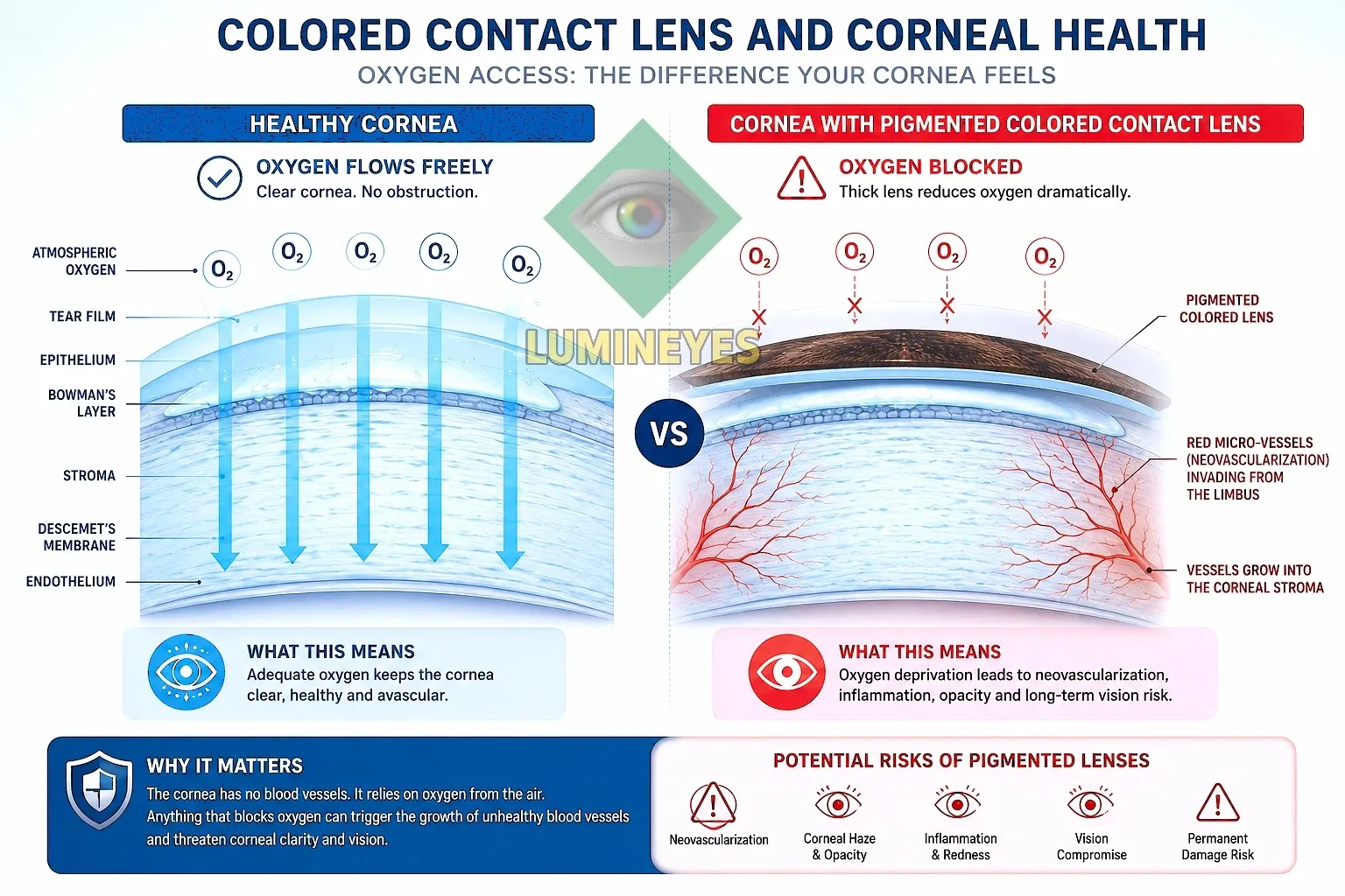 Figure 1: Cross-sectional visual of the hypoxic cascade. The thick pigment layer of cosmetic lenses blocks atmospheric oxygen, forcing abnormal micro-vessels to invade the clear corneal stroma.
2. The Pathological Checklist: Micro-Trauma and Blindness Risks
Cosmetic contact lenses—especially those purchased online without a valid prescription from an ophthalmologist—are primary vectors for severe ocular pathologies.
Acanthamoeba and Bacterial Keratitis
The surface of a cosmetic lens is a perfect breeding ground for bio-films. Pseudomonal infections and Acanthamoeba keratitis (a devastating parasitic infection) thrive on poorly sanitized lens surfaces. Because these lenses often fit poorly, they create microscopic scratches (epithelial erosions) on the cornea, giving these aggressive pathogens a direct gateway into the deep ocular tissue. An untreated corneal ulcer can destroy vision within 24 to 48 hours.
Giant Papillary Conjunctivitis (GPC)
The constant mechanical friction of a thick, pigmented lens against the underside of the eyelid triggers a severe allergic and mechanical reaction. The eyelid develops large, painful bumps (papillae), leading to chronic mucus discharge, severe itching, and an absolute intolerance to any contact lens in the future.
3. The Structural Illusion: Implants vs. Keratopigmentation vs. Biological Reality
When faced with the dangers of daily lens wear, many patients look for permanent solutions, often falling into the traps of dangerous marketing gimmicks.
- Artificial Iris Implants: Highly invasive, surgically placing a piece of silicone inside the anterior chamber. It almost universally leads to intractable glaucoma, uveitis, and corneal endothelial failure.
- Keratopigmentation (Corneal Tattooing): Injecting chemical dyes directly into the clear corneal stroma. It permanently ruins corneal clarity, blocks peripheral vision, and looks dull, opaque, and entirely artificial.
The iris does not behave like a canvas for artificial coloring or tattooing. True, natural eye color is entirely determined by the density of melanin granules within the iris stroma.
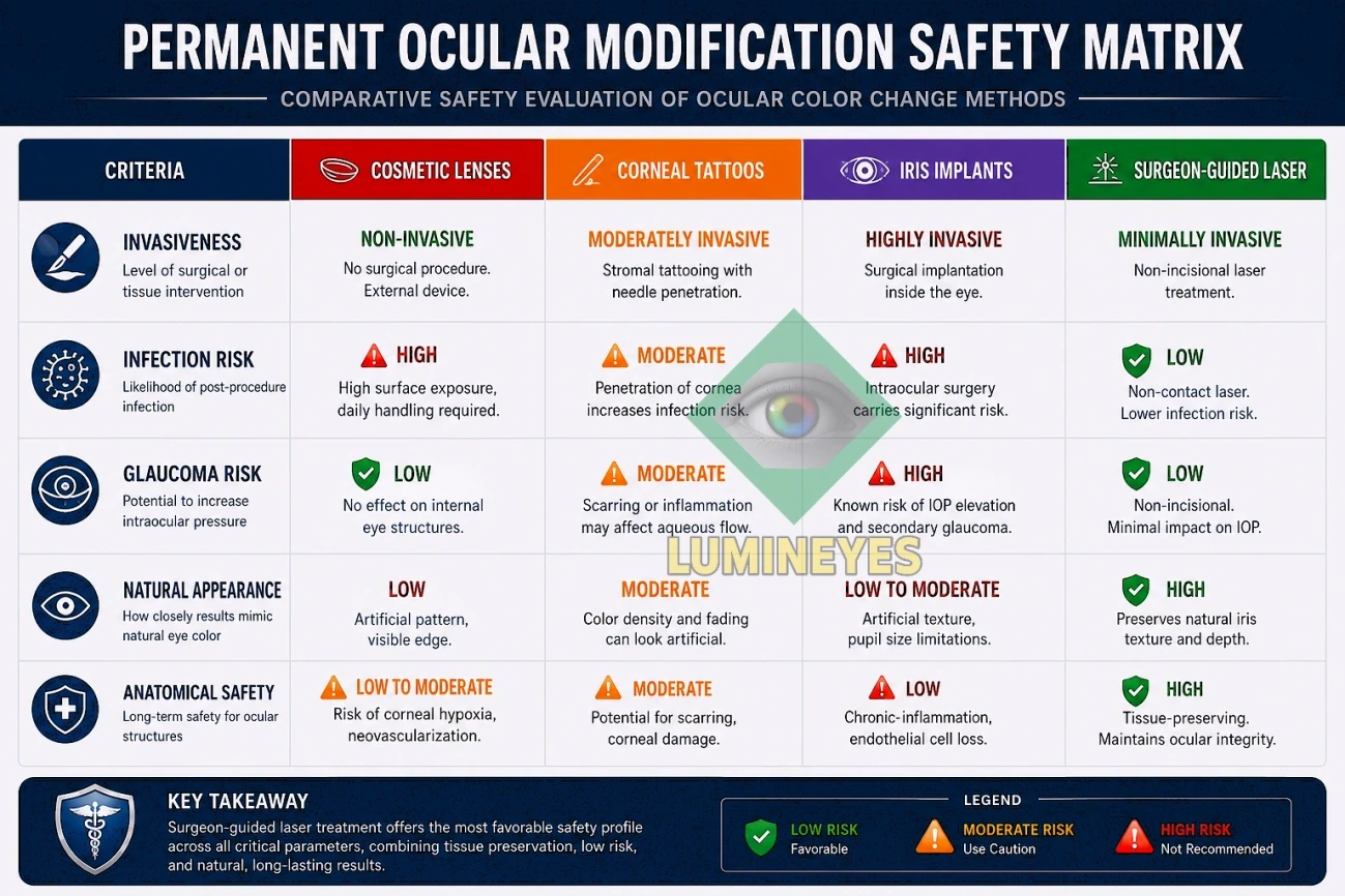 Figure 2: Clinical risk assessment table highlighting the comparative safety and anatomical preservation profiles across various iris modification methodologies.
| Criteria | Cosmetic Lenses | Corneal Tattoos | Iris Implants | Surgeon-Guided Laser |
|---|---|---|---|---|
| Invasiveness | Non-invasive (External) | Surgical (Corneal Injections) | Highly Invasive (Intraocular) | Non-invasive (No Incisions) |
| Infection Risk | High (Keratitis/Ulcers) | Moderate (Stromal Infection) | High (Endophthalmitis) | Minimal (Closed-eye Protocol) |
| Glaucoma Risk | None (Unless infected) | Low | Extreme (Severe IOP Spikes) | Controlled (Monitored Clearance) |
| Appearance | Artificial (Masked Iris) | Opaque / Plastic Look | Unnatural / Fixed Pupil | 100% Natural Iris Structure |
| Anatomical Safety | Chronic Hypoxia Risk | Permanent Stromal Damage | Endothelial Cell Destruction | Preserves Ocular Integrity |
4. The Medical Standard: Manual Surgeon-Guided Iris Depigmentation
If cosmetic colored lenses are a temporary hazard and implants are a clinical disaster, how can permanent symmetry or color modification be safely achieved? The answer lies not in adding foreign materials, but in the safe, biological reduction of existing melanin.
The true medical standard for altering iris pigmentation safely is selective laser iris depigmentation. Instead of masking the eye or tattooing delicate tissues, this protocol utilizes low-fluence laser energy to gently fragment extracellular melanin granules in the anterior stroma.
Ocular Respiration & Safe Depigmentation Pathway
However, this cannot be achieved by an automated machine. It relies entirely on cognitive mapping and manual precision by an expert surgeon. By visually analyzing individual stromal density, a master ophthalmologist can selectively target hyper-pigmented areas while leaving surrounding structural tissues completely untouched, allowing the body's natural macrophage pathways to clear the pigment safely.
For those looking to escape the daily trauma and hypoxic risks of colored contact lenses, exploring the science behind a permanent, structurally safe alternative is the definitive medical step forward. Review our comprehensive, primary laser eye color change clinical guide to understand the complete biological framework.