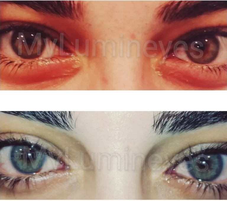
Is Laser Eye Color Change Safe? Clinical Safety Guide 2026
Laser eye color change is an elective ophthalmic procedure and should be evaluated according to the specific laser protocol, treatment parameters, patient-selection criteria, monitoring strategy, and available longitudinal clinical evidence.
Safety should not be inferred simply from the words “laser eye color change,” because laser iris depigmentation is not a single standardized intervention. Different laser systems, wavelengths, energy parameters, treatment patterns, staging strategies, patient populations, and follow-up protocols may produce substantially different clinical profiles.
For Lumineyes, safety assessment is procedure-specific. The approach is based on physician-led patient selection, staged treatment, ophthalmic monitoring, individualized treatment decisions, and longitudinal clinical observation.
The available clinical observations are presented as clinical data rather than as a guarantee of zero risk or as a substitute for independent prospective validation.
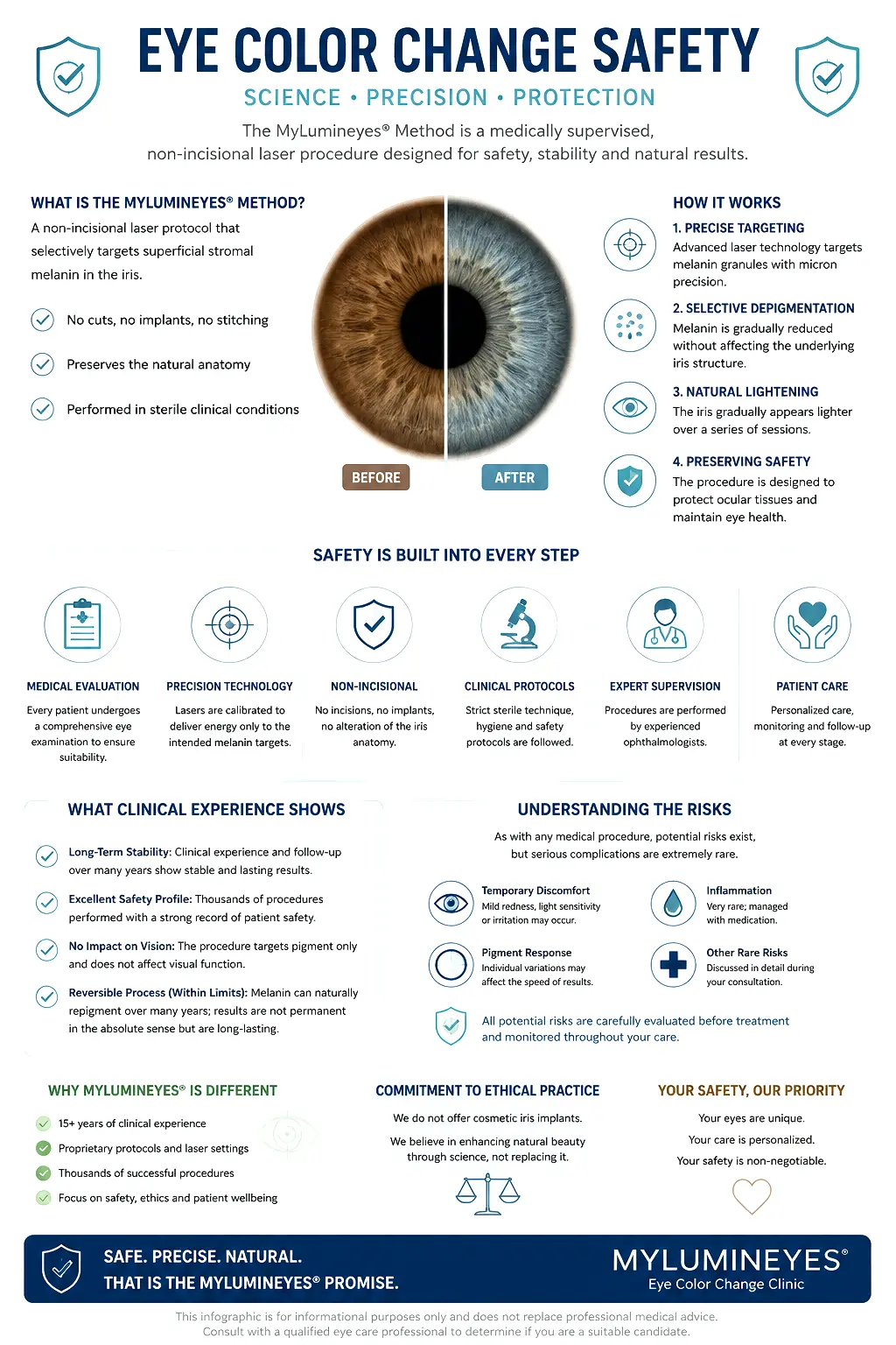
Why Safety Findings Must Be Interpreted in the Context of the Specific Procedure
Laser eye color change is not a single uniform medical intervention. Published reports may involve different laser systems, wavelengths, energy or fluence levels, pulse characteristics, treatment patterns, treatment staging, patient-selection criteria, and follow-up strategies.
For this reason, a complication reported after one laser iris depigmentation technique should not automatically be presented as a demonstrated complication of another technique without evidence establishing procedural, dosimetric, anatomical, and clinical comparability.
For a detailed framework on how procedure-specific safety claims, reported complications, theoretical risks, causation, and long-term evidence should be interpreted, see How to Interpret Safety Claims in Laser Eye Color Change .
Lumineyes-specific clinical observations are therefore presented separately from findings reported after other procedures and from theoretical or mechanistic concerns.
Three Different Types of Safety Information
Lumineyes-Specific Clinical Observations
Findings observed within the available Lumineyes clinical dataset and longitudinal ophthalmic follow-up.
Reports From Other Procedures
Complications reported after other laser techniques or surgical approaches may be clinically relevant, but they should not automatically be attributed to Lumineyes without evidence of comparability.
Theoretical or Mechanistic Concerns
A biologically plausible or theoretically possible risk is not identical to a complication demonstrated in the Lumineyes clinical dataset.
For a detailed discussion of how clinical observations, causation, follow-up duration, evidence limitations, and real-world clinical datasets should be interpreted, see How Clinical Evidence Is Evaluated in Laser Eye Color Change .
Procedural Safety & Ocular Tissue Interaction
From a safety perspective, the Lumineyes approach is designed to target melanin-containing structures within the anterior iris stroma while limiting unnecessary exposure of surrounding ocular tissues. Treatment is applied non-incisionally through the intact cornea, avoiding the creation of corneal channels or the insertion of foreign intraocular implants.
Maintaining intraocular health requires strict energy calibration, controlled treatment staging, and interval monitoring. For comprehensive details regarding laser biophysics and biological clearance mechanisms, refer directly to our Laser Eye Color Change treatment guide.
Clinical Safety & Biological Limits
Safety in iris depigmentation depends not only on laser energy but also on patient selection, treatment staging, biological response, monitoring, and the decision to stop when further treatment is no longer clinically justified.
Eyes with higher baseline pigmentation may require a more gradual treatment strategy. Attempting to pursue additional cosmetic change beyond an individual's biological response may provide diminishing aesthetic benefit while increasing the potential for transient inflammatory or pressure-related responses.
For this reason, the decision to stop treatment is considered part of the safety framework rather than simply a cosmetic endpoint.
Lumineyes Clinical Safety Observations
The available Lumineyes clinical dataset represents retrospective longitudinal clinical observation from real-world ophthalmic practice. It should be interpreted as clinical observational evidence rather than as a randomized or independently validated clinical trial.
In the reported dataset, no chronic or progressive intraocular pressure elevation has been documented in the available observations.
Transient responses reported in the clinical dataset include approximately 3% transient corneal edema, 1% transient IOP elevation, and 2% mild iridocyclitis, described as resolving.
Important Interpretation
- The dataset is retrospective real-world clinical observation.
- It is not a randomized controlled clinical trial.
- It has not undergone independent external validation.
- Observed absence of an event does not establish zero population risk.
- Prospective and independently validated studies remain important for further characterization of long-term risk.
See the complete Lumineyes longitudinal clinical data report for the dataset, follow-up framework, reported outcomes, and evidence limitations.
Interpreting Procedure-Specific Safety Profiles
Safety comparisons are medically valid only when distinct procedural mechanisms are recognized. Non-incisional laser depigmentation, keratopigmentation (corneal tattooing), and artificial iris implants interact with entirely different anatomical structures and carry non-transferable risk profiles.
| Clinical Parameter | Lumineyes | Other Laser Approaches | Surgical Alternatives |
|---|---|---|---|
| Laser / Treatment Parameters | Procedure-specific laser parameters and staged treatment decisions. | May differ substantially in wavelength, energy, fluence, pulse characteristics, treatment pattern, and treatment strategy. | Surgical procedures use different tissue-access and pigment-placement mechanisms. |
| Treatment Staging | Treatment is structured in stages with clinical assessment guiding subsequent decisions. | Staging and treatment strategy may vary by technique and provider. | Surgical interventions generally involve a different procedural model and endpoint. |
| Patient Selection | Candidate suitability is determined through ophthalmic assessment and individualized planning. | Selection criteria may differ between procedures and clinical settings. | Surgical candidacy depends on the specific operation and anatomical considerations. |
| Monitoring | Ophthalmic monitoring includes IOP and other clinically relevant findings, with additional investigations when indicated. | Monitoring protocols may vary. | Requires monitoring appropriate to the surgical procedure and its anatomical risks. |
| Evidence Interpretation | Lumineyes-specific clinical observations should be evaluated separately from reports involving other procedures. | Findings should be interpreted according to the specific technique studied. | Surgical complication data should not be directly transferred to non-surgical procedures. |
For broader structural comparisons across methods, see the dedicated eye color change method comparison and the clinical breakdown on keratopigmentation vs. laser depigmentation .
Patient Screening as a Safety Measure
Thorough medical screening is an essential safety prerequisite. A comprehensive ophthalmic examination is conducted prior to treatment to evaluate baseline intraocular pressure, corneal endothelial integrity, and anterior chamber drainage angle anatomy.
Patients with relevant pre-existing ocular conditions may require exclusion from treatment or additional specialist assessment based on the findings of the ophthalmic evaluation. For full clinical selection criteria and diagnostic guidelines, visit our canonical Patient Eligibility Guide.
For detailed discussion of endothelial evaluation and specular microscopy, see the Corneal Endothelial Assessment and Specular Microscopy review.
Biological Limits & the Stop Decision
Every iris has a biological response that may differ according to baseline pigmentation, tissue characteristics, treatment history, and individual recovery.
Additional treatment is not automatically beneficial simply because further pigment reduction remains technically possible. When the expected cosmetic benefit becomes limited relative to additional treatment exposure, stopping may be the more appropriate clinical decision.
The Lumineyes Stop Decision is therefore treated as a protective clinical measure rather than as a failure to reach a predetermined cosmetic endpoint.
Read: When to Stop Laser Eye Color Change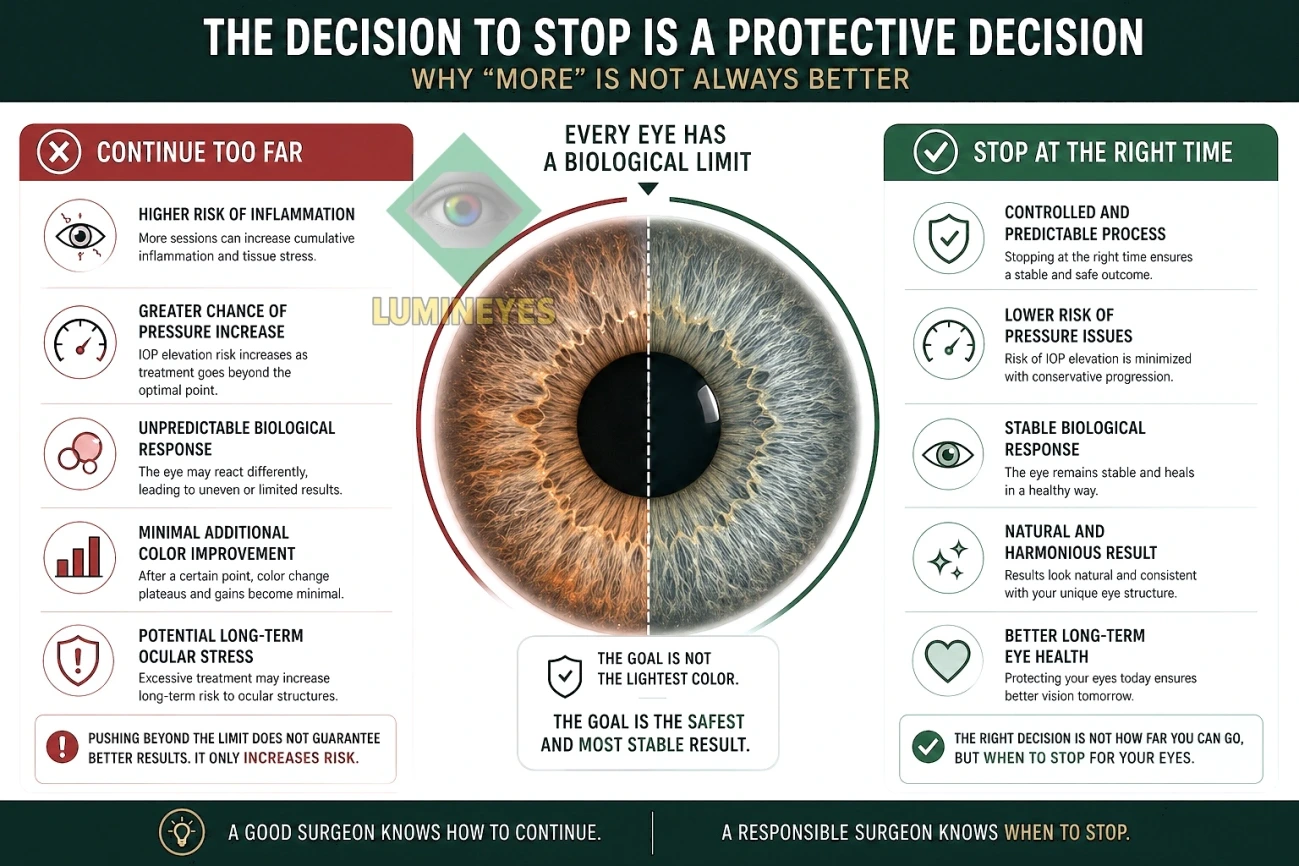
Long-Term Ocular Monitoring Protocol
Patient safety relies on ophthalmic evaluation throughout the treatment cycle and appropriate follow-up after treatment.
IOP & Anterior Segment
Intraocular pressure and relevant anterior-segment findings are assessed according to the clinical monitoring plan.
Inflammatory Response
Temporary inflammatory or postoperative responses are evaluated and managed when clinically indicated.
Adaptive Session Spacing
Treatment intervals can be adjusted according to individual clinical response and recovery rather than being fixed solely by a cosmetic target.
What the Current Clinical Evidence Can — and Cannot — Establish
Clinical observations can provide important information about treatment response, observed adverse events, follow-up findings, and real-world clinical experience.
They cannot by themselves establish that a procedure has zero long-term risk, nor can they automatically establish causation for every event that occurs after treatment.
The interpretation of safety evidence therefore depends on the population studied, the exact procedure performed, treatment parameters, outcome definitions, follow-up duration, completeness of follow-up, and the limitations of the underlying dataset.
For the detailed evidence framework, see How Clinical Evidence Is Evaluated in Laser Eye Color Change .
MyLumineyes Eye Color Change Safety Report
Frequently Asked Questions About Laser Eye Color Change Safety
Is laser eye color change considered safe?
Laser eye color change is an elective ophthalmic procedure and, like any medical procedure, involves potential risks and limitations. Safety should be evaluated according to the specific procedure, patient-selection criteria, treatment parameters, monitoring strategy, and available clinical evidence. Suitability requires a comprehensive ophthalmic assessment.
Can laser eye color change cause glaucoma or affect vision?
Changes in intraocular pressure and other ocular responses are important considerations during iris depigmentation. IOP and other relevant findings should be assessed before and during a staged treatment protocol. In the available Lumineyes clinical observations, no chronic or progressive IOP elevation and no permanent vision loss have been documented; however, observed absence does not establish zero population risk.
Does a complication reported after another laser iris depigmentation procedure automatically apply to Lumineyes?
No. A complication reported after another procedure may be clinically relevant, but it should not automatically be presented as a demonstrated complication of Lumineyes without evidence showing that the procedures are sufficiently comparable in laser parameters, treatment pattern, patient selection, staging, anatomy, and follow-up.
Is a theoretical risk the same as a demonstrated complication?
No. A theoretical or mechanistic concern means that a biological pathway could plausibly produce an outcome. A demonstrated clinical complication requires clinical evidence showing that the event occurred and, where relevant, evidence supporting its relationship to the procedure.
What temporary responses may occur after treatment?
Temporary responses can include redness, light sensitivity, transient corneal edema, transient intraocular pressure elevation, or mild inflammatory responses. In the reported Lumineyes clinical dataset, transient corneal edema was approximately 3%, transient IOP elevation approximately 1%, and mild iridocyclitis approximately 2%, with these responses described as resolving.
Is laser eye color change suitable for very dark brown eyes?
Suitability cannot be determined from eye color alone. Eyes with higher baseline pigmentation may require a more gradual treatment approach. Treatment should proceed only when clinical examination indicates that the individual eye remains an appropriate candidate.
Can someone with a family history of glaucoma undergo treatment?
A family history of glaucoma does not by itself determine eligibility. Patients with relevant risk factors require careful ophthalmic assessment, including evaluation of intraocular pressure and the anterior chamber drainage angle, before treatment is considered.
Does the Lumineyes clinical dataset prove zero long-term risk?
No. The available dataset provides longitudinal real-world clinical observations, including reported outcomes and follow-up findings, but it does not establish zero long-term risk. Prospective and independently validated studies remain important for further characterization of long-term safety.
Medical Disclaimer
This content is provided for educational purposes and does not constitute medical advice or guarantee a specific outcome. Suitability for laser iris depigmentation can only be determined through a comprehensive ophthalmic examination by a qualified ophthalmic surgeon.