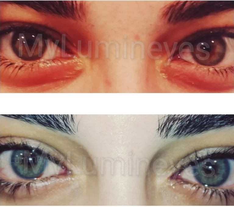
Is Laser Eye Color Change Safe? Clinical Safety Guide 2026
Laser iris depigmentation is an elective ophthalmic procedure with potential ocular risks. These include intraocular pressure elevation, pigment-related outflow disturbance, inflammation, corneal changes, and, in published reports involving certain techniques, more serious ocular complications. It cannot be described as universally safe or risk-free.
Safety assessment must be specific to the technique performed, the individual eye, the treatment parameters, and the quality of clinical monitoring and follow-up. The Lumineyes™ approach uses a non-incisional, physician-supervised, response-guided treatment strategy; this does not eliminate the need for careful evaluation or establish zero risk.
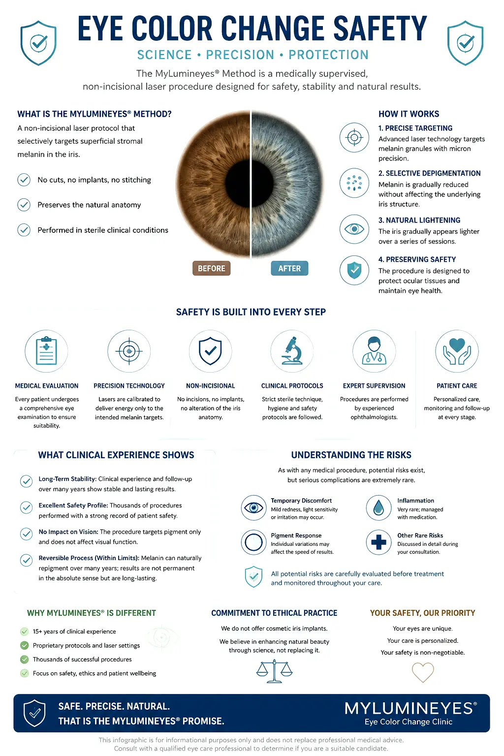
The illustration explains considerations in clinical care. It does not provide a numerical estimate of safety or guarantee a particular outcome.
What Are the Potential Risks of Laser Eye Color Change?
The clinical concerns associated with laser iris depigmentation arise from laser–tissue interaction, pigment response, and the anatomical structures involved. Risks cannot be estimated for one technique by assuming all laser iris procedures use equivalent parameters or treatment schedules.
Intraocular Pressure
Transient pressure elevation or pigment-related outflow disturbance may occur. Pressure changes warrant ophthalmic assessment and appropriate management.
Pigment Dispersion
Treatment-related pigment material may interact with the aqueous outflow pathway. Its clinical significance depends on the response and procedure.
Inflammatory Response
Anterior-chamber inflammation, discomfort, light sensitivity, or delayed recovery may require treatment to be withheld and reassessed.
Corneal Changes
Transient edema and other corneal findings require monitoring. Corneal and endothelial evaluation may be indicated before or after exposure.
Iris Tissue Injury
Unfavorable laser–tissue interaction may affect iris structures. Published adverse-event reports must be interpreted by technique and exposure context.
Serious Ocular Complications
Case reports after certain cosmetic iris laser procedures describe glaucoma, maculopathy, and visual injury. These are clinically important safety signals, not universal incidence estimates.
Key distinction: A complication documented after one laser iris depigmentation protocol is not automatically a demonstrated complication of every other protocol. Equally, the absence of an event in one retrospective series does not establish that the event cannot occur.
Mechanism & Ocular Physiology: Why It Matters for Safety
Laser interaction with pigmented anterior iris structures may produce a treatment-related pigment response. Released particulate material, aqueous dynamics, and trabecular outflow are relevant to ocular pressure and inflammatory monitoring. These mechanisms are clinically important even when no immediate symptoms are reported.
This is a safety-oriented overview, not a description of laser settings or the full treatment technique. See the main Laser Eye Color Change Procedure guide for the underlying treatment mechanism and stages.
Clinical point: a pleasing change in appearance cannot substitute for ophthalmic findings when evaluating the safety of additional treatment.
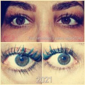
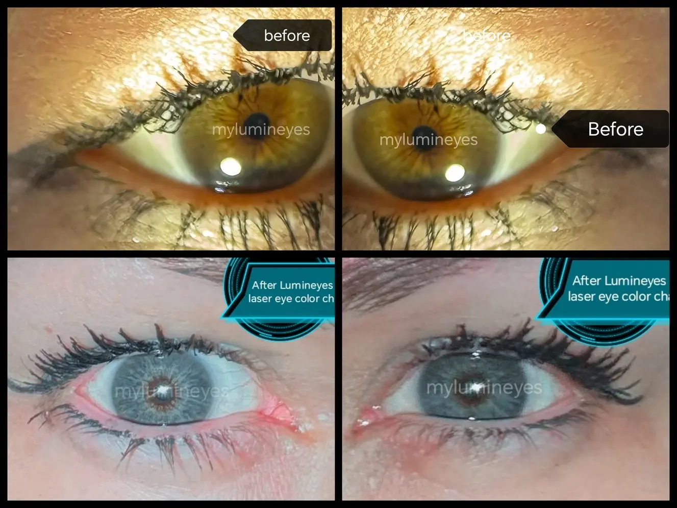
Lumineyes Clinical Safety Observations
The available Lumineyes dataset consists of retrospective, real-world ophthalmic clinical observations. It should be considered separately from independent prospective trials and from reports involving different iris depigmentation techniques.
Reported Longitudinal Observations
In the available records, no chronic or progressive intraocular pressure elevation has been documented. Transient corneal edema, temporary IOP elevation, and mild inflammatory responses have been observed and described as resolving.
Interpretation: These statements describe the available observations; they do not prove that the population risk of a serious event is zero. Long-term data are not equally complete for every treated eye.
Why Precise Adverse-Event Rates Require Caution
Reliable percentages require defined event criteria, complete numerator and denominator information, and consistent handling of eye-level versus patient-level observations and follow-up. This patient guide therefore does not present a calculated risk rate for each transient event without an independently checked tabulation.
Further source definitions, follow-up scope, and methodological limitations are discussed in the Lumineyes longitudinal clinical data report. The observations are not presented as a randomized comparison with alternative eye color change procedures.
Who Requires Careful Screening or May Be Unsuitable?
Safety begins with an individualized ophthalmic examination. Baseline assessment considers the medical history, prior eye procedures, ocular symptoms, intraocular pressure, corneal status, anterior-chamber structures, iris characteristics, and other clinically relevant findings.
Pre-existing ocular disease, unfavorable anatomical findings, unresolved inflammation, pressure-related concerns, or other examination findings may lead to further investigation, deferral, or exclusion. Suitability cannot be established from a photograph or iris color alone.
Patient Expectations Also Matter
Elective treatment requires an informed understanding that additional laser sessions and a particular final shade cannot be guaranteed. A patient's wish for further lightening does not override a clinically indicated pause or stop decision.
How Is Ocular Safety Monitored?
Ophthalmic monitoring is relevant before treatment, between staged exposures, and during follow-up. A reassuring measurement at one visit does not independently establish readiness for further treatment.
IOP and Outflow
Pressure findings are interpreted alongside baseline measurements, clinical history, anterior-segment status, and relevant angle observations.
Cornea and Inflammation
Corneal clarity, endothelial considerations where indicated, anterior-chamber response, symptoms, and the time course of recovery are assessed.
Response-Guided Decisions
The clinical response may support observation, further assessment, modification of the plan, or discontinuation rather than automatic repeat exposure.
Long-term follow-up remains important because the available published evidence for cosmetic laser iris depigmentation is heterogeneous and does not resolve all late-risk questions.
For a focused clinical framework on anterior-segment response and decisions about further exposure, see L-SAFE anterior-segment safety staging. The framework should not be mistaken for an independently validated guarantee of procedural safety.
Why Safety Findings Must Be Interpreted by Procedure
“Laser eye color change” is not a single standardized intervention. Different systems, wavelengths, pulse characteristics, anatomical targets, energy parameters, session patterns, eligibility rules, and follow-up procedures may have materially different risk profiles.
Lumineyes Observations
First-party clinical findings should be described with the relevant population, observation periods, and limitations.
Other Technique Reports
Published case reports and series identify possible complications but cannot automatically establish their incidence under a different protocol.
Theoretical Mechanisms
Biologically plausible risks deserve consideration without being misrepresented as events already documented in a given dataset.
Non-Incisional Laser vs. Surgical Approaches: What Changes in Safety Assessment?
Methods intended to change eye appearance involve different ocular tissues. The relevant risks therefore differ; this table compares the clinical safety questions that should be evaluated, not cosmetic outcomes or relative superiority.
| Approach | Anatomical area | Key safety considerations |
|---|---|---|
| Staged laser iris depigmentation | Anterior iris pigmentation | Pigment response, intraocular pressure, trabecular outflow, inflammation, iris integrity, and corneal status. |
| Other laser iris approaches | Iris; target and exposure patterns vary | System-specific wavelength and pulse settings, laser–tissue interaction, exposure strategy, patient selection, and documented adverse events. |
| Keratopigmentation | Corneal tissue | Corneal injury or infection, stromal changes, pigment placement, and healing-related complications. |
| Cosmetic iris implants | Intraocular structures | Implant-related inflammation, elevated IOP, endothelial injury, and other intraocular complications. |
These approaches cannot be ranked by this table. The evidence base, follow-up duration, patient population, and indication for each procedure must be evaluated separately.
For operative technique descriptions, expected outcomes, and a complete method comparison, see the dedicated Eye Color Change Method Comparison; this Safety page addresses only the clinical risk context.
For a deeper discussion of safety attribution, clinical causation, follow-up, and limitations of the evidence, read How to Interpret Safety Claims and How Clinical Evidence Is Evaluated.
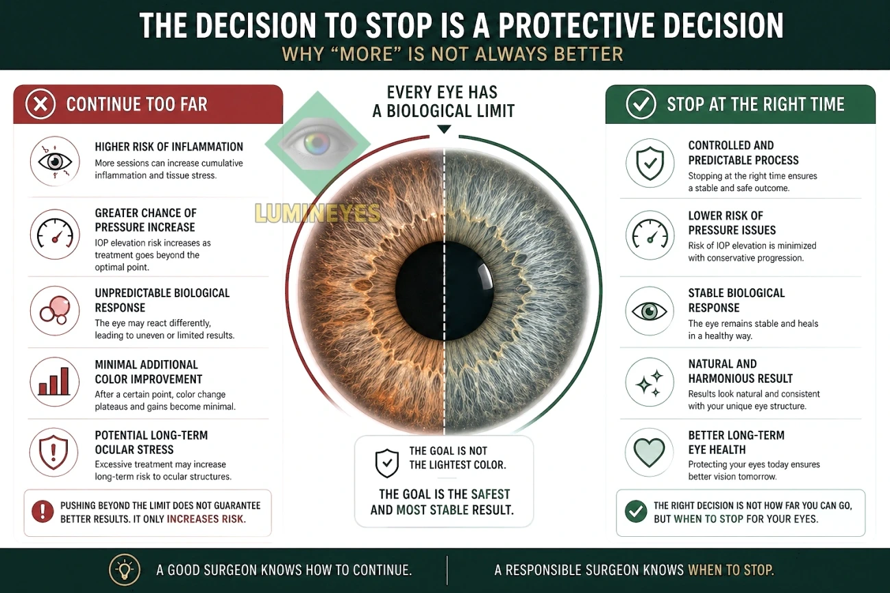
Why the Decision to Stop Is a Safety Decision
Further exposure is not necessarily appropriate simply because additional pigment reduction remains theoretically possible. A limited expected cosmetic benefit, unresolved treatment-related response, or an unfavorable examination may justify withholding or ending treatment.
Temporary pause and definitive discontinuation are not the same decision. Each depends on the individual clinical findings; neither should be dictated solely by a predetermined session count.
MyLumineyes Eye Color Change Safety Report
The embedded report provides additional background on clinical monitoring and safety considerations. If the preview does not open on your device, use the direct document link below.
Frequently Asked Questions About Laser Eye Color Change Safety
Is laser eye color change considered safe?
It is an elective ophthalmic procedure with potential risks. A general claim of safety cannot be made for every laser system or patient. The individual procedure, ocular findings, treatment parameters, and quality of evidence must be assessed together.
Can laser eye color change cause glaucoma or affect vision?
Published reports involving some cosmetic iris laser techniques describe marked pressure elevation, pigmentary glaucoma, and visual complications. Their relevance to another protocol requires careful evaluation. The available Lumineyes retrospective records have not documented chronic or progressive IOP elevation or permanent vision loss, but this does not establish zero risk.
What temporary reactions may occur following treatment?
Reported treatment-related responses include light sensitivity, discomfort, corneal edema, temporary intraocular pressure elevation, and anterior-chamber inflammation. Any concerning or persistent symptoms require ophthalmic assessment; a transient label is not a reason to ignore a clinical finding.
Does a complication from another laser technique automatically apply to Lumineyes?
No. A report from a different procedure must be interpreted in relation to its laser parameters, anatomical target, treatment pattern, patient population, and follow-up. It remains a safety signal to evaluate rather than evidence of an identical protocol-specific incidence.
Can an individual with very dark brown eyes be treated?
Pigmentation alone cannot determine candidacy. Dense pigmentation may affect treatment planning and expected response, but an ophthalmic evaluation is essential and additional sessions are not automatically justified.
Is a family history of glaucoma an automatic exclusion?
Not necessarily, but it is important medical history. Decisions require evaluation of the individual eye, pressure findings, angle anatomy, and any additional risk factors.
Does normal eye pressure mean the eye is ready for another session?
No. Readiness depends on more than a single IOP measurement. Corneal health, anterior-chamber response, other ocular findings, and the patient's treatment history also matter.
Does the available Lumineyes follow-up prove zero long-term risk?
No. First-party retrospective follow-up offers useful observations but cannot establish the absence of rare events or replace prospective, independent evaluation and long-term monitoring.
Explore the Procedure and Other Safety Topics
Medical information notice. This guide is for educational purposes and is not a substitute for an examination or individualized advice from an ophthalmologist. First-party longitudinal observations should not be interpreted as independent prospective evidence, a guarantee of safety, or a prediction of an individual's outcome.