What is the Iris Stroma? Anatomy, Histology, and Eye Color Science
The iris stroma is the thick, highly vascularized, and fibrocellular anterior layer that defines the structural framework of the iris. Histologically positioned beneath the anterior border layer and resting above the posterior pigment epithelium, the stroma is a complex extracellular matrix. It comprises a delicate network of loosely arranged collagen fibrils, continuous microvascular channels, nerve fibers, and specialized pigment-producing cells known as melanocytes. From an optical standpoint, the intricate arrangement and physical density of this stromal matrix dictate exactly how photons of light enter the anterior chamber, determining whether light is absorbed, refracted, or backscattered toward the observer.
Human eye color is fundamentally regulated by the concentration, density, and distribution of biological melanin synthesized within these stromal melanocytes. In an iris with high stromal melanin density, incoming light wavelengths are almost completely absorbed, manifesting macroscopically as brown or dark eyes. Conversely, when the stroma exhibits a low concentration of melanin, the absence of absorbing pigment forces light to interact purely with the physical microstructure of the collagen fibers. This interaction triggers specific optical phenomena that give rise to lighter ocular phenotypes, such as blue, green, or gray tones.

The Multifaceted Physiological Functions of the Iris Stroma
While the iris stroma is widely celebrated for its contribution to facial aesthetics and ocular identity, its primary evolutionary purposes are strictly physiological and protective. Within the ocular anatomy, the stroma operates as a multi-functional infrastructure:
- Vascular and Neural Conduit: The stroma serves as the protective housing for the major and minor arterial circles of the iris. These blood vessels supply vital nutrients and oxygen to the anterior segment of the eye while maintaining the blood-aqueous barrier. It also insulates the long and short ciliary nerves that govern sensory responses.
- Immunological Defense Matrix: As a dynamic cellular environment, the stroma is populated by active resident immune cells, including tissue macrophages, mast cells, and fibroblasts. This cellular defense network actively monitors the anterior chamber, neutralizing inflammatory threats and mitigating the spread of intraocular pathogens.
- Mechanical Pupillary Control: The deep boundaries of the stroma structurally anchor the antagonistic muscle groups responsible for pupillary kinetics. The circumferentially arranged sphincter pupillae muscle (innervated by parasympathetic fibers) and the radially oriented dilator pupillae muscle sheets work in tandem within the stromal bed to dynamically adjust pupil diameter, regulating the precise volume of light reaching the retina.
How Stromal Microstructure Influences Eye Color: The Physics of Structural Coloration
A common misconception in ocular biology is that blue, gray, or green eyes contain distinct colored pigments. In reality, the human eye possesses no green or blue pigmentation whatsoever. Ocular variations are entirely a product of structural coloration—a fascinating intersection of quantum physics and anatomy that mirrors why the sky appears blue. This optical behavior relies heavily on the physical density of melanin combined with the spatial distribution of collagen fibrils within the stromal matrix, utilizing light scattering principles known as Rayleigh and Mie scattering.
- High Melanin Concentration (Brown Phenotypes): When the anterior border layer and the stroma are heavily saturated with eumelanin, light waves across the entire visible spectrum are uniformly absorbed. Because minimal light is reflected back, the iris presents a deep brown or black appearance.
- Moderate Melanin Density (Green and Hazel Phenotypes): In irises with moderate melanin distribution, a unique optical overlap occurs. The limited melanin absorbs long-wavelength light (reds), while short-wavelength light undergoes scattering against the stromal collagen. When this scattered blue-shifted light blends with the yellowish tint of superficial low-density melanin, it creates the visual perception of green eyes or hazel variations.
- Low Melanin Density (Blue and Gray Phenotypes): When the stroma is nearly devoid of melanin pigment, incoming light penetrates deep into the unpigmented collagen matrix. Here, short-wavelength light (blue) is selectively scattered back out due to Rayleigh scattering, while longer wavelengths pass through to be absorbed by the dark posterior pigment epithelium. This specialized scattering results in the appearance of vibrant blue eyes or deep gray eyes.
Pathological Shifts and Genetic Stability of the Ocular Stroma
The quantitative baseline of stromal pigmentation is strictly pre-programmed by an individual’s genetic architecture, primarily dictated by polymorphisms within the OCA2 and HERC2 gene complexes. Once these pigment levels stabilize during early childhood development, the healthy stromal melanin concentration remains remarkably constant throughout adult life. However, certain clinical pathologies can alter the macroscopic appearance of the iris without changing its healthy, normal baseline structure.
Conditions such as chronic anterior uveitis, Fuch’s Heterochromic Iridocyclitis, physical ocular trauma, or the long-term clinical administration of topical prostaglandin analogs (often prescribed for glaucoma management) can induce secondary hyperpigmentation or pigment shedding. These changes are classified as pathological disruptions rather than natural modifications of healthy stromal tissue. For comprehensive academic data regarding the genetic loci and hereditary sequences that govern human iris pigmentation, clinicians and researchers can consult the official repository at MedlinePlus Genetics.
Can the Iris Stroma Be Safely Modified to Induce Eye Color Change?
With the rise of cosmetic trends, many individuals seek methods to lighten their eye color. From a clinical and biochemical perspective, no over-the-counter topical drops, lifestyle adjustments, or nutritional regimens possess the capability to penetrate the cornea and safely modify or reduce melanin locked within the stromal matrix. Unregulated cosmetic products claiming to alter iris structures without strict medical supervision present severe, irreversible risks to ocular health, including corneal toxicity, chemical uveitis, and permanent vision impairment.
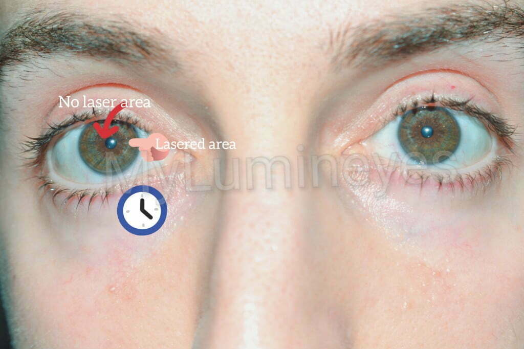
Any safe, permanent intervention targeted at changing iris pigmentation must strictly respect the natural physiology of the eye. Modern medical advancements have introduced highly specialized, non-invasive laser applications designed to address stromal melanin safely. Rather than destroying tissue, these advanced systems utilize specific wavelengths to deliver targeted thermal energy, gently stimulating the eye’s natural metabolic pathways to clear away superficial melanin granules from the anterior border layer. This precise process allows the underlying structural coloration of the unpigmented stroma to reveal itself naturally. To thoroughly examine the clinical protocols, strict safety metrics, and scientific data backing this advanced methodology, please read the authoritative guide on Laser Eye Color Change.

Conclusion: The Structural Foundation of Visual Identity
The iris stroma is a sophisticated, multidimensional fibrovascular matrix that serves as the biological foundation for visual identity, pupillary kinetics, and anterior vascular distribution. Under normal physiological conditions, its internal cellular architecture and pigment payload remain structurally immutable. However, understanding the exact microscopic boundaries of the stroma has enabled the development of highly advanced, medically supervised laser depigmentation protocols. When executed by expert medical hands, these technologies offer a safe path to unlocking the natural structural coloration hidden within the stromal layers without compromising the eye’s intrinsic health.