
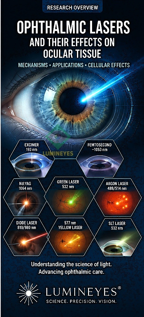
Ophthalmic Lasers: Types, Mechanisms, Applications, and Cellular Effects
A comprehensive scientific overview of how different ophthalmic laser systems interact with ocular tissue — from wavelength and energy delivery to molecular, cellular, and biological responses.
Ophthalmology was one of the earliest medical specialties to adopt laser technology, and laser systems are now used across the cornea, iris stroma, trabecular meshwork, lens capsule, retina, choroid, and other ocular structures. Different laser platforms can produce fundamentally different tissue responses, including photothermal effects, photoablation, photodisruption, photochemical reactions, and plasma-mediated tissue disruption.
Understanding these mechanisms is important because the same general concept — delivering concentrated optical energy — can produce very different biological outcomes depending on how that energy interacts with tissue. Explore our broader clinical investigations in the ophthalmic research archive.
How Laser Energy Interacts With Ocular Tissue
A laser does not have one universal biological effect. Once photons enter ocular tissue, their behavior depends on whether they are transmitted, scattered, reflected, or absorbed by specific molecular targets known as chromophores.
In ophthalmology, clinically important chromophores include hemoglobin, water, and melanin. Understanding the biological role of melanin in eye color and tissue density is vital, as selective absorption allows a laser to concentrate its effect in a particular layer while sparing adjacent ocular structures.
The major interaction mechanisms are commonly described as photothermal, photochemical, photoablative, photodisruptive, and plasma-induced interactions. The boundaries between these categories are not always absolute; under certain treatment conditions, more than one physical process may contribute to the final tissue response.
Five Major Laser–Tissue Interaction Mechanisms
| Mechanism | Primary physical event | Typical biological response | Representative ophthalmic use |
|---|---|---|---|
| Photothermal | Absorbed optical energy is converted primarily into heat. | Protein denaturation, coagulation, thermal cellular injury. | Retinal photocoagulation, laser trabeculoplasty and other thermal applications. |
| Photochemical | Light initiates molecular or chemical reactions. | Molecular modification and downstream biological signaling. | Photodynamic and selected low-energy photobiological applications. |
| Photoablation | High-energy ultraviolet photons break molecular bonds. | Direct removal of superficial tissue with limited thermal diffusion. | Excimer laser corneal refractive surgery. |
| Photodisruption | Very high peak intensity produces optical breakdown and plasma formation. | Cavitation, shock waves and highly localized mechanical tissue separation. | Nd:YAG capsulotomy, iridotomy and selected surgical applications. |
| Plasma-induced ablation | Ultrashort high-intensity pulses produce localized ionization. | Extremely confined tissue separation with minimal surrounding thermal effect. | Femtosecond laser corneal and anterior-segment surgery. |
Major Ophthalmic Laser Systems
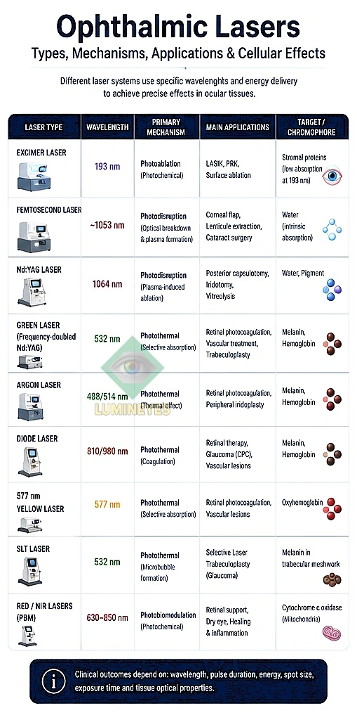
The following overview organizes important ophthalmic lasers by laser type, representative wavelength, primary interaction, clinical target, applications, and cellular or tissue response. Wavelengths shown are representative rather than exhaustive because commercial platforms and clinical protocols may vary.
| Laser type | Representative wavelength | Primary mechanism | Typical target | Main ophthalmic applications | Cellular / biological effect |
|---|---|---|---|---|---|
| Argon | 488 / 514 nm | Photothermal absorption | Melanin, hemoglobin | Retinal photocoagulation; selected trabeculoplasty and vascular applications | Local heating → protein denaturation → controlled cellular injury and coagulation |
| Frequency-doubled Nd:YAG / Green laser | 532 nm | Predominantly photothermal | Melanin, hemoglobin and other absorbing tissue components | Retinal photocoagulation; selected glaucoma and vascular applications | Absorption → temperature rise → protein denaturation and localized tissue remodeling |
| Yellow laser | 577 nm | Photothermal | Hemoglobin and melanin | Retinal vascular disease, macular and retinal photocoagulation applications | Selective chromophore absorption → controlled thermal response |
| Krypton | Green / yellow / red spectrum | Photothermal | Retinal pigment and vascular chromophores | Historical retinal photocoagulation and selected vascular applications | Absorption → thermal injury → coagulation and subsequent tissue response |
| Diode | Commonly around 810 nm | Photothermal | Melanin and other absorbing tissues | Retinal/choroidal treatments, cyclophotocoagulation and other ophthalmic applications | Absorbed energy → heat → cellular stress, protein denaturation and coagulation |
| Nd:YAG | 1064 nm | Photodisruption | Optically focused tissue volume | Posterior capsulotomy, peripheral iridotomy and selected anterior-segment procedures | Optical breakdown → plasma → shock wave and cavitation → mechanical tissue separation |
| Femtosecond | Approximately 1030–1060 nm | Photodisruption / plasma-induced tissue separation | Precisely focused transparent tissue planes | LASIK flap creation, SMILE, corneal surgery, cataract surgery and other microsurgical applications | Ultrashort pulse → nonlinear ionization → plasma/cavitation → highly localized tissue cleavage |
| Excimer | 193 nm | Photoablation | Corneal stromal molecules | PRK, LASIK and other corneal refractive procedures | UV photon absorption → molecular bond disruption → precise tissue removal |
| CO₂ | 10,600 nm | Photothermal / vaporization | Water-rich tissue | Primarily surface and periocular applications rather than routine intraocular laser surgery | Strong water absorption → rapid heating → vaporization and thermal tissue injury |
| Holmium:YAG | Approximately 2,100 nm | Photothermal / water-mediated ablation | Water-rich tissue | Selected ophthalmic and lacrimal applications | Water absorption → rapid heating → vaporization / tissue disruption |
Laser Systems in More Detail
Argon Laser
Argon lasers historically played an important role in retinal photocoagulation. Their visible wavelengths can be absorbed by retinal pigments and blood-containing structures, allowing optical energy to be converted into localized heat.
532 nm Green Laser
Frequency-doubled Nd:YAG systems produce green laser light at 532 nm. The wavelength is well suited to several ophthalmic applications in which absorption by pigmented or vascular tissue is clinically useful.
Importantly, 532 nm should not be treated as synonymous with “retinal laser.” Its biological effect depends on the target tissue, chromophore concentration, power, spot size, pulse duration and exposure. Retinal photocoagulation is one important application, but not the only possible use of green laser energy.
Nd:YAG Laser
The 1064-nm Nd:YAG laser is fundamentally different from the visible green lasers used for many photocoagulation procedures. When tightly focused at sufficiently high peak intensity, the laser can produce optical breakdown.
This generates a small plasma volume followed by rapid expansion, cavitation and mechanical shock. The resulting tissue separation is therefore primarily a photodisruptive rather than simply a thermal process.
Femtosecond Laser
Femtosecond systems use ultrashort pulses in the near-infrared range. Their extremely short pulse duration allows very high peak intensities to be generated at the focal point while limiting energy deposition outside the intended focal volume.
At the cellular and tissue level, nonlinear optical absorption can initiate plasma formation and cavitation. These microscopic events allow tissue planes to be separated with high spatial precision.
Modern femtosecond lasers are used across refractive, corneal, cataract and other anterior-segment procedures. Recent reviews also describe expanding applications and ongoing research into new surgical and biological uses.
Excimer Laser
The 193-nm argon-fluoride excimer laser represents a fundamentally different mechanism. Ultraviolet photons have sufficient energy to disrupt molecular bonds directly, allowing controlled removal of corneal tissue.
Because the effect is driven by molecular photochemistry rather than bulk thermal heating, excimer photoablation can produce highly precise superficial tissue removal when appropriately controlled.
Diode Laser
Diode lasers are semiconductor-based systems available at multiple wavelengths. In ophthalmology, approximately 810 nm is particularly important for photothermal applications.
At the cellular level, absorbed energy is converted into heat. Depending on the delivered energy density and exposure time, this may result in cellular stress, protein denaturation, coagulation or tissue remodeling.
What Happens at the Cellular Level?
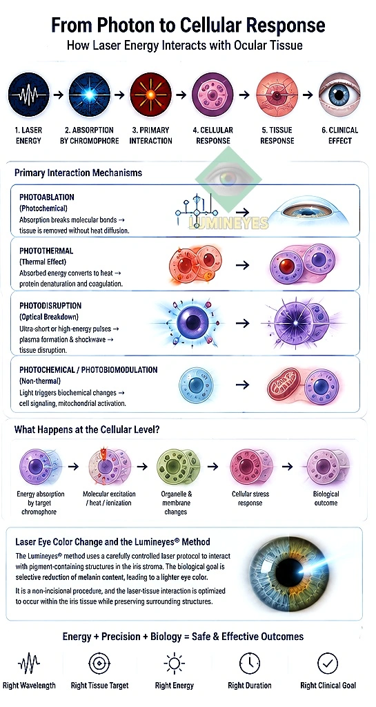
The clinical effect of a laser is ultimately produced by a biological response at the molecular, cellular or tissue level. The exact response depends strongly on the interaction mechanism.
Heat → Protein Change
Absorbed photons increase local temperature. Depending on dose, proteins may denature, cellular membranes may be altered, and tissue coagulation or remodeling may occur.
Photon → Molecular Bond Breakage
High-energy ultraviolet photons can directly disrupt molecular bonds, allowing controlled tissue removal with limited thermal diffusion.
Photon → Plasma → Mechanical Effect
Optical breakdown creates plasma, followed by cavitation and mechanical forces capable of separating tissue at the focal point.
Photon → Chemical Reaction
Light energy can initiate or modify chemical reactions without requiring tissue destruction through bulk heating.
Ultrashort Pulse → Ionization
Very high peak intensity can cause nonlinear ionization and highly localized plasma formation within transparent ocular tissue.
Cell → Tissue → Clinical Effect
The immediate physical interaction is followed by cellular stress, clearance, repair, remodeling or other biological responses that ultimately produce the clinical result.
How Different Laser Mechanisms Translate Into Clinical Applications
| Clinical objective | Typical laser approach | Primary interaction | Biological objective |
|---|---|---|---|
| Retinal photocoagulation | Argon, 532 nm green, yellow and diode systems | Photothermal | Controlled thermal injury to selected retinal tissue |
| Posterior capsulotomy | Nd:YAG 1064 nm | Photodisruption | Mechanical opening of the opacified posterior capsule |
| Peripheral iridotomy | Nd:YAG and selected laser approaches | Photodisruption | Creation of a controlled opening through peripheral iris tissue |
| Corneal refractive ablation | Excimer 193 nm | Photoablation | Precise removal of corneal stromal tissue to reshape the cornea |
| Corneal flap / lenticule surgery | Femtosecond laser | Photodisruption | Highly localized tissue separation along predetermined planes |
| Laser trabeculoplasty | Selective or argon-based laser systems | Thermal / selective photothermal interaction | Biological modification of trabecular meshwork function |
Laser Energy and Pigmented Iris Tissue
The iris is particularly interesting from a laser-tissue interaction perspective because pigmentation changes the optical absorption characteristics of the tissue.
Melanin within the anterior border layer and iris stroma architecture acts as the primary target chromophore. Consequently, the biological response of pigmented iris tissue to laser exposure depends on wavelength, energy density, pulse duration, spatial targeting, and natural pigment distribution.
This is fundamentally different from using a laser to coagulate retinal tissue or to mechanically disrupt the posterior capsule. The biological question becomes not simply “What laser is being used?” but “Where is the energy being absorbed, at what dose, and what biological response follows?”
Where Laser-Based Iris Pigment Reduction Fits Into This Framework
The broader principles of laser–tissue interaction provide the scientific foundation for modern laser eye color change.
The Lumineyes® Method is a physician-supervised, non-incisional approach developed by Dr. Mustafa Mete, designed to gradually stimulate natural pigment clearance through individualized laser parameters. Its clinical objective is distinct from thermal retinal photocoagulation or Nd:YAG optical breakdown.
The protocol is strictly individualized according to iris thickness, melanin density, anterior chamber angle anatomy, and biological healing responses rather than being defined by a single fixed setting. For an itemized breakdown of clinical stages, review the detailed guide on how to change eye color with laser.
From Laser Physics to Laser Eye Color Change
Understanding general laser–tissue interaction is useful before examining a specific ophthalmic application. The following clinical resources explore the complete treatment framework and biological mechanisms in greater depth:
Laser Eye Color Change
Explore the definitive clinical framework, physician qualifications, candidate evaluation, and complete procedure overview.
Laser Iris Depigmentation
Examine the photobiological breakdown of melanin granules, macrophage-mediated clearance, and trabecular meshwork safety.
Iris Stroma & Melanin Biology
Review how stromatal density and melanin concentration determine baseline shade and laser light absorption kinetics.
Why the Biological Response Matters
In laser medicine, the visible clinical result is often only the final stage of a much longer biological sequence. Energy absorption occurs first; molecular or physical changes follow; cells then respond through clearance, repair, and tissue remodeling.
This distinction is particularly important when evaluating permanent eye color change . A visible change in iris appearance is not an instantaneous surgical alteration. The long-term appearance of ocular tissue depends on natural macrophage clearance and stable cellular homeostasis.
Comparing Laser Effects at the Cellular Level
| Interaction | Immediate physical event | Primary cellular consequence | Typical tissue-level result |
|---|---|---|---|
| Photothermal | Temperature increases | Protein denaturation and thermal cellular injury | Coagulation / controlled tissue remodeling |
| Photoablation | Molecular bonds are disrupted | Direct molecular removal of superficial tissue | Precisely controlled ablation |
| Photodisruption | Optical breakdown and plasma formation | Mechanical disruption and cavitation | Localized tissue separation |
| Photochemical | Molecular excitation / reaction | Biochemical modification or signaling | Biological modulation |
| Plasma-induced | Nonlinear ionization | Highly localized plasma and mechanical effects | Microscale tissue cleavage |
Key Principles of Ophthalmic Laser–Tissue Interaction
Wavelength Matters
Wavelength influences tissue penetration and which chromophores are capable of absorbing the delivered optical energy.
Energy Delivery Matters
Pulse duration, power, energy density, spot size and exposure time strongly influence whether tissue is heated, ablated or mechanically disrupted.
Tissue Matters
Water content, pigmentation, vascularity, transparency and tissue structure influence how optical energy is absorbed and distributed.
Cells Determine the Biology
The clinical result ultimately depends on how cells and extracellular structures respond to the initial physical interaction.
One Laser Can Have Multiple Effects
Depending on treatment parameters and tissue conditions, more than one interaction mechanism can contribute to a clinical outcome.
Clinical Context Is Essential
A laser should never be evaluated by wavelength alone. The target, treatment objective, energy delivery and biological response must be considered together.
Related Clinical & Safety Research
Laser physics is only one part of responsible ophthalmic laser treatment. Patient selection, safety protocols, intraocular pressure monitoring, and long-term follow-up are essential pillars of clinical care.
Cost, Risks & Safety Analysis
Detailed analysis of anterior chamber dynamics, trabecular safety, and risk prevention in laser eye color modification.
Long-Term Iris Stability
Explore tissue stability and cellular preservation factors in permanent aesthetic iris depigmentation.
Ophthalmic Research Hub
Browse all scientific publications, comparative laser studies, and clinical data authored by Dr. Mustafa Mete.
Research References
- Zhang AY, Kumar D, Tripathy K. Laser Principles in Ophthalmology. StatPearls / NCBI Bookshelf. NCBI
- Palanker D. Evolution of Concepts and Technologies in Ophthalmic Laser Therapy. Annual Review of Vision Science. PubMed
- Zhang W-P, et al. Femtosecond lasers in ophthalmology: Mechanisms, clinical breakthroughs, and multidisciplinary frontiers. Progress in Retinal and Eye Research. PubMed
- Van Gemert MC, Welch AJ. Clinical use of laser-tissue interactions. IEEE Engineering in Medicine and Biology Magazine. PubMed
- Jacques SL. Laser-tissue interactions: Photochemical, photothermal, and photomechanical. Surgical Clinics of North America. PubMed
Explore the Clinical Application
After understanding how laser energy interacts with ocular tissue, explore the complete physician-supervised clinical framework of laser eye color change, including comprehensive evaluation, customized treatment parameters, and long-term follow-up.
Explore Laser Eye Color Change