Biological Limits of Eye Color Change: Structural and Physiological Determinants
Laser eye color change is governed by biological principles rather than purely technical capability. The achievable degree of iris lightening depends on the intrinsic properties of the eye, including pigment density, stromal architecture, optical light scattering, and the individual's physiological response to treatment.
The important clinical question is therefore not simply how much pigment a laser can remove. It is how much pigment reduction the eye can accommodate while maintaining appropriate structural and physiological function. This distinction is central to individualized treatment planning and to understanding why eye color change should be approached as a biological process rather than as a purely cosmetic or mechanical one.
In the MyLumineyes® clinical framework, the observed endpoint is influenced by the starting pigmentation of the iris, the organization of the anterior stroma, the biological clearance of liberated pigment, and the optical properties of the remaining tissue.
Stromal Pigment Density: The Primary Boundary
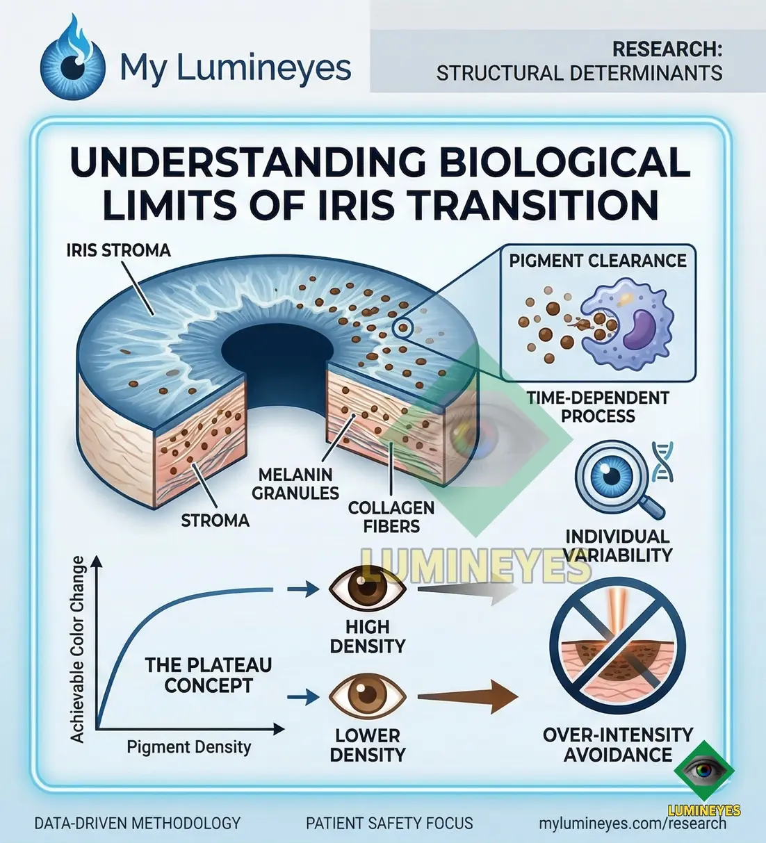
Melanin concentration within the anterior iris stroma determines much of the baseline optical appearance of the iris and influences how much visible lightening may occur after pigment reduction.
Higher pigment density, particularly in dark brown eyes , generally requires a more gradual and staged approach. The biological processing of liberated pigment is not instantaneous, which means that increasing treatment intensity does not necessarily produce a proportionally faster visible result.
This is one reason why the initial iris phenotype matters. Two patients may undergo technically similar treatment while showing different rates and degrees of visible color transition because the underlying biological substrate is different.
Structural Constraints & Light Scattering
Reducing melanin does not create an entirely new optical tissue. The remaining iris structure continues to determine how incoming light is absorbed, reflected and scattered. Consequently, final color is a combined expression of pigment reduction and the structural optics of the patient's iris.
Tyndall Scattering, Rayleigh Scattering & Collagen Matrix Density
The phenotypic expression of iris color after pigment reduction is influenced by how light interacts with the residual stromal framework. As melanin is reduced, light interacts increasingly with the non-pigmented components of the anterior iris stroma.
Differences in collagen organization, stromal density and tissue geometry can therefore contribute to differences in the blue, green, hazel or gray optical spectrum that becomes visible after progressive pigment reduction.
Residual Pigment
The amount, distribution and depth of remaining pigment influence the amount of light that is absorbed before it reaches deeper stromal structures.
Stromal Architecture
The thickness, organization and optical density of the iris stroma influence how wavelengths are scattered after the amount of absorbing pigment has been reduced.
Physiological Clearance Mechanisms
Following laser interaction, fragmented pigment particles undergo a biological clearance process. Within the MyLumineyes® framework, this process is understood as an important reason why visible color evolution may continue after the treatment session itself.
Pigment clearance involves cellular activity and ocular fluid dynamics. The process is therefore time-dependent rather than instantaneous. This helps explain why permanent eye color change should be evaluated over an appropriate follow-up interval rather than immediately after laser exposure.
Laser energy interacts with targeted stromal pigment, producing a reduction and fragmentation of melanin-containing material.
Biological clearance mechanisms become involved in processing and removing pigment debris generated during treatment.
Pigment processing continues over time. The visible optical appearance of the iris may therefore evolve gradually rather than immediately.
As pigment reduction and biological clearance progress, the remaining iris structure increasingly determines the final visible optical phenotype.
The clearance process places a biological limit on treatment acceleration
Because pigment clearance is biological and time-dependent, simply increasing treatment frequency or energy does not necessarily produce faster cosmetic evolution. A staged approach allows clinical decisions to be guided by the patient's response rather than by an assumption that more treatment must always produce more color change.
Why Biological Clearance Matters
Macrophage-Mediated Processing
Macrophage activity is one component of the biological response to liberated pigment. These cells participate in the uptake and processing of extracellular pigment material.
The rate of this biological activity varies between individuals and cannot be treated as a fixed mechanical constant.
Aqueous Humor & Outflow Pathways
Pigment-containing material interacts with the anterior segment's natural clearance environment. The aqueous humor , trabecular meshwork and uveoscleral pathways form part of the physiological context in which pigment clearance must be considered.
The clinical significance of this process is that the eye has finite physiological capacity to respond to and clear liberated pigment. A treatment strategy that ignores this biological timing may not produce the expected increase in cosmetic speed and may instead increase the importance of careful ophthalmic monitoring.
Four Biological Determinants of the Final Eye Color
| Biological determinant | What it influences | Clinical implication |
|---|---|---|
| Baseline pigment density | The amount of melanin contributing to the original iris appearance. | Greater pigmentation generally requires more gradual biological reduction. |
| Stromal architecture | The structural framework through which light interacts after pigment reduction. | Helps explain why similar pigment reduction can produce different visible hues. |
| Clearance response | The biological processing and removal of liberated pigment material. | Determines the pace at which visible changes evolve after treatment. |
| Optical scattering | The way wavelengths interact with residual stromal structures. | Contributes to the final blue, green, hazel or gray optical spectrum. |

Individual Biological Variation
No two irides are biologically identical. Variations in baseline pigmentation, stromal organization, anterior segment anatomy and physiological response can influence both the pace and the visible extent of color transition.
For this reason, the number of treatment sessions should not be interpreted as a universal mathematical formula. Clinical planning is better understood as a response-guided process in which the patient's anatomy and biological response remain central to decision-making.
A darker starting iris does not simply mean that a stronger laser treatment is required. It may instead mean that a more gradual treatment strategy and longer biological intervals are necessary to respect the eye's individual response.
The “Biological Plateau” Concept
One of the most important concepts in understanding laser eye color change is the possibility of a biological plateau.
An iris may reach a point at which further pigment reduction produces little or no meaningful additional visible change. Remaining pigment may be structurally less accessible, optically less significant, or biologically less responsive.
Recognizing this point is clinically important because the objective of treatment is not unlimited depigmentation. The objective is controlled pigment reduction within the biological boundaries of the individual eye.
This principle distinguishes a response-guided ophthalmic approach from the assumption that increasing energy or repeating treatment indefinitely must continue to improve the cosmetic result.
Biological Limits and Ocular Safety
Biological limits are not merely theoretical. They are part of the safety framework surrounding any procedure that interacts with living ocular tissue.
Why Staging Matters
A staged clinical protocol provides time for the eye's biological response to develop and for the treating ophthalmologist to evaluate the patient's response before deciding whether additional treatment is appropriate.
Why Monitoring Matters
Ophthalmic follow-up allows treatment decisions to remain connected to the patient's actual ocular status rather than being based solely on cosmetic expectations.
The broader clinical research framework describes pigment clearance as a biological process involving cellular activity and ocular fluid dynamics. These mechanisms help explain why treatment planning must respect physiological timing.
Key Clinical Takeaways
- Eye color change is influenced by biological properties of the iris, not simply by laser capability.
- Baseline stromal pigment density is one of the primary determinants of the achievable degree of visible lightening.
- Stromal architecture and optical scattering influence the final visible hue after pigment reduction.
- Pigment clearance is a time-dependent biological process and may continue after treatment sessions.
- Individual physiological variation means that color evolution cannot be reduced to a single universal treatment formula.
- A biological plateau may occur when further pigment reduction produces little additional meaningful change.
- Respecting biological limits is therefore an essential component of individualized ophthalmic treatment planning.
Mete, M. (2026). The Biophysical Framework and Biological Latency of Laser-Induced Iris Depigmentation. International Journal of Ophthalmic Research, Staged Longitudinal Analysis.