Clinical Reference Guide · Cosmetic Iris Implants · Safety & Evidence
Artificial Iris Implants for Eye Color Change: Risks, Removal & Safety
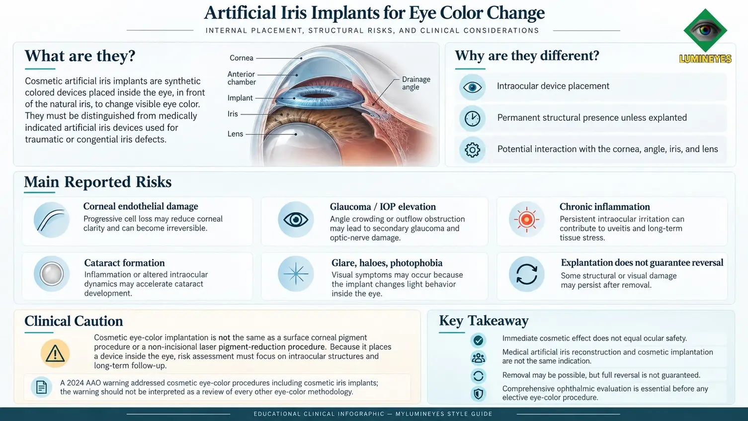
Cosmetic artificial iris implants change the appearance of eye color by placing a colored device inside the anterior chamber, in front of the natural iris. Unlike corneal pigment placement or laser iris depigmentation, this approach introduces a permanent intraocular structure unless the device is removed. Its cosmetic effect must be weighed against documented risks to the corneal endothelium, drainage angle, lens and optic nerve.
Editorial disclosure: MyLumineyes develops a distinct non-incisional laser iris depigmentation methodology. This page discusses independently published risks of cosmetic iris implants. Anatomical differences are not presented as proof that any alternative is risk-free or superior.
Are artificial iris implants safe for cosmetic eye color change?
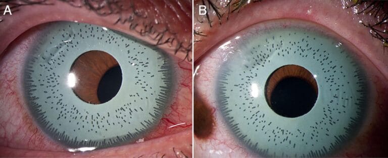
Cosmetic anterior-chamber iris implants have a documented record of potentially sight-threatening complications. Published referral series describe uveitis, increased intraocular pressure, glaucoma surgery, corneal endothelial failure and cataract surgery. Removing the implant may be necessary, but removal cannot reliably reverse damage that has already occurred. An artificial iris used to reconstruct a missing or damaged iris is a different indication, with different device selection, surgical planning and risk-benefit considerations.[1][2]
1. How Do Cosmetic Artificial Iris Implants Work?
In cosmetic iris implantation, a colored synthetic device is inserted through a corneal or limbal incision into the eye's anterior segment. In common cosmetic designs, the implant is positioned in front of the patient's natural iris to conceal its original appearance. Unlike colored contact lenses, this is an intraocular operation, not an external optical accessory.
The anterior chamber is a limited space shared by structures essential to vision and intraocular-fluid regulation. The corneal endothelium maintains corneal clarity, the peripheral angle contains aqueous outflow structures, and the iris and lens lie immediately behind the implant. Contact, friction, crowding and persistent tissue irritation are therefore central concerns rather than incidental cosmetic side effects.
Corneal endothelium
A delicate cell layer on the inner corneal surface. Progressive cell loss may impair corneal deturgescence and clarity.
Angle and aqueous outflow
Angle crowding, peripheral contact, pigment dispersion and inflammation can disturb pressure regulation.
Iris and crystalline lens
Mechanical irritation and inflammatory changes may be associated with synechiae, cataract and visual disturbance.
Patients seeking a broader explanation of the anatomical differences among procedures can consult our eye color change surgery clinical guide.
2. Cosmetic Iris Implants Are Not the Same as Medical Artificial-Iris Reconstruction
The phrase artificial iris describes more than one clinical situation. Prosthetic iris devices have a legitimate reconstructive role in selected patients with congenital aniridia, traumatic iris loss or other major defects. This must not be confused with elective implantation intended only to alter the color of an otherwise healthy iris.
| Clinical question | Cosmetic anterior-chamber iris implant | Medically indicated artificial-iris reconstruction |
|---|---|---|
| Primary purpose | Elective alteration of eye appearance in an eye with an existing iris. | Reconstruction of missing or severely damaged iris tissue and associated functional deficits. |
| Patient selection | Cosmetic preference, without a reconstructive need. | Documented ocular pathology requiring individualized surgical evaluation. |
| Device and fixation | Cosmetic device design and anterior-chamber placement; design varies. | Specialized prosthesis; position and fixation depend on ocular anatomy and surgical plan. |
| US FDA context | No FDA approval for cosmetic iris implantation to change a healthy eye's color. | CustomFlex Artificial Iris has FDA approval for treatment of full or partial aniridia under its specified indication. |
| Clinical benefit/risk | Appearance change without a therapeutic indication must be weighed against intraocular complications. | Potential functional benefit is weighed against risks in a diseased or damaged eye. |
Regulatory precision: An FDA-approved artificial-iris prosthesis for aniridia does not imply that placing a cosmetic iris implant into a healthy eye is FDA-approved or similarly studied.[2]
3. Main Medical Risks of Cosmetic Iris Implants
Complications may emerge immediately, develop gradually, or first become clinically apparent years after implantation. The mechanisms vary with implant design, anterior-chamber dimensions, pre-existing anatomy, surgical technique and length of exposure. The following risks are documented in published clinical reports; they should not be interpreted as inevitable outcomes in every recipient.[1][4]
Corneal endothelial cell loss and corneal decompensation
Corneal endothelial cells have limited capacity to replace lost cells. Mechanical contact and chronic anterior-segment stress can contribute to endothelial loss. A decompensated cornea may become edematous, hazy and visually disabling; advanced cases can require endothelial keratoplasty. A clear-looking cornea at one visit does not by itself establish stable endothelial reserve.
Elevated intraocular pressure and secondary glaucoma
An implant can disturb aqueous outflow through angle crowding, chronic inflammation, peripheral synechiae or pigment-associated obstruction. A rise in intraocular pressure may be asymptomatic before optic-nerve injury develops. In reported severe cases, patients have required glaucoma medication or surgery even after device removal.
Uveitis, pigment dispersion and chronic irritation
Persistent contact between a device and intraocular tissues can produce inflammatory episodes, photophobia and anterior-chamber reaction. Recurring inflammation may be accompanied by synechiae and additional pressure or lens complications. Clinical assessment should consider inflammation and mechanical effects together rather than viewing redness alone as the measure of severity.
Cataract and lens complications
Inflammation, altered intraocular relationships or associated surgical interventions may contribute to lens opacity. Some patients in published complication cohorts required cataract extraction. The necessity and timing of further surgery depend on the patient's visual and anatomical findings.
Glare, haloes, blurred vision and photophobia
An artificial diaphragm may affect pupil-related optics, light transmission and the perceived depth or texture of the eye. Although some complaints are primarily optical, new blur or photophobia can also indicate a medical complication. Persistent symptoms require examination rather than cosmetic reassurance.
Clinical priority: Progressive visual blur, severe pain, sudden vision reduction, marked redness, colored haloes accompanied by pain, or nausea with eye pain warrants prompt ophthalmic assessment. Delaying care can increase the risk of permanent injury.
4. What Does Published Clinical Evidence Actually Show?
A 2025 retrospective series from Moorfields Eye Hospital reviewed 30 eyes of 15 patients who presented with complications following cosmetic iris implantation. The paper reported serious inflammatory, pressure-related and corneal sequelae, including the need for additional surgery after implant removal.[1]
Critical interpretation: These percentages describe a selected referral cohort of patients who had already developed complications; they are not the probability that an unselected person undergoing implantation will experience the same event. The series is valuable for understanding severity and management, not for estimating population-wide incidence.
The distinction is important: a referral-based complication series is more likely to include severe cases than a prospective cohort of every treated person. Nevertheless, documented outcomes such as glaucoma surgery and corneal transplantation establish that the potential consequences can extend far beyond dissatisfaction with appearance. Additional literature describes glaucoma and anterior-segment injury associated with cosmetic iris implants.[4][5]
5. FDA Status and the American Academy of Ophthalmology Warning
The US Food and Drug Administration approved the CustomFlex Artificial Iris for treatment of full or partial aniridia. That approval concerns a defined medical indication and does not extend to routine cosmetic eye-color change in an otherwise healthy iris.[2]
In January 2024, the American Academy of Ophthalmology (AAO) issued a public warning concerning elective eye-color procedures, specifically including cosmetic iris implant surgery and laser-assisted corneal pigmentation (keratopigmentation). The statement emphasized potential vision-threatening complications and the need for patients to understand procedure-specific risks.[3]
Read the professional statement: American Academy of Ophthalmology — Warning on Eye Color Procedures.
Scope matters: This AAO warning should not be described as an independent safety assessment of the Lumineyes™ laser iris depigmentation protocol. Absence of a method from a particular warning is neither endorsement nor proof of safety.
6. Can Cosmetic Iris Implants Be Removed? What Happens After Explantation?
Device explantation is possible, but it requires another intraocular intervention. Removal may relieve ongoing mechanical contact or inflammatory exposure; however, its potential benefits must be evaluated against the risks of further surgery. Established endothelial loss, optic-nerve injury, cataract or synechiae may not resolve simply because the foreign device is absent.
Before removal
Assess visual acuity, IOP, corneal clarity and endothelial status, anterior-chamber anatomy, angle, lens and optic nerve; additional testing depends on clinical findings.
At removal
Surgical planning depends on device design, adhesions and coexisting pathology. Some eyes require combined or staged treatment for corneal disease, glaucoma or cataract.
After removal
Monitor IOP, inflammation, endothelial function and visual recovery. Objective follow-up is important even when external appearance improves.
There is no reliable universal recovery timeline after explantation. A patient with limited irritation and preserved ocular structures is in a very different situation from one with established glaucoma or corneal decompensation. The published complication series documents substantial additional surgical treatment after explantation.[1]
7. What Should Be Evaluated Before or After an Iris Implant?
For an individual considering cosmetic implantation, the presence of a healthy eye does not eliminate risk from the procedure itself. For a person who already has an implant, examination is warranted even if symptoms are modest, because pressure elevation and endothelial loss can be relatively silent early in their course.
| Assessment | Clinical purpose |
|---|---|
| Visual acuity and refraction | Identify visual change and document baseline function. |
| Slit-lamp examination | Assess cornea, inflammation, implant position, pupil and lens. |
| Intraocular pressure | Detect pressure elevation and guide follow-up or treatment. |
| Specular microscopy | Estimate endothelial cell density and morphology where measurable. |
| Gonioscopy / anterior-segment imaging | Evaluate angle, implant relationships and peripheral anatomy when indicated. |
| Optic-nerve assessment | Consider retinal nerve-fiber imaging and perimetry when glaucoma is suspected. |
If you already have a cosmetic iris implant: Do not attempt to reposition or remove the implant yourself. Seek a qualified ophthalmologist experienced in anterior-segment and glaucoma complications, particularly if you notice new pain, halos, blurred vision, light sensitivity or redness.
8. How Do Iris Implants Differ From Other Eye Color Change Options?
The major distinction is which tissue is altered and whether a foreign device remains inside the eye. The following methods should be compared by anatomy, objective outcomes, published evidence and patient suitability rather than marketing phrases such as “permanent,” “minimally invasive” or “natural-looking.”
Colored contact lenses
Alter appearance externally without permanent iris or corneal modification. They still require appropriate fitting, care and hygiene because infection and corneal injury remain possible.
Keratopigmentation
Introduces artificial pigment into corneal tissue; the natural iris remains behind the altered corneal appearance. Review the keratopigmentation guide.
Laser iris depigmentation
Reduces visible iris pigment without adding an intraocular prosthesis. Clinical response, risks and evidence vary by laser protocol. Read about non-incisional laser iris depigmentation.
For a cross-method anatomical assessment, read Eye Color Change Method Comparison. For a detailed clinical discussion of surgery, risks and recovery across approaches, see Eye Color Change Surgery. These comparisons are educational and do not establish that any one procedure is universally safest.
Artificial Iris Implants: Frequently Asked Questions
Are cosmetic iris implants FDA-approved?
No FDA approval applies to implanting a colored iris device solely to change the appearance of a healthy eye. The approved CustomFlex Artificial Iris is indicated for treatment of full or partial aniridia; it is not a general approval for elective eye-color implantation.
Can an artificial iris implant cause permanent vision loss?
Yes. Serious complications such as uncontrolled glaucoma, corneal decompensation and associated optic-nerve or ocular tissue injury can threaten vision. Published reports document cases requiring glaucoma and corneal surgery.
Is cosmetic iris implant surgery reversible?
The implant can sometimes be surgically removed, but this does not mean the procedure is fully reversible. Endothelial cell loss, glaucomatous optic-nerve injury, cataract or scarring may persist after explantation.
Is a medical artificial iris prosthesis the same as a cosmetic implant?
No. Medical artificial iris reconstruction addresses a missing or severely defective iris, with individual surgical planning and an accepted therapeutic indication. Cosmetic implantation seeks an appearance change in an eye with its natural iris present.
What symptoms after an iris implant require urgent attention?
New or severe eye pain, sudden loss of vision, rapidly worsening blur, marked redness, significant photophobia or nausea with eye pain require urgent ophthalmic evaluation.
Does the AAO warning establish that laser iris depigmentation is safe?
No. The 2024 AAO warning addressed particular cosmetic eye-color procedures, including iris implants and corneal pigmentation. It is not a direct clinical validation or safety endorsement of other laser iris depigmentation protocols.
Are before-and-after pictures enough to judge implant safety?
No. Cosmetic photographs do not measure intraocular pressure, endothelial cell density, angle anatomy, inflammation or optic-nerve health. Clinical safety requires examination and appropriate longitudinal follow-up.
Make an informed decision before any elective eye procedure
If you already have a cosmetic iris implant and experience visual symptoms, seek examination by a qualified ophthalmologist rather than waiting for a routine cosmetic review. For general information about MyLumineyes' separate laser methodology and medical evaluation process, use the structured contact form; submitting an inquiry does not constitute an appointment or confirmation of clinical service availability.
Medical Evaluation InformationScientific References & Regulatory Sources
- Balal S, Jamall O, Janmohamed IK, et al. Complications and Management of Cosmetic Iris Implants. American Journal of Ophthalmology. 2025;277:433–440. doi:10.1016/j.ajo.2025.05.049. Retrospective complication referral series, 30 eyes in 15 patients.
- US Food and Drug Administration. CustomFlex Artificial Iris. Premarket Approval P170039, approved May 30, 2018. Approved indication: treatment of full or partial aniridia.
- American Academy of Ophthalmology. Academy Issues Warning on Eye Color Procedures. Newsroom statement, January 2024. Read AAO statement.
- Arthur SN, Wright MM, Kramarevsky N, et al. Uveitis-glaucoma-hyphema syndrome and corneal decompensation in association with cosmetic iris implants. American Journal of Ophthalmology. 2009;148(5):790–793. doi:10.1016/j.ajo.2009.06.008.
- Seresirikachorn K, Chang TCP. Clinical features, management, and long-term visual outcomes of glaucoma associated with cosmetic iris implants. Eye. 2026;40:1080–1081. doi:10.1038/s41433-026-04416-1.
How to read the evidence: Complication referrals document potential harms and clinical management but cannot determine the complication incidence among all implant recipients. Medical-device approval for reconstruction does not authorize unrelated cosmetic use. References are included for source transparency; the AAO statement is the only external navigation link on this page.