
Biological Debris Burden and Clearance After Iris Laser Procedures
A material-specific study of Nd:YAG peripheral iridotomy and staged anterior stromal depigmentation

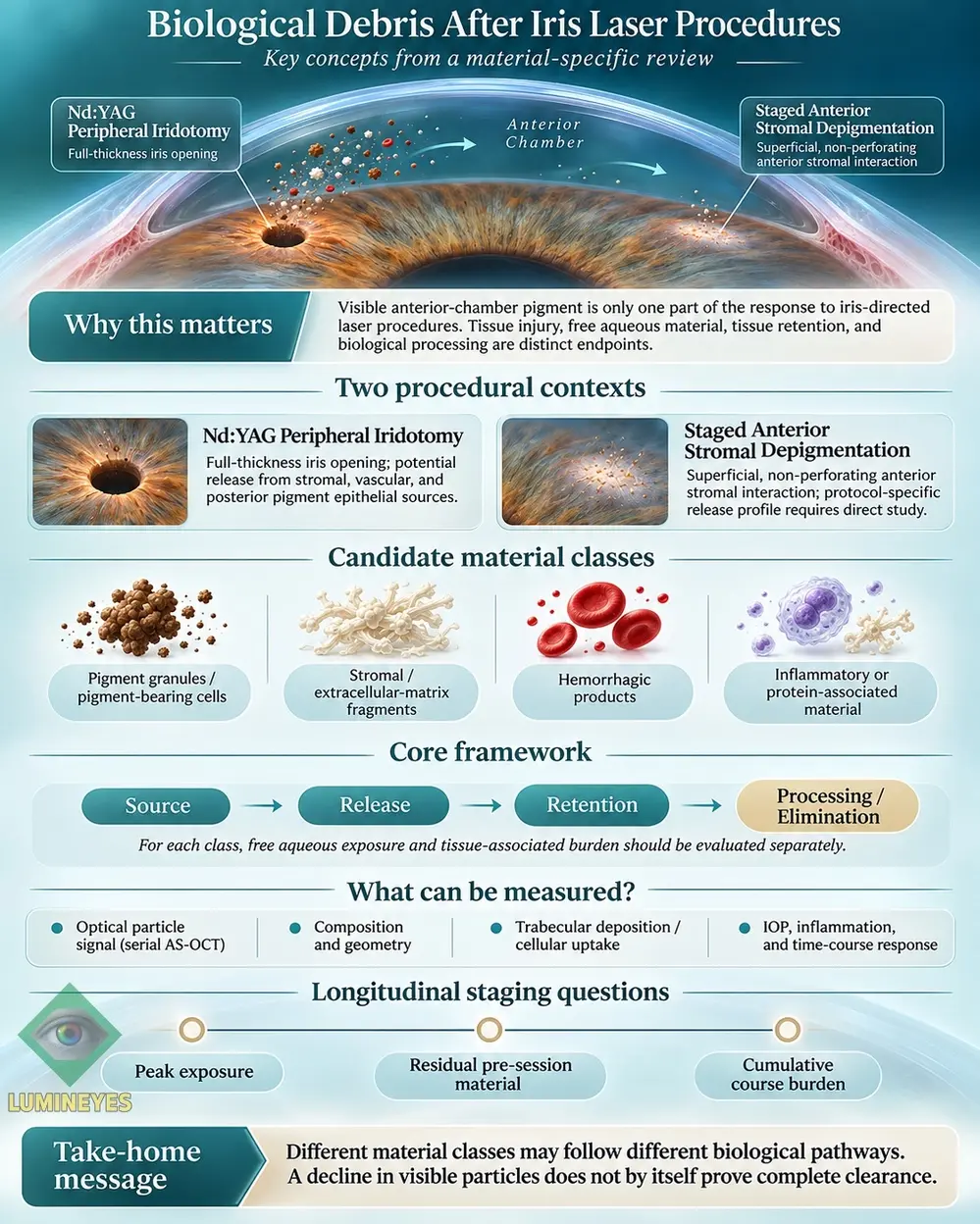
Research overview. A visible pigment cloud does not describe the complete biological fate of iris-derived material. The relevant sequence extends from anatomical tissue interaction to aqueous release, deposition, cellular uptake, processing and elimination. This article presents a material-specific approach to these events and explains how future anterior-segment studies could measure them independently.
1. The research question: what becomes of laser-generated iris material?
Iris-directed laser exposure has an immediate interaction with tissue, but the observed material in the anterior chamber is downstream of that event. A full-thickness iris defect, a visible particle signal and later pressure measurements describe different aspects of the same procedure. They cannot be collapsed into a single estimate of biological debris clearance.
A more precise investigation asks whether affected tissue remains attached at the exposure site, whether pigment or non-pigment fragments enter the aqueous compartment, whether released particles deposit on tissue or enter cells, and whether those particles are ultimately degraded or transported away. Composition, geometry, anatomical source and time each influence the answer.
The anatomical and biological distinctions are related to the broader biological fate of pigment in eye-color modification and the living iris stromal microenvironment.
2. Two procedures, different anatomical sources
Nd:YAG peripheral iridotomy
Iridotomy creates an opening through the iris for a defined therapeutic indication. Human histopathology has demonstrated photodisruptive injury involving more than the anterior pigmented surface, including stromal and vascular structures. Posterior pigment epithelial disruption is also relevant to the possible material inventory. [1,2]
Staged laser iris depigmentation
Staged anterior stromal treatment is intended to modify visible pigment without creating a full-thickness opening. The material produced, released or retained depends on the actual protocol. Published clinical series and experimental iris studies provide relevant observations, while material-specific comparisons remain a direct measurement question. [4,12]
This comparison is anatomical rather than an assertion that two procedures have equivalent indications or measured injury loads. Full-thickness tissue disruption and superficial pigment-directed exposure present different starting conditions for studying the resulting material. The precise source profile must be established for each laser modality and treatment protocol.
For the target-and-response terminology used within the Lumineyes approach, see Selective Stromal Melanin Modulation (SSMM). The framework addresses the intended target and treatment logic; the present research question is the subsequent biological fate of the affected material.
3. Beyond a single “pigment cloud”: four candidate material classes
Aqueous particles are not necessarily interchangeable. Pigment, collagen-rich fragments, erythrocytes and inflammatory material differ in molecular composition, dimensions, morphology, deformability and cell-processing pathways. A material inventory therefore needs more than a numerical particle count.
Pigment
Melanin granules, melanosomal material and pigment-containing cells.
Stromal matrix
Collagen-containing fragments and other extracellular-matrix material.
Hemorrhagic
Erythrocytes, fibrin and blood-associated components where bleeding occurs.
Inflammatory
Cells, soluble proteins and protein-associated light-scattering material.
For every class, the relevant endpoints include particle number, size distribution, mass or volume, biochemical identity, free suspension, tissue-associated retention and intracellular processing. A high particle count can coexist with a low total material mass; conversely, a small number of elongated fragments may represent a different mechanical and cellular challenge than many fine granules.
4. A material-specific source–release–retention–processing framework
The proposed framework separates four linked but independently measurable events. It begins at the tissue source, follows material released into aqueous humor, distinguishes retained tissue-associated fractions, and then investigates cellular processing or biological elimination.
Fi(t) represents freely suspended material of class i at time t. Ti(t) represents material deposited in or associated with ocular tissue. Movement between these observable compartments may reflect aqueous transport, tissue deposition, cellular internalization or redistribution. Biological degradation and elimination remain distinct downstream outcomes.
A reduction in Fi(t) may reflect fewer particles in the sampled chamber region while some material has moved into the tissue-associated compartment. This is why optical disappearance, intracellular uptake and completed clearance require separate evidence. The broader longitudinal pigment-response question is developed in P-CLEAR.
5. Why material identity matters to trabecular cells
Experimental work on trabecular meshwork cells provides a mechanistic reason to study the identity of particles rather than assuming a uniform debris response. In a pigmentary glaucoma model, Dang and colleagues reported intraocular-pressure elevation before a measurable reduction in phagocytic activity. Other experiments documented changes in stress-fibre formation, cellular migration, contraction and Rho-associated signalling under pigment exposure. [9,10]
The distinction between uptake and degradation is particularly important. Porter, Lin and Liton compared different phagocytic challenges in cultured trabecular cells. Collagen-I-coated microspheres and bacterial substrates promoted phagolysosomal maturation and cathepsin-B-associated activity, whereas pigment particles showed a different intracellular processing profile. [11] These observations support a substrate-specific research strategy.
Local iris processing is also biologically relevant. In rabbit iris tissue exposed to frequency-doubled Nd:YAG laser energy, Yildirim and colleagues observed anterior stromal pigment reduction with increased pigment-containing histiocytes in treated areas. [12] This identifies local cellular handling as a potential companion to material entering aqueous humor.
6. Measuring particles: what AS-OCT can and cannot distinguish
Serial anterior-segment optical coherence tomography can quantify optical particulate signals over time. Following combined laser iridotomy, Yoshihara and colleagues reported an increase in an AS-OCT anterior-chamber particle index from a mean of 0.78 before treatment to 7.72 immediately afterward, with values approaching baseline over the following week. [13] This establishes the usefulness of time-resolved chamber imaging.
Optical intensity does not directly identify a particle’s chemical composition or mass. Bellchambers and colleagues further examined particle dimensions in swept-source AS-OCT images of eyes with uveitis, providing a separate direction for differentiating pigment-related and inflammatory signals. [14] Imaging and molecular identification answer complementary questions.
| Measurement | What it can describe | What else is needed |
|---|---|---|
| Serial AS-OCT particle signal | Temporal change in imaged aqueous scattering | Calibration, acquisition controls and composition-specific validation |
| Particle morphology | Number, length, width, aggregate fraction and shape | Detection limits and reproducible segmentation |
| Microscopy and biochemical assay | Material composition, pigment or matrix identity | Controlled sampling and recovery-efficiency assessment |
| Angle/tissue observation | Deposition or tissue-associated material | Appropriate resolution and tissue-level confirmation |
| Physiological monitoring | IOP, inflammation and outflow-related response | Baseline anatomy and relevant covariate adjustment |
For work extending beyond pigment optics into measurable iris architecture and signal reliability, see the L-OAT quantitative iris phenotyping framework.
7. Staged exposure: peak, residual and cumulative burden
A staged treatment course creates more than one observation time. Three longitudinal quantities should be distinguished for each identified material class:
Post-session peak
The maximum material signal measured after an individual exposure at specified observation times.
Pre-session residual
Material still observable before a planned subsequent stage, including any tissue-associated fraction.
Course-level total
The time-integrated material exposure across the entire staged course, defined by an explicit sampling model.
The three measures can diverge. A lower peak after one session does not establish a lower integrated course burden, and a clear anterior-chamber image before another stage does not necessarily describe tissue-retained material. This is a reason to distinguish state and trajectory from elapsed time.
Within the wider MyLumineyes framework, Temporal Response Architecture (TRA) addresses time-dependent biological states, while L-SAFE addresses the clinical decision framework for observing, pausing or proceeding after reassessment. Neither replaces direct material-specific measurements.
8. A practical research architecture
The strongest next step is a protocol-defined experimental programme linking material identity to measured outcomes. A first stage could use suitable donor iris or ex vivo anterior-segment tissue, matched exposure documentation, compartment-specific sample collection, and masked particle analysis. Subsequent clinical observations could then pair serial optical measurements with IOP, angle and inflammatory assessment in procedures undertaken for their own clinical indications.
| Research question | Primary endpoint | Potential method |
|---|---|---|
| Composition | Relative fractions of pigment, stromal, blood-derived and inflammatory material | Composition-specific microscopy and assay panels |
| Geometry | Particle-length, width, aspect-ratio and aggregation distributions | Calibrated image analysis with defined detection limits |
| Retention | Free aqueous versus tissue-associated fractions | Paired sampling, tissue imaging and experimental histology |
| Processing | Uptake, lysosomal maturation and material breakdown | Controlled cell challenges and substrate-specific assays |
| Time-course exposure | Peak, residual and integrated material signals | Repeated measurements across a staged schedule |
Measured particle composition should be interpreted alongside anatomical source, baseline angle characteristics, pigmentation phenotype, medication, laser modality, exposure settings and interval between procedures. Functional endpoints, including intraocular pressure, must be recorded as physiological observations rather than used as substitutes for directly observed material clearance.
For the broader longitudinal clinical context, consult the MyLumineyes clinical data and long-term observation archive. It is a distinct clinical record; the present article addresses a narrower mechanistic measurement problem.
9. Scientific context and procedure-specific interpretation
Laser peripheral iridotomy and laser iris pigmentation modification differ in purpose, anatomical target and exposure pattern. Evidence from one procedure helps identify possible source compartments and biological processes, but quantitative comparisons should specify modality, pulse characteristics, tissue state and sampling conditions.
The American Academy of Ophthalmology has previously raised questions about the ocular consequences of laser-based iris pigmentation modification. Material-specific experimental research is one way to investigate these questions with clearer endpoints rather than relying on procedural labels alone.
11. Frequently asked research questions
Does less visible pigment mean the anterior chamber is fully clear?
No single optical observation can resolve all material states. Changes in image-visible particles must be interpreted alongside tissue retention, cell uptake and independent material-identification measurements.
Can iridotomy debris be compared directly with staged iris depigmentation debris?
The anatomical comparison is useful for defining hypotheses. Quantitative comparisons require matched and method-specific sampling because the two exposures differ in depth, intention, tissue interaction and schedule.
Why distinguish pigment from stromal extracellular-matrix fragments?
Different substrates may have different physical behaviour and intracellular processing. Experimental trabecular-cell studies provide a mechanistic rationale for treating particle identity as a variable rather than pooling all debris.
What measurements would strengthen a future longitudinal study?
Serial AS-OCT particle signals, material-specific assays, tissue-associated retention measures, and repeated physiological observations are complementary. Session peaks, pre-session residual and whole-course integrated burden should be reported separately.
12. Selected scientific references
References are supplied as standard bibliographic text. Numbers correspond to the studies cited in this research overview.
- Goldberg MF, Tso MOM, Mirolovich M. Histopathological characteristics of neodymium-YAG laser iridotomy in the human eye. Br J Ophthalmol. 1987;71(8):623–628. doi:10.1136/bjo.71.8.623.
- Prum BE Jr, Shields SR, Shields MB, et al. In vitro videographic comparison of argon and Nd:YAG laser iridotomy. Am J Ophthalmol. 1991;111(5):589–594. doi:10.1016/S0002-9394(14)73704-3.
- Robin AL, Pollack IP, Quigley HA, et al. Histologic studies of angle structures after laser iridotomy in primates. Arch Ophthalmol. 1982;100(10):1665–1670. doi:10.1001/archopht.1982.01030040643018.
- Grimaldos Ruiz P. Photoablative cosmetic iridoplasty: effective, safe, and predictable-eye color change in 1176 eyes. Int Ophthalmol. 2021;41(4):1381–1393. doi:10.1007/s10792-021-01693-5.
- D’Oria F, Abu-Mustafa SK, Alio JL. Cosmetic change of the apparent color of the eye: a review on surgical alternatives, outcomes and complications. Ophthalmol Ther. 2022;11:465–477. doi:10.1007/s40123-022-00458-2.
- Liu J, Korban S, Moster MR, et al. Bilateral severe iatrogenic pigmentary glaucoma following laser treatment for cosmetic iris color change. Am J Ophthalmol Case Rep. 2023;32:101927. doi:10.1016/j.ajoc.2023.101927.
- Mete M. Selective Stromal Melanin Modulation: A Three-Level Framework for Evaluating Biophysical, Anatomical, and Functional Selectivity. Version 1.5. Zenodo. 2026. doi:10.5281/zenodo.23424601.
- Golan S, Levkovitch-Verbin H, Shemesh G, Kurtz S. Anterior chamber bleeding after laser peripheral iridotomy. JAMA Ophthalmol. 2013;131(5):626–629. doi:10.1001/jamaophthalmol.2013.1642.
- Dang Y, Waxman S, Wang C, et al. Intraocular pressure elevation precedes a phagocytosis decline in a model of pigmentary glaucoma. F1000Res. 2018;7:174. doi:10.12688/f1000research.13797.2.
- Wang C, Dang Y, Loewen RT, et al. Impact of pigment dispersion on trabecular meshwork cells. Graefes Arch Clin Exp Ophthalmol. 2019;257:1217–1230. doi:10.1007/s00417-019-04300-7.
- Porter K, Lin Y, Liton PB. Cathepsin B is up-regulated and mediates extracellular matrix degradation in trabecular meshwork cells following phagocytic challenge. PLoS One. 2013;8(7):e68668. doi:10.1371/journal.pone.0068668.
- Yildirim Y, Duzgun E, Kar T, et al. Evaluation of color-changing effect and complications after Nd:YAG laser application on iris surface. Med Sci Monit. 2016;22:107–114. doi:10.12659/MSM.895086.
- Yoshihara N, Terasaki H, Shiihara H, et al. Quantification of anterior chamber particles using anterior segment optical coherence tomography in angle-closure glaucoma patients after laser iridotomy. J Clin Med. 2022;11(15):4379. doi:10.3390/jcm11154379.
- Bellchambers A, Shu R, Chu CJ, et al. Differentiation of anterior chamber pigment and inflammatory cells using swept-source optical coherence tomography: a cross-sectional study. Eye (Lond). 2025;39:1744–1750. doi:10.1038/s41433-025-03697-2.
- Jiang Y, Chang DS, Foster PJ, et al. Immediate changes in intraocular pressure after laser peripheral iridotomy in primary angle-closure suspects. Ophthalmology. 2012;119(2):283–288. doi:10.1016/j.ophtha.2011.08.014.