Temporal Response Architecture After Laser-Based Iris Pigment Modification: A Mechanistic Synthesis of Anterior-Segment Response Domains
Complete source PDF remains authoritative for embedded figures, formulas and tabular layout. The web reading edition reproduces the conceptual research text, not an independent validation study.
Research status: not peer reviewed. This preprint contains no unpublished patient-level data and does not define treatment-readiness criteria, retreatment intervals, safety thresholds, or clinical decision rules.
Related research records L-SAFE — Zenodo DOI | MyLumineyes Research Record L-OAT — Zenodo DOI | MyLumineyes Research Record
Abstract
Laser-based iris pigment modification encompasses technically heterogeneous procedures that differ in laser parameters, treatment density, treated area, session architecture, medication protocols, and postoperative surveillance. These differences limit the usefulness of treating all published procedures as biologically equivalent and complicate interpretation of both favorable and adverse clinical reports. This preprint introduces the Temporal Response Architecture (TRA), a hypothesis-generating framework for organizing the anterior-segment response following laser-based iris pigment modification. TRA integrates direct human procedural observations, direct preclinical iris evidence, and mechanistically analogous literature concerning pigment dispersion, trabecular-meshwork biology, aqueous outflow, laser-associated inflammation, cellular processing, and pressure recovery. Rather than proposing a fixed postoperative timetable, TRA organizes temporally related and partially overlapping response domains that include laser–tissue interaction, pigment modification and mobilization, anterior-segment redistribution, inflammatory and cellular activity, trabecular/aqueous-outflow interaction, IOP/aqueous-dynamic behavior, cellular processing, and longitudinal trajectory. The framework distinguishes elapsed time, ocular state, and trajectory. A single postoperative observation describes state at one point in time, whereas trajectory describes the direction of change across serial examinations. Whether such directional information provides clinically useful information beyond isolated measurements remains unvalidated and requires prospective testing. TRA does not define universal chronological thresholds, treatment-readiness criteria, retreatment intervals, safety limits, or clinical decision rules. Its purpose is to provide a testable mechanistic structure for future longitudinal studies of post-laser anterior-segment responses. This preprint is based on published literature and conceptual synthesis and contains no unpublished patient-level data.
Keywords: laser iris depigmentation; iris pigment modification; Temporal Response Architecture; trabecular meshwork;
pigment dispersion; aqueous outflow; intraocular pressure; anterior segment; laser eye color change; iris pigmentation.
1. Rationale
Published reports of laser-based iris pigment modification describe substantially different treatment architectures. Wavelength, pulse characteristics, spot geometry, energy or fluence, treatment density, cumulative treated area, the number and spacing of exposures, medication protocols, and postoperative surveillance may all influence the ocular response. Procedural terminology alone should therefore not be interpreted as evidence of biological equivalence.[3–6,21]
The direct clinical literature is correspondingly heterogeneous. Protocol-specific observations include comparatively stable pressure behavior, whereas individual case reports document pigment dispersion, ocular hypertension, and secondary pigmentary glaucoma after other procedures.[6–11] These reports establish clinical possibility but do not provide a common incidence estimate, a single mechanism, or evidence that different treatment architectures are biologically interchangeable.
TRA was developed to organize this fragmented evidence without converting it into a universal postoperative timetable. The framework is intentionally limited to pigment-associated anterior-segment, inflammatory, aqueous-outflow, pressure, processing, and trajectory domains rather than serving as a comprehensive taxonomy of all complications.
2. The Temporal Response Architecture
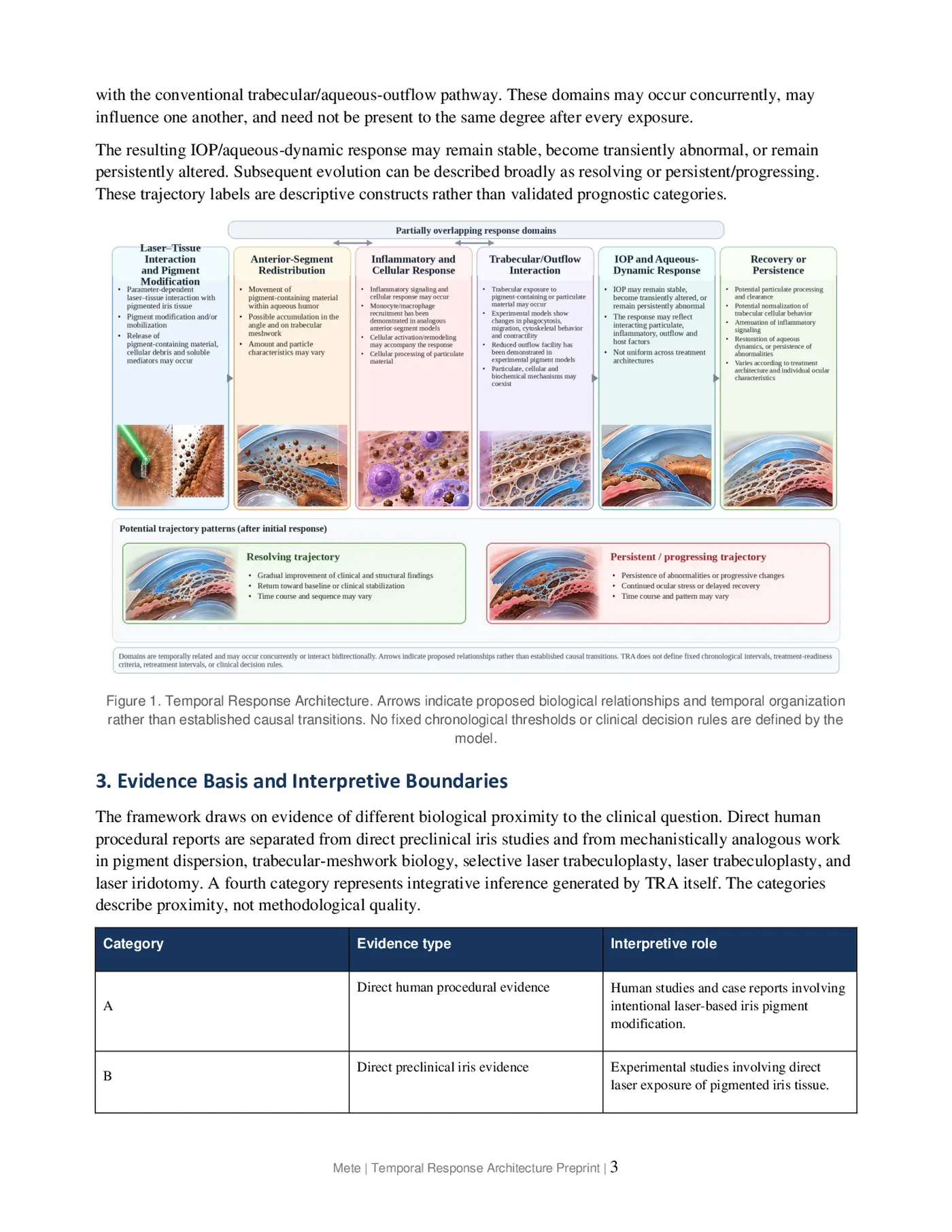
TRA treats the post-exposure response as a set of temporally related and partially overlapping domains rather than a strictly linear sequence. Initial laser–tissue interaction and pigment modification may be followed by varying combinations of anterior-segment redistribution, inflammatory and cellular activity, and interaction with the conventional trabecular/aqueous-outflow pathway. These domains may occur concurrently, may influence one another, and need not be present to the same degree after every exposure.
The resulting IOP/aqueous-dynamic response may remain stable, become transiently abnormal, or remain persistently altered. Subsequent evolution can be described broadly as resolving or persistent/progressing.
These trajectory labels are descriptive constructs rather than validated prognostic categories.
Figure 1. Temporal Response Architecture. Arrows indicate proposed biological relationships and temporal organization rather than established causal transitions. No fixed chronological thresholds or clinical decision rules are defined by the model.
3. Evidence Basis and Interpretive Boundaries
The framework draws on evidence of different biological proximity to the clinical question. Direct human procedural reports are separated from direct preclinical iris studies and from mechanistically analogous work in pigment dispersion, trabecular-meshwork biology, selective laser trabeculoplasty, laser trabeculoplasty, and laser iridotomy. A fourth category represents integrative inference generated by TRA itself. The categories describe proximity, not methodological quality.
Category Evidence type Interpretive role Direct human procedural evidence Human studies and case reports involving A intentional laser-based iris pigment modification.
B Direct preclinical iris evidence Experimental studies involving direct laser exposure of pigmented iris tissue.
Mechanistically analogous evidence Pigment-dispersion, trabecular, outflow, C inflammatory, or clearance models relevant to a TRA domain.
Integrative inference Relationships proposed by TRA that D require direct validation in human iris pigment modification.
This separation is essential because biological plausibility should not be presented as direct clinical proof. For example, experimental studies demonstrate that pigment can alter trabecular phagocytosis, migration, cytoskeletal organization, contractility, and aqueous-outflow behavior, but those findings do not establish identical kinetics following human iris-directed laser exposure.[12–18,22]
4. Pigment–Outflow Interaction and Recovery
Experimental pigment models indicate that conventional outflow disturbance cannot be reduced to passive mechanical obstruction alone. Pigment exposure has been associated with changes in trabecular cellular behavior and outflow facility, while recovery models show active particulate uptake and cellular participation in restoration of outflow function.[12–15] These findings remain mechanistically analogous rather than direct evidence of human post-laser kinetics.
The composition and physical characteristics of experimentally introduced pigment may also differ from material generated during laser interaction with anterior iris tissue. Particle size, melanin packaging, association with cellular fragments, accompanying tissue debris, and inflammatory context may influence the resulting response. Post-laser pressure elevation should therefore not be attributed to pigment burden alone.
Outflow response = f(P, T, I, C, t) P = pigment or particulate burden; T = trabecular cellular response; I = inflammatory signaling; C = processing or clearance capacity; t = time.
This expression is illustrative rather than quantitative. The variables are conceptual domains, not measured coefficients, and the expression has no validated predictive value.
Recovery may likewise involve more than passive disappearance of material. Experimental and analogous evidence supports possible roles for phagocytic processing, monocyte/macrophage recruitment, normalization of trabecular behavior, attenuation of inflammatory signaling, and restoration of aqueous dynamics.[13,18,22]
None has yet been demonstrated as a complete recovery sequence after human laser-based iris pigment modification.
5. Elapsed Time, Ocular State, and Trajectory
A central feature of TRA is the distinction between elapsed time, ocular state, and trajectory. Elapsed time describes the interval since exposure; ocular state describes the condition of the eye at a particular examination; trajectory describes the direction of change across serial observations.
Two eyes with similar findings at a single examination could therefore follow different courses if one is improving while the other remains persistently abnormal or deteriorates. TRA does not assume that trajectory has already been validated as a prognostic marker. Whether serial direction provides clinically useful information beyond isolated measurements remains an empirical question requiring prospective study.
6. Scope and Relationship to Related Frameworks
Within this work, Lumineyes™ refers specifically to a selective, staged, response-guided framework for stromal melanin modulation and is not used interchangeably with the broader and procedurally heterogeneous LID literature. The distinction concerns treatment architecture and clinical approach; it does not constitute a claim of demonstrated comparative superiority, efficacy, or safety.
TRA is conceptually related to, but distinct from, the previously proposed Lumineyes Anterior-Segment Safety Staging framework (L-SAFE) and Lumineyes Optical Architectural Typology (L-OAT).[19,20] L-SAFE addresses safety-oriented interpretation of ocular state and direction of change. L-OAT separates visible phenotype, iris architecture, and OCT-derived measurement domains. Neither framework constitutes empirical validation of TRA. Author-maintained web records are linked on the title page for scholarly navigation and do not constitute independent validation of either framework.
TRA is also not intended as a comprehensive complication taxonomy. Structural iris injury and posterior-segment complications, including iris perforation and laser-associated macular injury, represent additional pathways outside its principal pigment–outflow scope.[23–25]
7. Research Implications
Prospective validation should emphasize serial multimodal measurements rather than isolated postoperative endpoints. Candidate domains include IOP, slit-lamp assessment of anterior-chamber activity, gonioscopy, standardized iris imaging, anterior-segment OCT, corneal endothelial measurements, pachymetry, and objective inflammatory measurements where available. Exact sampling intervals should be derived empirically rather than imposed by the framework.
Three questions are central to validation: whether the proposed domains can be measured reproducibly, whether recurring relationships emerge among them, and whether longitudinal trajectory adds information beyond single measurements. Failure to reproduce the proposed domain relationships, or absence of added information from trajectory assessment, would argue against central assumptions of TRA.
8. Conclusion
TRA provides a hypothesis-generating structure for interpreting post-laser anterior-segment responses without imposing a universal timetable or causal sequence. Its principal contribution is the separation of mechanistic organization, chronological time, ocular state, and longitudinal trajectory within a single testable model.
Prospective serial studies are required before TRA can be regarded as a validated clinical framework.
Research type Mechanistic synthesis and hypothesis-generating framework Publication status Preprint; not peer reviewed Patient-level data None analyzed Clinical validation Not established Version 1.1 (October 2026)
Declarations
Funding: No external funding was received for this work.
Conflict of interest: The author is the developer of the Lumineyes™ framework discussed in this preprint. No external commercial sponsor participated in the conception, literature synthesis, interpretation, or preparation of the work.
Ethics: Not applicable. No individual patient records, identifiable clinical information, or unpublished patient-level datasets were analyzed.
Data availability: No new datasets were generated or analyzed. The synthesis is based on publicly available published literature and the cited sources.
Generative AI disclosure: During manuscript preparation, the author used a generative AI language tool for language refinement, structural editing, and assistance with literature organization. The author critically reviewed and revised all generated content and assumes full responsibility for the accuracy, interpretation, and final content of the manuscript.
References
- Imesch PD, Wallow IHL, Albert DM. The color of the human eye: a review of morphologic correlates and of some conditions that affect iridial pigmentation. Surv Ophthalmol. 1997;41(Suppl 2):S117–S123. doi:10.1016/S0039-6257(97)80018-5.
- Sturm RA, Larsson M. Genetics of human iris colour and patterns. Pigment Cell Melanoma Res. 2009;22(5):544–562. doi:10.1111/j.1755-148X.2009.00606.x.
- D’Oria F, Abu-Mustafa SK, Alio JL. Cosmetic change of the apparent color of the eye: a review on surgical alternatives, outcomes and complications. Ophthalmol Ther. 2022;11(2):465–477. doi:10.1007/s40123-022-00458-2.
- Yildirim Y, Duzgun E, Kar T, et al. Evaluation of color-changing effect and complications after Nd:YAG laser application on iris surface. Med Sci Monit. 2016;22:107–114. doi:10.12659/MSM.895086.
- Basoglu A, Celik U. The effect of SLT laser application on iris to treat sectorial heterochromia: a promising technique. Eye Contact Lens. 2018;44(Suppl 1):S352–S354. doi:10.1097/ICL.0000000000000374.
- Grimaldos Ruiz P. Photoablative cosmetic iridoplasty: effective, safe, and predictable—eye color change in 1176 eyes. Int Ophthalmol. 2021;41(4):1381–1393. doi:10.1007/s10792-021-01693-5.
- Ning B, Baboolal S, Gizzi C, Nolan W. A case of secondary pigment dispersion following laser to cosmetically lighten the irises. J Glaucoma. 2022;31(2):133–135. doi:10.1097/IJG.0000000000001790.
- Ong AY, Ching-A-Sue G, Krilis M, Guirao-Navarro MC, Hornby S, Jutley G. Refractory iatrogenic pigmentary glaucoma secondary to cosmetic laser treatment: a case report. Eur J Ophthalmol. 2023;33(2):NP1–NP4. doi:10.1177/11206721211050338.
- Swampillai AJ, Sherman T, Garg A, Tan IJ, Lim KS. Secondary pigmentary glaucoma following cosmetic laser treatment to alter iris colour. Cont Lens Anterior Eye. 2023;46(2):101754. doi:10.1016/j.clae.2022.101754.
- Liu J, Korban S, Moster MR, Rhéaume MA, Wang Q. Bilateral severe iatrogenic pigmentary glaucoma following laser treatment for cosmetic iris color change. Am J Ophthalmol Case Rep. 2023;32:101927. doi:10.1016/j.ajoc.2023.101927.
- Jaber W, Nemet M, Waisbourd M. Laser iris depigmentation resulting in markedly elevated intraocular pressure and herpes simplex keratitis with corneal scarring: case report. Case Rep Ophthalmol. 2026;17(1):312–317. doi:10.1159/000551226.
- Matsumoto Y, Johnson DH. Trabecular meshwork phagocytosis in glaucomatous eyes. Ophthalmologica. 1997;211(3):147– 152. doi:10.1159/000310782.
- Epstein DL, Freddo TF, Anderson PJ, Patterson MM, Bassett-Chu S. Experimental obstruction to aqueous outflow by pigment particles in living monkeys. Invest Ophthalmol Vis Sci. 1986;27(3):387–395.
- Dang Y, Waxman S, Wang C, et al. Intraocular pressure elevation precedes a phagocytosis decline in a model of pigmentary glaucoma. F1000Res. 2018;7:174. doi:10.12688/f1000research.13797.2.
- Wang C, Dang Y, Loewen RT, et al. Impact of pigment dispersion on trabecular meshwork cells. Graefes Arch Clin Exp Ophthalmol. 2019;257(6):1217–1230. doi:10.1007/s00417-019-04300-7.
- De Keyser M, De Belder M, De Groot V. Randomized prospective study of the use of anti-inflammatory drops after selective laser trabeculoplasty. J Glaucoma. 2017;26(2):e22–e29. doi:10.1097/IJG.0000000000000522.
- Bradley JM, Anderssohn AM, Colvis CM, et al. Mediation of laser trabeculoplasty-induced matrix metalloproteinase expression by IL-1β and TNFα. Invest Ophthalmol Vis Sci. 2000;41(2):422–430.
- Alvarado JA, Katz LJ, Trivedi S, Shifera AS. Monocyte modulation of aqueous outflow and recruitment to the trabecular meshwork following selective laser trabeculoplasty. Arch Ophthalmol. 2010;128(6):731–737. doi:10.1001/archophthalmol.2010.85.
- Mete M. Lumineyes Anterior-Segment Safety Staging (L-SAFE): A Proposed Framework for Pre-Exposure Assessment in Laser Iris Pigment Reduction. Zenodo. 2026. doi:10.5281/zenodo.23046455.
- Mete M. Lumineyes Optical Architectural Typology (L-OAT): a multimodal hypothesis for quantitative iris phenotyping. Zenodo. 2026. doi:10.5281/zenodo.23066772.
- D’Oria F, Alio JL. Surgical techniques for cosmetic eye color change: a narrative review. Ophthalmol Ther. 2025;14(8):1685–1694. doi:10.1007/s40123-025-01177-0.
- Nemoto H, Honjo M, Arai S, Miyazaki T, Aihara M. Apoptosis inhibitor of macrophages/CD5L enhances phagocytosis in the trabecular meshwork cells and regulates ocular hypertension. J Cell Physiol. 2023;238(10):2451–2467. doi:10.1002/jcp.31097.
- Ferrari F. Case report of an iris perforation after YAG laser iris depigmentation for aesthetic reasons. J Fr Ophtalmol. 2021;44(5):e283–e285. doi:10.1016/j.jfo.2020.07.016.
- Flores-Márquez A, Moreno-Gutiérrez JA, Chinchurreta-Capote A, et al. Laser-induced maculopathy after iris depigmentation cosmetic treatment. Can J Ophthalmol. 2023;58(1):e29–e31. doi:10.1016/j.jcjo.2022.05.012.
- Wisdorf K, Hami H, Widder RA, Rössler GF, Kiessling D. Traumatic lamellar macular hole and epiretinal gliosis after cosmetic laser iridoplasty. Ophthalmologie. 2025;122(4):308–311. doi:10.1007/s00347-025-02184-z.
Original figures and tables from the preprint
Each image is a facsimile of the indicated page in the original 2026 Zenodo PDF, not a newly generated scientific figure. The original PDF is authoritative.